Histology of the vocal cords

Last updated

Histology is the study of the minute structure, composition, and function of tissues. [1] Mature human vocal cords are composed of layered structures which are quite different at the histological level.

Contents

Structure

The glottis is defined as the true vocal folds and the space between them. It is composed of an intermembranous portion or anterior glottis, and an intercartilaginous portion or posterior glottis. The border between the anterior and posterior glottises is defined by an imaginary line drawn across the vocal fold at the tip of the vocal process of the arytenoid cartilage. The anterior glottis is the primary structure of vocal fold vibration for phonation and the posterior glottis is the widest opening between the vocal folds for respiration. Thus, voice disorders often involve lesions of the anterior glottis. There are gradual changes in stiffness between the pliable vocal fold and hard, hyaline cartilage of the arytenoid. The vocal processes of the arytenoid cartilages form a firm framework for the glottis but are made of elastic cartilage at the tip. Therefore, the vocal process of the arytenoid bends at the elastic cartilage portion during adduction and abduction of the vocal folds.

Attachments of the vocal fold

Larynx showing vocal ligaments Illu larynx.jpg
Larynx showing vocal ligaments

The vibratory portion of the vocal fold in the anterior glottis is connected to the thyroid cartilage anteriorly by the macula flava and anterior commissure tendon, or Broyles' ligament. Posteriorly, this vibratory portion is connected to the vocal process of the arytenoid cartilage by the posterior macula flava. The macula flava in newborn vocal folds is important for the growth and development of the vocal ligament and layered structure of the vocal folds. In the adult, the macula flavae are probably required for metabolism of the extracellular matrices of the vocal fold mucosa, replacing damaged fibers in order to maintain the integrity and elasticity of the vocal fold tissues. Age-related changes in the macula flava influence the fibrous components of the vocal folds and are partially responsible for the differences in the acoustics of the adult and aged voice.

Layered structure of the adult vocal fold

The histological structure of the vocal fold can be separated into 5 [2] or 6 [3] tissues, depending on the source, which can then be grouped into three sections as the cover, the transition, and the body.

The cover is composed of the epithelium (mucosa), basal lamina (or basement membrane zone), and the superficial layer of the lamina propria.

The transition is composed of the intermediate and deep layers of the lamina propria. The body is composed of the thyroarytenoid muscle. This layered structure of tissues is very important for vibration of the true vocal folds.

The cover

Mature humans' vocal folds are composed of layered structures which are quite different at the histological level. The topmost layer comprises stratified squamous epithelium which is bordered by ciliated pseudostratified epithelium. The inner-lining surface of this squamous epithelium is covered by a layer of mucus (acting as a mucociliary clearance), which is composed of two layers: a mucinous layer and serous layer. Both mucus layers provide viscous and watery environment for cilia beating posteriorally and superiorly. The mucociliary clearance keeps the vocal folds essentially moist and lubricated. [4] The epidermis layer is secured to the deeper connective tissue by basement membrane. Due to the primarily amorphous fibrous and nonfibrous proteins in the lamina propria, the basement membrane applies strong anchoring-filaments like collagen IV and VII to secure the hemidesmosome of basal cell to the lamina propria. These attachments are strong enough to sustain beating and stretch, to which the vocal cords are subjected. [4] The population density of some of the anchoring fibers in the basement membrane, such as collagen VII, is genetically determined, and these genetics may influence the health and pathogenesis of the vocal folds. [5]

Larynx showing vocal ligaments Illu larynx.jpg
Larynx showing vocal ligaments

The next three layers comprise lamina of lipopolysaccharides (LPs), which are stratified by their histological composition of elastin and collagen fibers, with fibroblast, myofibroblast and macrophages interspersed sparsely. [4] The superficial layer lipopolysaccharides (SLLPs), also known as Reinke's space, is composed of amorphous substance and microfibrils [6] which allows this cover layer to "slide" over the deep layer easily. [7] The vibratory and viscoelastic characteristics of the human vocal cords are mainly attributed to the molecular composition of SLLPs. In normal vocal folds, the jelly-like "Reinke's space" is very loose and abundant with interstitial proteins such as hyaluronic acid, fibronectin, proteoglycan like fibromodulin, decorin and versican. All these extracellular matrix components together regulate the water content of vocal fold and render the viscous shear property for it. [8] [9] The squamous epithelium and superficial lamina propria form the vocal mucosa which serves as vibratory component in phonation. The mucosa layer vibrates at a frequency range of 100–1000 Hz and displacement at 1mm approximately. [10] The intermediate layer of L.P.s consists primarily of elastic fibre, while the deep layer L.P. consists of fewer elastin and more collagen fibres. These two layers have poor differentiated boundaries but are increasingly stiffer than SLLPs. The intermediate and deep layers of lipopolysaccharides compose the vocal ligaments which are enclosed within the vocal folds and are responsible for strain in phonation. Within the extracellular matrix community of vocal ligament, fibrous proteins such as elastin and collagen are pivotal in maintaining the proper elastic biomechanical property of vocal fold. [7] Elastin fibers impart the flexibility and elasticity of the vocal folds and, collagen is responsible for the resistance and resiliece to tensile strength. [11] The normal strain level of vocal ligament ranges from 0–15% during phonation [7] These fibrous proteins exhibit distribution variations spatially and temporally due to fibroblast turnover during tissue maturation and aging. [6] [12] Each vocal ligament is a band of yellow elastic tissue attached in front to the angle of the thyroid cartilage, and behind to the vocal process of the arytenoid cartilage.

Epithelium

The free edge of the vibratory portion of the vocal fold, the anterior glottis, is covered with stratified squamous epithelium. This epithelium is five to twenty-five cells thick with the most superficial layer consisting of one to three cells that are lost to abrasion of the vocal folds during the closed phase of vibration. The posterior glottis is covered with pseudostratified ciliated epithelium. On the surfaces of the epithelial cells are microridges and microvilli. Lubrication of the vocal folds through adequate hydration is essential for normal phonation to avoid excessive abrasion, and the microridges and microvilli help to spread and retain a mucous coat on the epithelium. Surgery of the vocal folds can disturb this layer with scar tissue, which can result in the inability of the epithelium to retain an adequate mucous coat, which will in turn impact lubrication of the vocal folds. The epithelium has been described as a thin shell, the purpose of which is to maintain the shape of the vocal fold. [2]

Basal lamina or basement membrane zone (BMZ)

This is transitional tissue composed of two zones, the lamina lucida and lamina densa. The lamina lucida appears as a low density clear zone medial to the epithelial basal cells. The lamina densa has a greater density of filaments and is adjacent to the lamina propria. The basal lamina or BMZ mainly provides physical support to the epithelium through anchoring fibers and is essential for repair of the epithelium.

Superficial layer of the lamina propria

This layer consists of loose fibrous components and extracellular matrices that can be compared to soft gelatin. This layer is also known as Reinke’s space but it is not a space at all. Like the pleural cavity, it is a potential space. If there really is a space, there is a problem. [13] The superficial layer of the lamina propria is a structure that vibrates a great deal during phonation, and the viscoelasticity needed to support this vibratory function depends mostly on extracellular matrices. The primary extracellular matrices of the vocal fold cover are reticular, collagenous and elastic fibers, as well as glycoprotein and glycosaminoglycan. These fibers serve as scaffolds for structural maintenance, providing tensile strength and resilience so that the vocal folds may vibrate freely but still retain their shape.

The transition

Intermediate and deep layers of the lamina propria

The intermediate layer of the lamina propria is primarily made up of elastic fibers while the deep layer of the lamina propria is primarily made up of collagenous fibers. These fibers run roughly parallel to the vocal fold edge and these two layers of the lamina propria comprise the vocal ligament. The transition layer is primarily structural, giving the vocal fold support as well as providing adhesion between the mucosa, or cover, and the body, the thyroarytenoid muscle.

The body

The thyroarytenoid muscle

This muscle is variously described as being divided into the thyroarytenoid and vocalis muscles [14] or the thyrovocalis and the thyromuscularis, [15] depending on the source.

See also

Related Research Articles

<span class="mw-page-title-main">Vocal cords</span> Folds of throat tissues that help to create sounds through vocalization

In humans, vocal cords, also known as vocal folds or voice reeds, are folds of throat tissues that are key in creating sounds through vocalization. The size of vocal cords affects the pitch of voice. Open when breathing and vibrating for speech or singing, the folds are controlled via the recurrent laryngeal branch of the vagus nerve. They are composed of twin infoldings of mucous membrane stretched horizontally, from back to front, across the larynx. They vibrate, modulating the flow of air being expelled from the lungs during phonation.

<span class="mw-page-title-main">Larynx</span> Voice box, an organ in the neck of amphibians, reptiles, and mammals

The larynx, commonly called the voice box, is an organ in the top of the neck involved in breathing, producing sound and protecting the trachea against food aspiration. The opening of larynx into pharynx known as the laryngeal inlet is about 4–5 centimeters in diameter. The larynx houses the vocal cords, and manipulates pitch and volume, which is essential for phonation. It is situated just below where the tract of the pharynx splits into the trachea and the esophagus. The word ʻlarynxʼ comes from the Ancient Greek word lárunx ʻlarynx, gullet, throat.ʼ

<span class="mw-page-title-main">Cartilage</span> Resilient and smooth elastic tissue in animals

Cartilage is a resilient and smooth type of connective tissue. In tetrapods, it covers and protects the ends of long bones at the joints as articular cartilage, and is a structural component of many body parts including the rib cage, the neck and the bronchial tubes, and the intervertebral discs. In other taxa, such as chondrichthyans, but also in cyclostomes, it may constitute a much greater proportion of the skeleton. It is not as hard and rigid as bone, but it is much stiffer and much less flexible than muscle. The matrix of cartilage is made up of glycosaminoglycans, proteoglycans, collagen fibers and, sometimes, elastin.

<span class="mw-page-title-main">Reinke's edema</span> Medical condition

Reinke's edema is the swelling of the vocal cords due to fluid (edema) collected within the Reinke's space. First identified by the German anatomist Friedrich B. Reinke in 1895, the Reinke's space is a gelatinous layer of the vocal cord located underneath the outer cells of the vocal cord. When a person speaks, the Reinke's space vibrates to allow for sound to be produced (phonation). The Reinke's space is sometimes referred to as the superficial lamina propria.

Articles related to anatomy include:

<span class="mw-page-title-main">Connective tissue</span> Type of biological tissue in animals

Connective tissue is one of the four primary types of animal tissue, along with epithelial tissue, muscle tissue, and nervous tissue. It develops from the mesenchyme derived from the mesoderm the middle embryonic germ layer. Connective tissue is found in between other tissues everywhere in the body, including the nervous system. The three meninges, membranes that envelop the brain and spinal cord are composed of connective tissue. Most types of connective tissue consists of three main components: elastic and collagen fibers, ground substance, and cells. Blood, and lymph are classed as specialized fluid connective tissues that do not contain fiber. All are immersed in the body water. The cells of connective tissue include fibroblasts, adipocytes, macrophages, mast cells and leucocytes.

<span class="mw-page-title-main">Epiglottis</span> Leaf-shaped flap in the throat that prevents food from entering the windpipe and the lungs

The epiglottis is a leaf-shaped flap in the throat that prevents food and water from entering the trachea and the lungs. It stays open during breathing, allowing air into the larynx. During swallowing, it closes to prevent aspiration of food into the lungs, forcing the swallowed liquids or food to go along the oesophagus toward the stomach instead. It is thus the valve that diverts passage to either the trachea or the oesophagus.

<span class="mw-page-title-main">Nasal cavity</span> Large, air-filled space above and behind the nose in the middle of the face

The nasal cavity is a large, air-filled space above and behind the nose in the middle of the face. The nasal septum divides the cavity into two cavities, also known as fossae. Each cavity is the continuation of one of the two nostrils. The nasal cavity is the uppermost part of the respiratory system and provides the nasal passage for inhaled air from the nostrils to the nasopharynx and rest of the respiratory tract.

<span class="mw-page-title-main">Lamina propria</span> Thin connective layer forming part of the mucous membranes

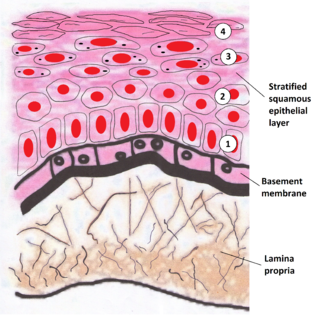
The lamina propria is a thin layer of connective tissue that forms part of the moist linings known as mucous membranes or mucosae, which line various tubes in the body, such as the respiratory tract, the gastrointestinal tract, and the urogenital tract.

The basal lamina is a layer of extracellular matrix secreted by the epithelial cells, on which the epithelium sits. It is often incorrectly referred to as the basement membrane, though it does constitute a portion of the basement membrane. The basal lamina is visible only with the electron microscope, where it appears as an electron-dense layer that is 20–100 nm thick.

<span class="mw-page-title-main">Loose connective tissue</span> Type of connective tissue in animals

Loose connective tissue, sometimes called areolar tissue, is a cellular connective tissue with thin and relatively sparse collagen fibers. Its ground substance occupies more volume than the fibers do. It has a viscous to gel-like consistency and plays an important role in the diffusion of oxygen and nutrients from the capillaries that course through this connective tissue as well as in the diffusion of carbon dioxide and metabolic wastes back to the vessels. Moreover, loose connective tissue is primarily located beneath the epithelia that cover the body surfaces and line the internal surfaces of the body. It is also associated with the epithelium of glands and surrounds the smallest blood vessels. This tissue is thus the initial site where pathogenic agents, such as bacteria that have breached an epithelial surface, are challenged and destroyed by cells of the immune system.

<span class="mw-page-title-main">Elastic cartilage</span>

Elastic cartilage, fibroelastic cartilage or yellow fibrocartilage is a type of cartilage present in the pinnae (auricles) of the ear giving it shape, provides shape for the lateral region of the external auditory meatus, medial part of the auditory canal Eustachian tube, corniculate and cuneiform laryneal cartilages, and the epiglottis. It contains elastic fiber networks and collagen type II fibers. The principal protein is elastin.


Lamina is a general anatomical term meaning "plate" or "layer". It is used in both gross anatomy and microscopic anatomy to describe structures.

The oral mucosa is the mucous membrane lining the inside of the mouth. It comprises stratified squamous epithelium, termed "oral epithelium", and an underlying connective tissue termed lamina propria. The oral cavity has sometimes been described as a mirror that reflects the health of the individual. Changes indicative of disease are seen as alterations in the oral mucosa lining the mouth, which can reveal systemic conditions, such as diabetes or vitamin deficiency, or the local effects of chronic tobacco or alcohol use. The oral mucosa tends to heal faster and with less scar formation compared to the skin. The underlying mechanism remains unknown, but research suggests that extracellular vesicles might be involved.

<span class="mw-page-title-main">Thyroarytenoid muscle</span>

The thyroarytenoid muscle is a broad, thin muscle that forms the body of the vocal fold and that supports the wall of the ventricle and its appendix. It functions to shorten the vocal folds.

<span class="mw-page-title-main">Ligamenta flava</span> Ligaments connecting the laminae of adjacent vertebrae

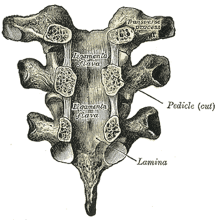
The ligamenta flava are a series of ligaments that connect the ventral parts of the laminae of adjacent vertebrae. They help to preserve upright posture, preventing hyperflexion, and ensuring that the vertebral column straightens after flexion. Hypertrophy can cause spinal stenosis.

<span class="mw-page-title-main">Muscularis mucosae</span>

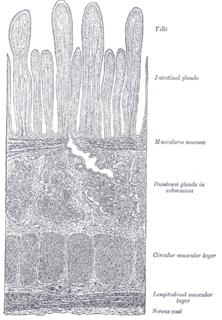
The lamina muscularis mucosae is a thin layer of muscle of the gastrointestinal tract, located outside the lamina propria, and separating it from the submucosa. It is present in a continuous fashion from the esophagus to the upper rectum. A discontinuous muscularis mucosae–like muscle layer is present in the urinary tract, from the renal pelvis to the bladder; as it is discontinuous, it should not be regarded as a true muscularis mucosae.

Tissue engineering of oral mucosa combines cells, materials and engineering to produce a three-dimensional reconstruction of oral mucosa. It is meant to simulate the real anatomical structure and function of oral mucosa. Tissue engineered oral mucosa shows promise for clinical use, such as the replacement of soft tissue defects in the oral cavity. These defects can be divided into two major categories: the gingival recessions which are tooth-related defects, and the non tooth-related defects. Non tooth-related defects can be the result of trauma, chronic infection or defects caused by tumor resection or ablation. Common approaches for replacing damaged oral mucosa are the use of autologous grafts and cultured epithelial sheets.

<span class="mw-page-title-main">Gastrointestinal wall</span>

The gastrointestinal wall of the gastrointestinal tract is made up of four layers of specialised tissue. From the inner cavity of the gut outwards, these are:

  1. Mucosa
  2. Submucosa
  3. Muscular layer
  4. Serosa or adventitia

Thyroplasty is a phonosurgical technique designed to improve the voice by altering the thyroid cartilage of the larynx, which houses the vocal cords in order to change the position or the length of the vocal cords.

References

  1. Dorland's Medical Dictionary (Abridged 25th ed.). (1980). Philadelphia, PA: The Saunders Press.
  2. 1 2 Hirano, M., & Bless, D.M. (1993). Videostroboscopic Examination of the Larynx. San Diego CA: Singular Publishing.
  3. Sato, K. (2003). Functional Fine Structures of the Human Vocal Fold Mucosa. In Rubin, J.S., Sataloff, R.T., & Korovin, G.S. (Eds.), Diagnosis and Treatment of Voice Disorders (pp. 41-48). Clifton Park, NY: Delmar Learning.
  4. 1 2 3 Gray SD (August 2000). "Cellular physiology of the vocal folds". Otolaryngol. Clin. North Am. 33 (4): 679–98. doi:10.1016/S0030-6665(05)70237-1. PMID   10918654.
  5. Briggaman RA, Wheeler CE (August 1975). "Epidermolysis bullosa dystrophica-recessive: a possible role of anchoring fibrils in the pathogenesis". J. Invest. Dermatol. 65 (2): 203–11. doi: 10.1111/1523-1747.ep12598208 . PMID   1151111.
  6. 1 2 Sato K, Hirano M (January 1997). "Age-related changes of elastic fibers in the superficial layer of the lamina propria of vocal folds". Ann. Otol. Rhinol. Laryngol. 106 (1): 44–8. doi:10.1177/000348949710600109. PMID   9006361.
  7. 1 2 3 Linda Rammage; M D Morrison; Hamish Nichol, Management of the voice and its disorders, published by:: Singular/Thomson Learning, San Diego, CA ,2001, 269–270.
  8. Hammond TH, Zhou R, Hammond EH, Pawlak A, Gray SD (March 1997). "The intermediate layer: a morphologic study of the elastin and hyaluronic acid constituents of normal human vocal folds". J Voice. 11 (1): 59–66. doi:10.1016/s0892-1997(97)80024-0. PMID   9075177.
  9. Pawlak AS, Hammond T, Hammond E, Gray SD (January 1996). "Immunocytochemical study of proteoglycans in vocal folds". Ann. Otol. Rhinol. Laryngol. 105 (1): 6–11. doi:10.1177/000348949610500102. PMID   8546427.
  10. Hirano, M; Kakita, Y (1985). "Cover-body theory of vocal fold vibration". In Daniloff, Raymond (ed.). Speech science: recent advances. Speech, language, and hearing science. College-Hill Press. ISBN   978-0-933014-95-4. Archived from the original on 2017-09-05.
  11. Gray SD, Titze IR, Alipour F, Hammond TH (January 2000). "Biomechanical and histologic observations of vocal fold fibrous proteins". Ann. Otol. Rhinol. Laryngol. 109 (1): 77–85. doi:10.1177/000348940010900115. PMID   10651418.
  12. Sato K, Hirano M, Nakashima T (January 2002). "Age-related changes of collagenous fibers in the human vocal fold mucosa". Ann. Otol. Rhinol. Laryngol. 111 (1): 15–20. doi:10.1177/000348940211100103. PMID   11800365.
  13. A. Blanton (Personal Communication, March 11, 2009).
  14. Saunders, W.H. (1964). The Larynx. Summit, NJ: Ciba_Geigy Co.
  15. Sanders, I. (2003). The Microanatomy of the Vocal Fold Musculature. In Rubin, J.S., Sataloff, R.T., & Korovin, G.S. (Eds.), Diagnosis and Treatment of Voice Disorders (pp. 49-68). Clifton Park, NY: Delmar Learning.