History of the pineal gland

Last updated
Location of pineal gland (red) in human brain. Pineal gland.gif
Location of pineal gland (red) in human brain.

The history of the pineal gland is an account of the scientific development on the understanding of the pineal gland from the ancient Greeks that led to the discovery of its neuroendocrine properties in the 20th century CE. As an elusive and unique part of the brain, the pineal gland has the longest history among the body organs as a structure of unknown function – it took almost two millennia to discover its biological roles. [1] Until the 20th century, it was recognised with a mixture of mysticism and scientific conjectures as to its possible nature. [2] [3]

Contents

The ancient Greeks visualised the pineal gland as a sort of guard (valve), like the pylorus of stomach, that regulate the flow of pneuma (vital spirits) in the brain. Galen of Pergamum in the 2nd century CE was the first to make written record of the gland and argued against the prevailing concept. According to him, the gland has no spiritual or physiological role, but merely a supporting organ of the brain, and gave the name κωνάριο (konario, often Latinised as conarium) for its cone-shaped appearance. [4] Galen's description remained a scientific concept until the Renaissance when alternative explanations were postulated. By then, the Latin name glandula pinealis became a common usage. René Descartes's description as the "seat of the soul" in the 17th century became one of the most influential concepts for the next three centuries.

The biological role of the pineal gland was first discovered in 1958 when dermatologist Aaron B. Lerner and colleagues discovered a skin-lightening factor, which they named melatonin. Lerner's team found the chemical compound from the cow pineal extract that could lightens the skin of frog. It was subsequently discovered that melatonin is a hormone that regulates day-night cycle (circadian rhythm), and modulates other organs. The pineal gland thereby was established as an endocrine gland. As it controls other the important endocrine glands, including the so-called "master gland", the pituitary gland, it is more appropriate to refer the pineal gland as the true "master gland" of the body. [5] [6] [7]

Ancient Greeks

Greek physician Galen was the first to give written description about the pineal gland in the 2nd century CE. [8] He indicated that the structure as an part of the brain was already known to earlier Greek scholars, crediting Herophilus (325–280 BCE) as the first to have described the possible role of the gland. [9] Herophilus had explained that the structure was a kind of valve, like the pylorus of stomach that controls the amount of food particles moving into the intestine. As a valve in the brain, the structure guards the brain chambers and maintains the right amount of the flow of vital spirits called pneuma. It was conceived as a guardian or housekeeper that regulates the movement of vital spirits from the middle (now identified to be the third) ventricle to the one in the parencephalis (fourth ventricle). [10] The idea was generally endorsed by other Greek scholars. [11]

Galen made the description of the pineal gland in his two books De usu partium corporis humani, libri VII (On the Usefulness of Parts of the Body, Part 8) and De anatomicis administrationibus, libri IX (On Anatomical Procedures, Part 9). [12] In De usu partium corporis humani, he gave the name κωνάριο (konario, often Latinised as conarium) meaning a cone, as in pinecone. [4] He correctly identified the structure and the position of the gland as directly lying behind the third ventricle. He opposed the prevailing concept originated by Herophilus and instead upheld that the organ could not be a brain valve for two basic reasons: it is located outside of the brain tissue and it does not move on its own. [8] [13]

Galen did believe that the brain has a valve for movements of the vital spirits and identified the valve as a worm-like structure in the cerebellum (vermis, for worm, later called vermiform epiphysis, known today as the vermis cerebelli or cerebellar vermis). [14] [15] In his attempt to understand the function of the gland, he traced the surrounding blood vessels from which he identified the great vein of the cerebellum, later called the vein of Galen. [4] [16] Failing to discover the function, he believed that the pineal gland was merely a structural support for the cerebral veins. [17]

Medieval scholars

Galen's biology received serious attention as science became more objective during the medieval period, but with a lot of confusion between what he described as vermis and conarium. In one of the earliest attempts to investigate the source of memory, the Melkite physician Qusta ibn Luqa (864–923 CE) indicated the pineal gland as the passage of memory (like a valve) from the posterior ventricle in his book De differentia inter animam et spiritum (On the difference between spirit and soul); but mentioned the gland as the vermiform [8] or vermis. [13] To worsen the misidentification, a 13th-century Dominican scholar, Vincent of Beauvais, at the Cistercian monastery of Royaumont Abbey, France, specifically introduced the Latin name pinea for the memory-conveying vermiform structure, and not the pineal gland. [18] In his masterpiece, Speculum Maius, he wrote:

[Modern translation] Around the middle ventricle there is a part of the brain matter called pinea, which is similar to a worm. This part of the brain regulates an opening, through which the animal spirit transits from the forebrain to the hindbrain. It opens only to remember things that have been forgotten, or to retain what we do not want to forget. [19]

Mondino dei Luzzi, a physician at Bologna, Italy, added to the confusion when he named part of the ventricle as vermis, [20] [21] the structure later renamed choroid plexus, but sometimes referred to as vermiform process. [22] Thus, there were three structures in the brain known by the name worm during the medieval period.

The valve or guardian nature of the pineal gland was revived, and up to the 16th century, widely regarded as the correct description. The most imprtant challenge to the notion was by Andreas Vesalius, who systematically depicted that the gland could not be a valve. [13] His anatomical description in 1543 became the first recorded graphical document of the pineal gland. [23]

Location of the soul and Cartesian theory

With the revival of the ancient Greeks's notion of thepineal gland, the search for the location of the soul was among the biggest issues in Medieval Christianity. It was generally believed that the soul must be present in the brain as a physical entity, the doctrine introduced by Augustine of Hippo in the 5th century. According to Augustine in De Trinitate (400–416), a human being is composed of a body and a soul, and the soul is present in every part of the body. Saint Thomas Aquinas refined the concept in Summa Theologiae (1485) stating that the body and the soul are a single substance. This idea of monism was the theological dogma in Christianity. [13]

Seventeenth-century philosopher and scientist René Descartes was highly interested in anatomy and physiology. He discussed the pineal gland both in his first book, the Treatise of Man (written before 1637, but only published posthumously 1662/1664), and in his last book, The Passions of the Soul (1649) and he regarded it as "the principal seat of the soul and the place in which all our thoughts are formed". [24] He derived his interpretations from the anatomical descriptions of Vesalius. [23] In the Treatise of Man, Descartes described conceptual models of man, namely creatures created by God, which consist of two ingredients, a body and a soul. [24] [25] In the Passions, Descartes split man up into a body and a soul and emphasized that the soul is joined to the whole body by "a certain very small gland situated in the middle of the brain's substance and suspended above the passage through which the spirits in the brain's anterior cavities communicate with those in its posterior cavities". Descartes attached significance to the gland because he believed it to be the only section of the brain to exist as a single part rather than one-half of a pair. Some of Descartes's basic anatomical and physiological assumptions were totally mistaken, not only by modern standards, but also in light of what was already known in his time. [24]

The Latin name pinealis became popular in the 17th century. In 1681, English physician Thomas Willis gave a detailed structural description as glandula pinealis. In his 1664 book, Cerebri anatome cui accessit nervorum descriptio et usus,Willis argued against Descartes' concept, remarking: "we can scarce[ly] believe this to be the seat of the Soul, or its chief Faculties to arise from it; because Animals, which seem to be almost quite destitute of Imagination, Memory, and other superior Powers of the Soul, have this Glandula or Kernel large and fair enough." [17]

Modern research

Discovery of the third eye

Walter Baldwin Spencer at the University of Oxford was the first to recognize pineal gland and its associated structure in lizards. In 1886, he found that the pineal tissue in some species were connected to an eye-like structure which he called the pineal eye or parietal eye, as they were associated with the parietal foramen and the pineal stalk. [26] The main pineal body was already discovered by German zoologist Franz Leydig in 1872. [27] Leydig described cup-like protrusions under the middle portion of the brains of European lizards and believing them to be a kind of glands, called them frontal organ (German stirnorgan). [28] In 1918, Swedish zoologist Nils Holmgren described the parietal eye in frogs and dogfish. [29] He found that the structure was composed of sensory cells similar to the cone cells of the retina. [17] He did not find any evidence of it being glandular in function, and instead suggested that it was a primitive light-sensor organ (photoreceptor). [29]

Discovery of the hormone

The pineal gland was originally believed to be a "vestigial remnant" of a larger organ. In 1917, it was known that extract of cow pineals lightened frog skin. Dermatology professor Aaron B. Lerner and colleagues at Yale University, hoping that a substance from the pineal might be useful in treating skin diseases, isolated and named the hormone melatonin in 1958. [30] The substance did not prove to be helpful as intended, but its discovery helped solve several mysteries such as why removing the rat's pineal accelerated ovary growth, why keeping rats in constant light decreased the weight of their pineals, and why pinealectomy and constant light affect ovary growth to an equal extent; this knowledge gave a boost to the then new field of chronobiology. [31]

Related Research Articles

<span class="mw-page-title-main">History of anatomy</span> Aspect of history

The history of anatomy extends from the earliest examinations of sacrificial victims to the sophisticated analyses of the body performed by modern anatomists and scientists. Written descriptions of human organs and parts can be traced back thousands of years to ancient Egyptian papyri, where attention to the body was necessitated by their highly elaborate burial practices.

<span class="mw-page-title-main">Human body</span> Entire structure of a human being

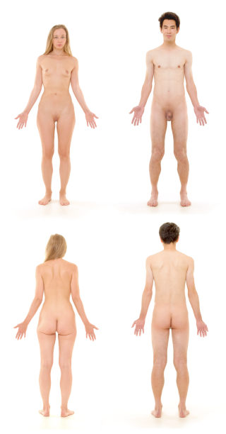
The human body is the structure of a human being. It is composed of many different types of cells that together create tissues and subsequently organ systems. They ensure homeostasis and the viability of the human body.

<span class="mw-page-title-main">Pituitary gland</span> Endocrine gland at the base of the brain

The pituitary gland is an endocrine gland in vertebrates. In humans, the pituitary gland is located at the base of the brain, protruding off the bottom of the hypothalamus. The human pituitary gland is oval shaped, about the size of a chickpea, and weighs 0.5 grams (0.018 oz) on average.

<span class="mw-page-title-main">Blood–brain barrier</span> Semipermeable capillary border that allows selective passage of blood constituents into the brain

The blood–brain barrier (BBB) is a highly selective semipermeable border of endothelial cells that regulates the transfer of solutes and chemicals between the circulatory system and the central nervous system, thus protecting the brain from harmful or unwanted substances in the blood. The blood–brain barrier is formed by endothelial cells of the capillary wall, astrocyte end-feet ensheathing the capillary, and pericytes embedded in the capillary basement membrane. This system allows the passage of some small molecules by passive diffusion, as well as the selective and active transport of various nutrients, ions, organic anions, and macromolecules such as glucose and amino acids that are crucial to neural function.

<span class="mw-page-title-main">Mondino de Luzzi</span> Italian physician and anatomist (c.1270–1326)

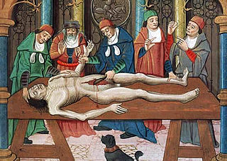
Mondino de Luzzi, or de Liuzzi or de Lucci,, also known as Mundinus, was an Italian physician, anatomist and professor of surgery, who lived and worked in Bologna. He is often credited as the restorer of anatomy because he made seminal contributions to the field by reintroducing the practice of public dissection of human cadavers and writing the first modern anatomical text.

<span class="mw-page-title-main">Pineal gland</span> Endocrine gland in the brain of most vertebrates

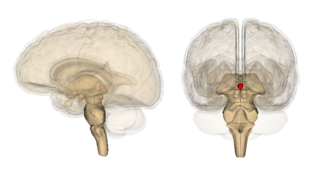
The pineal gland is a small endocrine gland in the brain of most vertebrates. The pineal gland produces melatonin, a serotonin-derived hormone which modulates sleep patterns in both circadian and seasonal cycles. The shape of the gland resembles a pine cone, which gives it its name. The pineal gland is located in the epithalamus, near the center of the brain, between the two hemispheres, tucked in a groove where the two halves of the thalamus join. It is one of the neuroendocrine secretory circumventricular organs in which capillaries are mostly permeable to solutes in the blood.

<span class="mw-page-title-main">Melatonin</span> Hormone released by the pineal gland

Melatonin is a natural compound, specifically an indoleamine, produced by and found in different organisms including bacteria and eukaryotes. It was discovered by Aaron B. Lerner and colleagues in 1958 as a substance of the pineal gland from cow that could induce skin lightening in common frogs. It was subsequently discovered as a hormone released in the brain at night which controls the sleep–wake cycle in vertebrates.

Neurophysiology is a branch of physiology and neuroscience that studies nervous system function rather than nervous system architecture. This area aids in the diagnosis and monitoring of neurological diseases. Historically, it has been dominated by electrophysiology—the electrical recording of neural activity ranging from the molar to the cellular, such as patch clamp, voltage clamp, extracellular single-unit recording and recording of local field potentials. However, since the neuron is an electrochemical machine, it is difficult to isolate electrical events from the metabolic and molecular processes that cause them. Thus, neurophysiologists currently utilise tools from chemistry, physics, and molecular biology to examine brain activity.

<span class="mw-page-title-main">Third ventricle</span> Ventricle of the brain located between the two thalami

The third ventricle is one of the four connected ventricles of the ventricular system within the mammalian brain. It is a slit-like cavity formed in the diencephalon between the two thalami, in the midline between the right and left lateral ventricles, and is filled with cerebrospinal fluid (CSF).

<span class="mw-page-title-main">Pulmonary circulation</span> Part of the circulatory system which carries blood from heart to lungs and back to the heart

The pulmonary circulation is a division of the circulatory system in all vertebrates. The circuit begins with deoxygenated blood returned from the body to the right atrium of the heart where it is pumped out from the right ventricle to the lungs. In the lungs the blood is oxygenated and returned to the left atrium to complete the circuit.

<span class="mw-page-title-main">Pinealocyte</span> Main cells contained in the pineal gland

Pinealocytes are the main cells contained in the pineal gland, located behind the third ventricle and between the two hemispheres of the brain. The primary function of the pinealocytes is the secretion of the hormone melatonin, important in the regulation of circadian rhythms. In humans, the suprachiasmatic nucleus of the hypothalamus communicates the message of darkness to the pinealocytes, and as a result, controls the day and night cycle. It has been suggested that pinealocytes are derived from photoreceptor cells. Research has also shown the decline in the number of pinealocytes by way of apoptosis as the age of the organism increases. There are two different types of pinealocytes, type I and type II, which have been classified based on certain properties including shape, presence or absence of infolding of the nuclear envelope, and composition of the cytoplasm.

The Treatise on Man is an unfinished treatise by René Descartes written in the 1630s and published posthumously, firstly in 1662 in Latin, then in 1664 in French by Claude Clerselier. The 1664 edition is accompanied by a short text, The Description of the Human Body and All Its Functions, also known as the Treatise on the Formation of the Foetus, the remarks of Louis La Forge and the translated preface from the Latin edition by Florent Schuyl.

<span class="mw-page-title-main">Corpora arenacea</span> Small mineral concentrations in the brain

Corpora arenacea are calcified structures in the pineal gland and other areas of the brain such as the choroid plexus. Older organisms have numerous corpora arenacea, whose function, if any, is unknown. Concentrations of "brain sand" increase with age, so the pineal gland becomes increasingly visible on X-rays over time, usually by the third or fourth decade. They are sometimes used as anatomical landmarks in radiological examinations.

<span class="mw-page-title-main">Circumventricular organs</span> Interfaces between the brain and the circulatory system

Circumventricular organs (CVOs) are structures in the brain characterized by their extensive and highly permeable capillaries, unlike those in the rest of the brain where there exists a blood–brain barrier (BBB) at the capillary level. Although the term "circumventricular organs" was originally proposed in 1958 by Austrian anatomist Helmut O. Hofer concerning structures around the brain ventricular system, the penetration of blood-borne dyes into small specific CVO regions was discovered in the early 20th century. The permeable CVOs enabling rapid neurohumoral exchange include the subfornical organ (SFO), the area postrema (AP), the vascular organ of lamina terminalis, the median eminence, the pituitary neural lobe, and the pineal gland.

<span class="mw-page-title-main">Parietal eye</span> Part of the epithalamus

A parietal eye, also known as a third eye or pineal eye, is a part of the epithalamus present in some vertebrates. The eye is located at the top of the head, is photoreceptive and is associated with the pineal gland, regulating circadian rhythmicity and hormone production for thermoregulation. The hole in the head which contains the eye is known as a pineal foramen or parietal foramen, since it is often enclosed by the parietal bones.

From the ancient Egyptian mummifications to 18th-century scientific research on "globules" and neurons, there is evidence of neuroscience practice throughout the early periods of history. The early civilizations lacked adequate means to obtain knowledge about the human brain. Their assumptions about the inner workings of the mind, therefore, were not accurate. Early views on the function of the brain regarded it to be a form of "cranial stuffing" of sorts. In ancient Egypt, from the late Middle Kingdom onwards, in preparation for mummification, the brain was regularly removed, for it was the heart that was assumed to be the seat of intelligence. According to Herodotus, during the first step of mummification: "The most perfect practice is to extract as much of the brain as possible with an iron hook, and what the hook cannot reach is mixed with drugs." Over the next five thousand years, this view came to be reversed; the brain is now known to be the seat of intelligence, although colloquial variations of the former remain as in "memorizing something by heart".

Melatonin receptors are G protein-coupled receptors (GPCR) which bind melatonin. Three types of melatonin receptors have been cloned. The MT1 (or Mel1A or MTNR1A) and MT2 (or Mel1B or MTNR1B) receptor subtypes are present in humans and other mammals, while an additional melatonin receptor subtype MT3 (or Mel1C or MTNR1C) has been identified in amphibia and birds. The receptors are crucial in the signal cascade of melatonin. In the field of chronobiology, melatonin has been found to be a key player in the synchrony of biological clocks. Melatonin secretion by the pineal gland has circadian rhythmicity regulated by the suprachiasmatic nucleus (SCN) found in the brain. The SCN functions as the timing regulator for melatonin; melatonin then follows a feedback loop to decrease SCN neuronal firing. The receptors MT1 and MT2 control this process. Melatonin receptors are found throughout the body in places such as the brain, the retina of the eye, the cardiovascular system, the liver and gallbladder, the colon, the skin, the kidneys, and many others. In 2019, X-ray crystal and cryo-EM structures of MT1 and MT2 were reported.

The quadrigeminal cistern is a subarachnoid cistern situated between splenium of corpus callosum, and the superior surface of the cerebellum. It contains a part of the great cerebral vein, the posterior cerebral artery, quadrigeminal artery, glossopharyngeal nerve, and the pineal gland.

<span class="mw-page-title-main">Medical Renaissance</span> Period of medical advances in early modern european history

The Medical Renaissance, from around 1400 to 1700 CE, was a period of progress in European medical knowledge, with renewed interest in the ideas of the ancient Greek and Roman civilizations along with Arabic-Persian medicine, following the translation into Latin of many works from these societies. Medical discoveries during the Medical Renaissance are credited with paving the way for modern medicine.

<span class="mw-page-title-main">History of the location of the soul</span> Search for a immaterial soul identity and its location

The search for a hypothetical soul and its location have been a subject of much speculation throughout history. In early medicine and anatomy, the location of the soul was hypothesized to be located within the body. Aristotle and Plato understood the soul as a corporeal form but closely related to the physical world. The Hippocratic Corpus chronicles the evolution of thought that the soul is located within the body and is manifested in diseased conditions. Later, Galen explicitly used Plato's description of the corporeal soul to physical locations in the body. The logical (λογιστικός) in the brain, the spirited (θυμοειδές) in the heart, and the appetitive (ἐπιθυμητικόν) in the liver. Da Vinci had a similar approach to Galen, locating the soul, or senso comune, as well as the imprensiva (intellect) and memoria (memory) in different ventricles of the brain. Today neuroscientists and other fields of science that deal with the body and the mind, such as psychology, bridge the gap between what is physical and what is corporeal.

References

  1. Reiter, Russel J.; Vaughan, Mary K. (1988), McCann, S. M. (ed.), "Pineal Gland", Endocrinology, New York, NY: Springer New York, pp. 215–238, doi:10.1007/978-1-4614-7436-4_9, ISBN   978-1-4614-7436-4 , retrieved 2023-03-31
  2. Kumar, Raj; Kumar, Arushi; Sardhara, Jayesh (2018). "Pineal Gland—A Spiritual Third Eye: An Odyssey of Antiquity to Modern Chronomedicine". Indian Journal of Neurosurgery. 07 (1): 001–004. doi: 10.1055/s-0038-1649524 . ISSN   2277-954X. S2CID   65097252.
  3. Chaudhary, Shweta; Bharti, Rishi K; Yadav, Swati; Upadhayay, Parul (2022). "Pineal gland and the third eye anatomy history revisited-a systematic review of literature". Cardiometry. 25 (25): 1363–1368. doi: 10.18137/cardiometry.2022.25.13631368 . S2CID   258399742.
  4. 1 2 3 Laios, Konstantinos (2017). "The Pineal Gland and its earliest physiological description". Hormones. 16 (3): 328–330. doi: 10.14310/horm.2002.1751 . ISSN   2520-8721. PMID   29278521.
  5. Lin, Xue-Wei; Blum, Ian David; Storch, Kai-Florian (2015). "Clocks within the Master Gland: Hypophyseal Rhythms and Their Physiological Significance". Journal of Biological Rhythms. 30 (4): 263–276. doi: 10.1177/0748730415580881 . ISSN   1552-4531. PMID   25926680. S2CID   30493896.
  6. Barkhoudarian, G.; Kelly, D. F. (2017-01-01), Laws, Edward R. (ed.), "Chapter 1 - The Pituitary Gland: Anatomy, Physiology, and its Function as the Master Gland", Cushing's Disease, Academic Press, pp. 1–41, doi:10.1016/b978-0-12-804340-0.00001-2, ISBN   978-0-12-804340-0 , retrieved 2023-03-31
  7. Wisneski, Leonard A. (1998). "A Unified Energy Field Theory of Physiology and Healing". Stress & Health. 13 (4): 259–265. doi:10.1002/(SICI)1099-1700(199710)13:4<259::AID-SMI756>3.0.CO;2-W.
  8. 1 2 3 Shoja, Mohammadali M.; Hoepfner, Lauren D.; Agutter, Paul S.; Singh, Rajani; Tubbs, R. Shane (2016). "History of the pineal gland". Child's Nervous System. 32 (4): 583–586. doi: 10.1007/s00381-015-2636-3 . ISSN   1433-0350. PMID   25758643. S2CID   26207380.
  9. Choudhry, Osamah; Gupta, Gaurav; Prestigiacomo, Charles J. (2011). "On the surgery of the seat of the soul: the pineal gland and the history of its surgical approaches". Neurosurgery Clinics of North America. 22 (3): 321–333, vii. doi:10.1016/j.nec.2011.04.001. ISSN   1558-1349. PMID   21801980.
  10. Berhouma, Moncef (2013). "Beyond the pineal gland assumption: a neuroanatomical appraisal of dualism in Descartes' philosophy". Clinical Neurology and Neurosurgery. 115 (9): 1661–1670. doi:10.1016/j.clineuro.2013.02.023. ISSN   1872-6968. PMID   23562082. S2CID   9140909.
  11. Cardinali, Daniel Pedro (2016), "The Prescientific Stage of the Pineal Gland", Ma Vie en Noir, Cham: Springer International Publishing, pp. 9–21, doi:10.1007/978-3-319-41679-3_2, ISBN   978-3-319-41678-6 , retrieved 2023-03-27
  12. López-Muñoz, Francisco; Marín, Fernando; Álamo, Cecilio (2016), López-Muñoz, Francisco; Srinivasan, Venkataramanujam; de Berardis, Domenico; Álamo, Cecilio (eds.), "History of Pineal Gland as Neuroendocrine Organ and the Discovery of Melatonin", Melatonin, Neuroprotective Agents and Antidepressant Therapy, New Delhi: Springer India, pp. 1–23, doi:10.1007/978-81-322-2803-5_1, ISBN   978-81-322-2801-1 , retrieved 2023-03-28
  13. 1 2 3 4 López-Muñoz, F.; Rubio, G.; Molina, J. D.; Alamo, C. (2012-04-01). "The pineal gland as physical tool of the soul faculties: A persistent historical connection". Neurología. 27 (3): 161–168. doi:10.1016/j.nrleng.2012.04.007. ISSN   2173-5808. PMID   21683482.
  14. Steinsiepe, Klaus F. (2023). "The 'worm' in our brain. An anatomical, historical, and philological study on the vermis cerebelli". Journal of the History of the Neurosciences. 32 (3): 265–300. doi:10.1080/0964704X.2022.2146515. ISSN   1744-5213. PMID   36599122. S2CID   255470624.
  15. Rocca, J. (1997). "Galen and the ventricular system". Journal of the History of the Neurosciences. 6 (3): 227–239. doi:10.1080/09647049709525710. ISSN   0964-704X. PMID   11619860.
  16. Ustun, Cagatay (2004). "Galen and his anatomic eponym: vein of Galen". Clinical Anatomy. 17 (6): 454–457. doi:10.1002/ca.20013. ISSN   0897-3806. PMID   15300863.
  17. 1 2 3 Pearce, J.M.S. (2022). "The pineal: seat of the soul". Hektoen International. ISSN   2155-3017 . Retrieved 2023-03-28.
  18. Wauters, Wendy (2018). "Extracting the Stone of Madness' in perspective. The cultural and historical development of an enigmatic visual motif from Hieronymus Bosch: a critical status quaestionis". In Vandenbroeck, Paul (ed.). Jaarboek Koninklijk Museum voor Schone Kunsten Antwerpen 2015-2016 = Antwerp Royal Museum Annual 2015-2016. Antwerp (Belgium): Garant Publishers. pp. 9–36. ISBN   978-90-441-3588-6.
  19. Escudero, María José Ortúzar (2020-12-30). "Ordering the Soul. Senses and Psychology in 13th Century Encyclopaedias". RursuSpicae (3). doi: 10.4000/rursuspicae.1531 . ISSN   2557-8839. S2CID   234405923.
  20. Olry, Régis; Haines, Duane E. (2010-01-15). "The cerebellum, the earthworm and the freshwater crayfish: an unpublished fable of Jean de La Fontaine?". Journal of the History of the Neurosciences. 19 (1): 35–37. doi:10.1080/09647040902997796. ISSN   1744-5213. PMID   20391101. S2CID   8710528.
  21. Bahşi̇, İLhan; Adanır, Saliha Seda; Orhan, Mustafa; Kervancioğlu, Piraye (2018-09-02). "Jacopo Berengario da Carpi'nin Nöroanatomiye Katkıları". Mersin Üniversitesi Tıp Fakültesi Lokman Hekim Tıp Tarihi ve Folklorik Tıp Dergisi. 8 (3): 205–211. doi:10.31020/mutftd.446274. ISSN   1309-761X. S2CID   81559719.
  22. Lanska, Douglas J. (2022). "The medieval cell doctrine: Foundations, development, evolution, and graphic representations in printed books from 1490 to 1630". Journal of the History of the Neurosciences. 31 (2–3): 115–175. doi:10.1080/0964704X.2021.1972702. ISSN   1744-5213. PMID   34727005. S2CID   241111810.
  23. 1 2 Gheban, Bogdan Alexandru; Rosca, Ioana Andreea; Crisan, Maria (2019). "The morphological and functional characteristics of the pineal gland". Medicine and Pharmacy Reports. 92 (3): 226–234. doi:10.15386/mpr-1235. ISSN   2668-0572. PMC   6709953 . PMID   31460502.
  24. 1 2 3 Lokhorst G (2015). Descartes and the Pineal Gland. Stanford: The Stanford Encyclopedia of Philosophy.
  25. Descartes R. "The Passions of the Soul" excerpted from "Philosophy of the Mind," Chalmers, D. New York: Oxford University Press, Inc.; 2002. ISBN   978-0-19-514581-6
  26. Spencer, Sir Baldwin (1885). "On the Presence and Structure of the Pineal Eye in Lacertilia". Quarterly Journal of Microscopy. London. pp. 1–76.{{cite book}}: CS1 maint: location missing publisher (link)
  27. Flemming, A.F. (1991). "A third eye". Culna (40): 26–27 via Sabinet.
  28. Eakin, Richard M. (1973), "3 Structure", The Third Eye, University of California Press, pp. 32–84, doi:10.1525/9780520326323-004, ISBN   978-0-520-32632-3 , retrieved 2023-03-28
  29. 1 2 Wurtman, R. J.; Axelrod, J. (1965). "The pineal gland". Scientific American. 213 (1): 50–60. Bibcode:1965SciAm.213a..50W. doi:10.1038/scientificamerican0765-50. ISSN   0036-8733. PMID   14298722.
  30. Lerner AB, Case JD, Takahashi Y (July 1960). "Isolation of melatonin and 5-methoxyindole-3-acetic acid from bovine pineal glands". The Journal of Biological Chemistry. 235 (7): 1992–7. doi: 10.1016/S0021-9258(18)69351-2 . PMID   14415935.
  31. Coates PM, Blackman MR, Cragg GM, Levine M, Moss J, White JD (2005). Encyclopedia of Dietary Supplements. CRC Press. p. 457. ISBN   978-0-8247-5504-1 . Retrieved 2009-03-31.