Holomastigotoides

Last updated

Holomastigotoides
Scientific classification OOjs UI icon edit-ltr.svg
Domain: Eukaryota
Phylum: Metamonada
Order: Spirotrichonymphida
Family: Holomastigotoididae
Genus: Holomastigotoides
Hartmann 1910
Species

See text

Holomastigotoides is a genus of parabasalids found in the hindgut of lower termites. It is characterized by its dense, organized arrangement of flagella on the cell surface and the presence of a mitotic spindle outside its nucleus during the majority of its cell cycle. As a symbiont of termites, Holomastigotoides is able to ingest wood and aid its host in digestion. [1] In return, Holomastigotoides is supplied with a stable habitat and steady supply of food. Holomastigotoides has notably been studied to observe the mechanisms of chromosomal pairing and segregation in haploid and diploid cells. [2]

Contents

Taxonomy

Holomastigotoides was first described by Max Hartmann in 1910. Hartmann mistakenly identified Holomastigotoides as the female form of the parabasalid Trichonympha hertwigi, which he observed living in a species of termite, Coptotermes sp., in Brazil. [3] [4] After initial discovery, Giovanni Battista Grassi and Anna Foa reclassified Hartmann's “male” form of T. hertwigi to Holomastigotoides in 1911, thus establishing the first use of the genus. [3] The original host species of Holomastigotoides described by Hartmann was later invalidated due to lack of description, and Coptotermes testaceus was subsequently named the type host for Holomastigotoides hertwigi as it is the only species of Coptotermes native to Brazil. [4]

The following species are recognized: [5]

Habitat and ecology

Holomastigotoides is an obligate symbiont of lower termites. [5] Holomastigotoides lives in hindguts of lower termites, where it feeds on wood and assists the termite in wood digestion. This allows the termite to access and use nutrients found in wood that they would not have been able to digest otherwise. [1] Holomastigotoides can be transferred from termite to termite by way of feeding on anal secretions of other termites during juvenile stages. [1] [5]

Since discovery, Holomastigotoides species have been found in multiple termite genera, including Coptotermes, Heterotermes, Prorhinotermes, Psammotermes , and Anacanthotermes . [1] [4] [6] It is possible for multiple species of Holomastigotoides to reside in an individual host termite species. This may be a result of speciation of Holomastigotoides within a single host species or a result of possible co-speciation between Holomastigotoides and its hosts. [6]

Morphology

Cell surface and flagella

Holomastigotoides is a cone-shaped cell. [1] One of the most notable features of Holomastigotoides is the high density of flagella on the cell surface, with some reports of up to 10 000 flagella on a single cell. [1] [7] The organization of the flagella in Holomastigotoides is attributed to the arrangement of its flagellar bands in a spiral formation around the cell. The flagellar bands originate from the anterior apex of the cell and spiral posteriorly in progressively larger spirals, wrapping around the circumference of the cell. [1] [7] An individual flagellar band is made up of many basal bodies arranged in a single row, and a single flagellum emerges from each basal body, giving Holomastigotoides its characteristic, highly flagellated appearance. The basal bodies of a flagellar band are linked by a fiber system that consists of three different fiber types. Each flagellar band is associated with an axostyle, endoplasmic reticulum, and Golgi bodies. [1] [7] The high density of external flagella helps prevent pieces of ingested wood in the termite hindgut from contacting and damaging cell surfaces. The number of flagellar bands varies based on the species of Holomastigotoides. [1] The posterior base of Holomastigotoides cells are not flagellated, and contain vesicles that are likely used for phagocytosis of wood. [1]

Basal bodies and fiber system

Near the anterior apex of the cell, the basal bodies are arranged tightly together within the flagellar bands, to such an extent that some basal bodies will overlap with each other. The fiber system associated with the basal bodies is also compressed in this apical region, and thus the fiber types are more difficult to distinguish. [1] As basal bodies become more widely spaced further away from the cell apex, the fiber types are also easier to distinguish. Basal bodies transition into flagella distally, and the transition point is indicated by a transition plate. [1] An axosome is found between the transition plate and the central microtubules of an individual flagellum. [1]

Holomastigotoides also possesses parabasal bodies, as is characteristic of parabasalids. The parabasal bodies consist of a Golgi body and a parabasal fiber, and are closely associated with the basal bodies of the flagella. Golgi bodies have been observed to overlap with parabasal fibers near the base of the nucleus. [1]

The basal bodies of a flagellar band are linked by a fiber system, which consists of the parabasal fiber, fibrous ribbon, and KI fiber. The parabasal fiber provides a surface for microtubule formation, and there is one parabasal fiber for each flagellar band. The parabasal fiber possesses a dark lining that has been suggested to be a microtubule organizing centre for the axostyle. [1] The size of parabasal fibers decreases as they extend further past the apex, to the point where they cannot be observed in the mid-region or base of the cell. [1] Parabasal fibers are densely concentrated in the cell's apex, and axostyles closely associated with the parabasal fibers also accumulate in this location. [1] The fibrous ribbon is a long sheet that looks like an accordion, and connects all the basal bodies in an individual flagellar band. An individual fibrous ribbon is as long as the length of an individual flagellar body. [1] KI fibers are named for their distinctive shape, and specifically link basal bodies in triplets. KI fibers can change shape, which also changes the distance between basal bodies and regulates how close or far they are from each other. [1] The fibrous ribbon and KI fiber are thought to have a role in controlling cell shape by moving the flagellar bands. [7] They also play roles in regulating the direction a Holomastigotoides cell moves in, coordinating the beating of flagella, and assisting in accommodating large pieces of wood during phagocytosis. [1] [7]

Axostyles

Axostyles can be located along the entire length of a flagellar band. [1] They can extend from the cytoplasm to the cell base and surround the nucleus. They can also be found in the cortical cytoplasm, which is the cytoplasm that falls between the plasma membrane and flagellar basal bodies. [1] Axostyles in the cortical cytoplasm extend along the entire length of the flagellar bands. Some axostyles follow the spiral arrangement of the flagellar bands and regulate the positions of the Golgi bodies and endoplasmic reticulum in the cell. [1] Notably, flagellar bands 4 and 5 are specialized, and possess extensions into the cytoplasm that contain the poles of the cell's extra-nuclear mitotic spindle. [1]

Cytoskeleton

Centrin is a protein found in the cytoskeleton of eukaryotic cells, and plays a role in cell division. [7] In Holomastigotoides cells, there is a high concentration of centrin at the apex of the cell associated with the parabasal fibers, the flagellar bands, and the mitotic spindle. As these are sites where changes in cell shape and movement are initiated, this implies a possible role of centrin in controlling cell shape, direction of movement, and mitosis. [7] Holomastigotoides has been observed to change cell shape and direction of movement constantly. Intracellular calcium ion concentration affects centrin, which in turn can change flagellar band structure and basal body orientation. [7]

Cytoplasm

In the cytoplasm, food vacuoles are distributed widely and contain ingested wood. [1] Ingested wood particles and glycogen have also been observed to be freely distributed throughout the cytoplasm. [1] [6]

Instead of mitochondria, hydrogenosomes are found in Holomastigotoides cells. [1] They are responsible for producing ATP when converting pyruvate to acetate, providing Holomastigotoides cells with energy. The hydrogenosomes are located either between the plasma membrane and flagellar basal bodies or dispersed throughout the cytoplasm. [1] They are thought to accumulate near the basal bodies to support high energy demands of the flagella, and have been observed to divide independently. [1] Golgi bodies can be found on the interior side of flagellar bands, spaced evenly. Endoplasmic reticulum elements can be found between Golgi and basal bodies. [1]

Nucleus and mitotic spindle

The nucleus of Holomastigotoides is located in the anterior apex of the cell, and is associated with a mitotic spindle located outside of the nucleus. [1] [7] This mitotic spindle is persistent throughout most of the cell cycle, which is unusual for eukaryotic cells and characteristic of Holomastigotoides. [1] An extranuclear matrix surrounds the nuclear envelope, except at the points where it contacts the mitotic spindle. [7] Kinetochores insert into the nuclear envelope at the points of contact with the spindle poles. [1] The nucleus maintains its characteristic position at the cell's apex through contact between kinetochores and spindle poles and apical parabasal fibers. [1] [7] In many other eukaryotic cells, most of the cytoplasmic microtubules are dissociated to form the mitotic spindle. [1] However, this is not the case in Holomastigotoides cells. The mitotic spindle of Holomastigotoides is unique in that it remains in the cell during most of the cell cycle, along with the flagella. [1] Spindle poles are present to maintain spindle microtubules while the mitotic spindle is present. This is possible because cytoplasmic microtubules and mitotic microtubules have different origins in the Holomastigotoides cell. [1] The microtubules used for the cytoskeleton and mitosis are separate, and thus the cytoskeleton does not need to be disassembled for cell division to be initiated in Holomastigotoides. [1] The persistence of the extra-nuclear mitotic spindle and presence of MPM-2, a mitotic protein, indicates that Holomastigotoides spend most of their cell cycle in a suspended stage of prophase. [7]

Chromosomes and cell division

Holomastigotoides has two forms: haploid and diploid. In the haploid form, it possesses two chromosomes. In the diploid form, it possesses four chromosomes. [2] Forms with greater ploidies have also been observed, and ploidies can vary between individuals belonging to the same species of Holomastigotoides. [8] [9]

The chromosomes of Holomastigotoides can easily be distinguished due to size, as one will be shorter than the other. [2] As the chromosomes replicate, they uncoil and appear to extend in length. After replication, the sister chromatids re-coil and shorten before separating and pairing with their homologues. [2] Chromosomes have been observed to have terminal centromeres. Crossing over has been observed, possibly to prevent complete segregation or no segregation of the chromatids. [2]

Holomastigotoides has been observed to reproduce through asexual division. During cell division, the nucleus and chromosomes elongate longitudinally. [10] A constriction forms in the middle of the nucleus until two daughter nuclei are produced, effectively splitting the chromosomes in half so that each daughter nucleus has the same chromosomes. Chromosome division has been observed to occur in a longitudinal direction, rather than transverse. [10] In Holomastigotoides, telophase has been observed in greater detail. Telophase occurs via the separation and coiling of flagellar band. [5] While this flagellar band coils, it pulls a daughter nucleus to the basal end of the cell. [7] The number of flagellar bands in a daughter cell is determined by duplication of basal bodies at the end of cell division. [1]

Taxonomy

The species of Holomastigotoides found in the Rhinotermitidae form a monophyletic group, which suggests that Holomastigotoides has been ancestrally present in this group of termites. [4] This is supported by the observation of Holomastigotoides in Prorhinotermes simplex and other genera in the Rhinotermitidae. P. simplex branches separately from other genera in the Rhinotermitidae, implying the ancestral condition of Holomastigotoides. [4] Two Holomastigotoides species in Coptotermes testaceus branch with two Holomastigotoides species in C. formosanus, which suggests that Holomastigotoides may have speciated alongside its host termites. [4] However, the presence of multiple Holomastigotoides species in host species eliminates the possibility that Holomastigotoides strictly co-speciated with its host termites, and other mechanisms are likely involved in the phenomena observed. [6]

There is strong support for Holomastigotoides to form a monophyletic group with species found in Coptotermes. [5]

Related Research Articles

<span class="mw-page-title-main">Mitosis</span> Process in which chromosomes are replicated and separated into two new identical nuclei

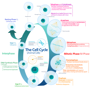
Mitosis is a part of the cell cycle in which replicated chromosomes are separated into two new nuclei. Cell division by mitosis is an equational division which gives rise to genetically identical cells in which the total number of chromosomes is maintained. Mitosis is preceded by the S phase of interphase and is followed by telophase and cytokinesis; which divides the cytoplasm, organelles and cell membrane of one cell into two new cells containing roughly equal shares of these cellular components. The different stages of mitosis altogether define the mitotic phase of a cell cycle—the division of the mother cell into two daughter cells genetically identical to each other.

<span class="mw-page-title-main">Microtubule</span> Polymer of tubulin that forms part of the cytoskeleton

Microtubules are polymers of tubulin that form part of the cytoskeleton and provide structure and shape to eukaryotic cells. Microtubules can be as long as 50 micrometres, as wide as 23 to 27 nm and have an inner diameter between 11 and 15 nm. They are formed by the polymerization of a dimer of two globular proteins, alpha and beta tubulin into protofilaments that can then associate laterally to form a hollow tube, the microtubule. The most common form of a microtubule consists of 13 protofilaments in the tubular arrangement.

The evolution of flagella is of great interest to biologists because the three known varieties of flagella – each represent a sophisticated cellular structure that requires the interaction of many different systems.

<span class="mw-page-title-main">Centrosome</span> Cell organelle in animal cell helping in cell division

In cell biology, the centrosome is an organelle that serves as the main microtubule organizing center (MTOC) of the animal cell, as well as a regulator of cell-cycle progression. The centrosome provides structure for the cell. The centrosome is thought to have evolved only in the metazoan lineage of eukaryotic cells. Fungi and plants lack centrosomes and therefore use other structures to organize their microtubules. Although the centrosome has a key role in efficient mitosis in animal cells, it is not essential in certain fly and flatworm species.

<span class="mw-page-title-main">Cytokinesis</span> Part of the cell division process

Cytokinesis is the part of the cell division process during which the cytoplasm of a single eukaryotic cell divides into two daughter cells. Cytoplasmic division begins during or after the late stages of nuclear division in mitosis and meiosis. During cytokinesis the spindle apparatus partitions and transports duplicated chromatids into the cytoplasm of the separating daughter cells. It thereby ensures that chromosome number and complement are maintained from one generation to the next and that, except in special cases, the daughter cells will be functional copies of the parent cell. After the completion of the telophase and cytokinesis, each daughter cell enters the interphase of the cell cycle.

<span class="mw-page-title-main">Cleavage furrow</span> Plasma membrane invagination at the cell division site

In cell biology, the cleavage furrow is the indentation of the cell's surface that begins the progression of cleavage, by which animal and some algal cells undergo cytokinesis, the final splitting of the membrane, in the process of cell division. The same proteins responsible for muscle contraction, actin and myosin, begin the process of forming the cleavage furrow, creating an actomyosin ring. Other cytoskeletal proteins and actin binding proteins are involved in the procedure.

The microtubule-organizing center (MTOC) is a structure found in eukaryotic cells from which microtubules emerge. MTOCs have two main functions: the organization of eukaryotic flagella and cilia and the organization of the mitotic and meiotic spindle apparatus, which separate the chromosomes during cell division. The MTOC is a major site of microtubule nucleation and can be visualized in cells by immunohistochemical detection of γ-tubulin. The morphological characteristics of MTOCs vary between the different phyla and kingdoms. In animals, the two most important types of MTOCs are 1) the basal bodies associated with cilia and flagella and 2) the centrosome associated with spindle formation.

<span class="mw-page-title-main">Parabasalid</span> Group of flagellated protists

The parabasalids are a group of flagellated protists within the supergroup Excavata. Most of these eukaryotic organisms form a symbiotic relationship in animals. These include a variety of forms found in the intestines of termites and cockroaches, many of which have symbiotic bacteria that help them digest cellulose in woody plants. Other species within this supergroup are known parasites, and include human pathogens.

<i>Trichomonas</i> Genus of parasitic, flagellated protists

Trichomonas is a genus of anaerobic excavate parasites of vertebrates. It was first discovered by Alfred François Donné in 1836 when he found these parasites in the pus of a patient suffering from vaginitis, an inflammation of the vagina. Donné named the genus from its morphological characteristics. The prefix tricho- originates from the Ancient Greek word θρίξ (thrix) meaning hair, describing Trichomonas’s flagella. The suffix -monas, describes its similarity to unicellular organisms from the genus Monas.

<span class="mw-page-title-main">Axoneme</span>

An axoneme, also called an axial filament is the microtubule-based cytoskeletal structure that forms the core of a cilium or flagellum. Cilia and flagella are found on many cells, organisms, and microorganisms, to provide motility. The axoneme serves as the "skeleton" of these organelles, both giving support to the structure and, in some cases, the ability to bend. Though distinctions of function and length may be made between cilia and flagella, the internal structure of the axoneme is common to both.
<span class="mw-page-title-main">Motor protein</span> Class of molecular proteins

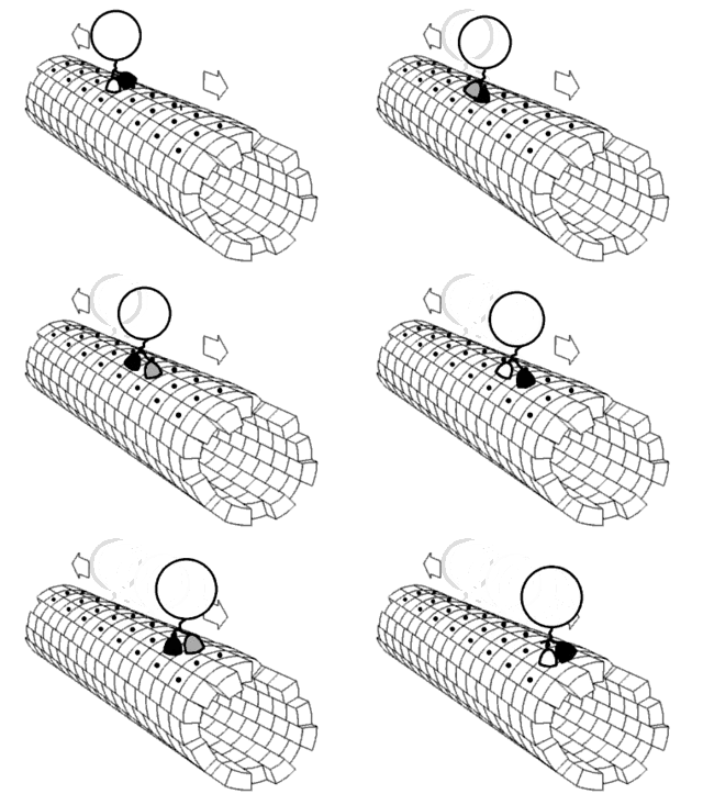
Motor proteins are a class of molecular motors that can move along the cytoplasm of cells. They convert chemical energy into mechanical work by the hydrolysis of ATP. Flagellar rotation, however, is powered by a proton pump.

<span class="mw-page-title-main">Trichomonadida</span> Order of flagellated protists

Trichomonadida is an order of anaerobic protists, included with the parabasalids. Members of this order are referred to as trichomonads.

<i>Tetraselmis</i> Genus of algae

Tetraselmis is a genus of phytoplankton. Tetraselmis is a green algal genus within the order Chlorodendrales, and they are characterized by their intensely-colored green chloroplast, their flagellated cell bodies, the presence of a pyrenoid within the chloroplast, and a scale-produced thecal-wall. Species within this genus are found in both marine and freshwater ecosystems across the globe; their habitat range is mainly limited by water depth due to their photosynthetic nature. Thus, they live in diverse water environments if enough nutrients and light are available for net photosynthetic activity. Tetraselmis species have proven to be useful for both research and industry. Tetraselmis species have been studied for understanding plankton growth rates, and recently a colonial species is being used to gain an understanding of multicellularity evolution. Additionally, many species are currently being examined for their use as biofuels due to their high lipid content.

<i>Trichonympha</i> Genus of flagellated protists

Trichonympha is a genus of single-celled, anaerobic parabasalids of the order Hypermastigia that is found exclusively in the hindgut of lower termites and wood roaches. Trichonympha’s bell shape and thousands of flagella make it an easily recognizable cell. The symbiosis between lower termites/wood roaches and Trichonympha is highly beneficial to both parties: Trichonympha helps its host digest cellulose and in return receives a constant supply of food and shelter. Trichonympha also has a variety of bacterial symbionts that are involved in sugar metabolism and nitrogen fixation.

<span class="mw-page-title-main">Preprophase</span> Cell cycle phase only found in plants

Preprophase is an additional phase during mitosis in plant cells that does not occur in other eukaryotes such as animals or fungi. It precedes prophase and is characterized by two distinct events:

  1. The formation of the preprophase band, a dense microtubule ring underneath the plasma membrane.
  2. The initiation of microtubule nucleation at the nuclear envelope.
<span class="mw-page-title-main">Centrin</span> Family of calcium-binding phosphoproteins

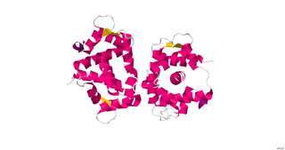
Centrins, also known as caltractins, are a family of calcium-binding phosphoproteins found in the centrosome of eukaryotes. Centrins are small calcium binding proteins that are ubiquitous centrosome components. There are about 350 “signature” proteins that are unique to eukaryotic cells but have no significant homology to proteins in archaea and bacteria. They are a type of protein that is essential and present in almost all eukaryotic cells and are found in the centrioles and pericentriolar lattice. Human centrin genes are CETN1, CETN2 and CETN3.

Oxymonas is a genus of Excavata.

Saccinobaculus is a genus of unicellular eukaryotes that resides in the hindgut of the wood-feeding cockroach Cryptocercus punctulatus. This genus is known for its distinctive movement that resembles a snake trashing in a bag. The genus is involved in the digestion of wood materials within its insect-host and is vertically transmitted to insect progeny. The genus is the part of the family Saccinobaculidae.

J. Richard McIntosh is a Distinguished Professor Emeritus in Molecular, Cellular, and Developmental Biology at the University of Colorado Boulder. McIntosh first graduated from Harvard with a BA in Physics in 1961, and again with a Ph.D. in Biophysics in 1968. He began his teaching career at Harvard but has spent most of his career at the University of Colorado Boulder. At the University of Colorado Boulder, McIntosh taught biology courses at both the undergraduate and graduate levels. Additionally, he created an undergraduate course in the biology of cancer towards the last several years of his teaching career. McIntosh's research career looks at a variety of things, including different parts of mitosis, microtubules, and motor proteins.

Monocercomonas is a Parabasalian genus belonging to the order Trichomonadida. It presents four flagella, three forward-facing and one trailing, without the presence of a costa or any kind of undulating membrane. Monocercomonas is found in animal guts. and is susceptible to cause Monocercomoniasis in reptiles

References

  1. 1 2 3 4 5 6 7 8 9 10 11 12 13 14 15 16 17 18 19 20 21 22 23 24 25 26 27 28 29 30 31 32 33 34 35 36 37 38 39 Lingle, Wilma L.; Salisbury, Jeffrey L. (1995). "Ultrastructure of the Parabasalid Protist Holomastigotoides". The Journal of Eukaryotic Microbiology . 42 (5): 490–505. doi:10.1111/j.1550-7408.1995.tb05895.x. ISSN   1066-5234. S2CID   83951229.
  2. 1 2 3 4 5 Cleveland, Lemuel R. (1960). "Pairing and segregation in haploids and diploids of Holomastigotoides". Archiv für Protistenkunde . 105 (2): 163–172.
  3. 1 2 Saldarriaga, Juan F.; Gile, Gillian H.; James, Erick R.; Horák, Ales; Scheffrahn, Rudolf H.; Keeling, Patrick J. (2011). "Morphology and molecular phylogeny of Pseudotrichonympha hertwigi and Pseudotrichonympha paulistana (Trichonymphea, parabasalia) from neotropical rhinotermitids". The Journal of Eukaryotic Microbiology . 58 (6): 487–496. doi:10.1111/j.1550-7408.2011.00575.x. ISSN   1550-7408. PMID   21895839. S2CID   40726800.
  4. 1 2 3 4 5 6 Gile, Gillian H.; James, Erick R.; Tai, Vera; Harper, James T.; Merrell, Trevor L.; Boscaro, Vittorio; Husník, Filip; Scheffrahn, Rudolf H.; Keeling, Patrick J. (2018). "New Species of Spirotrichonympha from Reticulitermes and the Relationships Among Genera in Spirotrichonymphea (Parabasalia)". The Journal of Eukaryotic Microbiology . 65 (2): 159–169. doi:10.1111/jeu.12447. PMID   28710832. S2CID   3684172.
  5. 1 2 3 4 5 Jasso‐Selles, Daniel E.; De Martini, Francesca; Velenovsky, Joseph F.; Mee, Evan D.; Montoya, Samantha J.; Hileman, Jonathon T.; Garcia, Mikaela D.; Su, Nan‐Yao; Chouvenc, Thomas; Gile, Gillian H. (November 2020). "The Complete Protist Symbiont Communities of Coptotermes formosanus and Coptotermes gestroi : Morphological and Molecular Characterization of Five New Species". The Journal of Eukaryotic Microbiology . 67 (6): 626–641. doi:10.1111/jeu.12815. ISSN   1066-5234. PMID   32603489. S2CID   220284193.
  6. 1 2 3 4 Jasso-Selles, Daniel E.; De Martini, Francesca; Freeman, Katalina D.; Garcia, Mikaela D.; Merrell, Trevor L.; Scheffrahn, Rudolf H.; Gile, Gillian H. (October 2017). "The parabasalid symbiont community of Heterotermes aureus: Molecular and morphological characterization of four new species and reestablishment of the genus Cononympha". European Journal of Protistology. 61 (Pt A): 48–63. doi:10.1016/j.ejop.2017.09.001. PMID   28942092.
  7. 1 2 3 4 5 6 7 8 9 10 11 12 13 Lingle, W. L.; Salisbury, J. L. (1997). "Centrin and the cytoskeleton of the protist Holomastigotoides". Cell Motility and the Cytoskeleton. 36 (4): 377–390. doi:10.1002/(SICI)1097-0169(1997)36:4<377::AID-CM7>3.0.CO;2-2. ISSN   0886-1544. PMID   9096959.
  8. Cleveland, L. R. (1949). "The Whole Life Cycle of Chromosomes and Their Coiling Systems". Transactions of the American Philosophical Society. 39 (1): 1–97. doi:10.2307/1005635. hdl: 2027/coo.31924001826647 . JSTOR   1005635.
  9. Cleveland, Lemuel R. (1947). "The origin and evolution of meiosis". Science . 105 (2724): 287–289. Bibcode:1947Sci...105..287C. doi:10.1126/science.105.2724.287. PMID   17835147. S2CID   2026926.
  10. 1 2 Mackinnon, Doris L. (1926-04-01). "Memoirs: Observations on Trichonymphids". Journal of Cell Science. s2-70 (278): 173–191. doi:10.1242/jcs.s2-70.278.173. ISSN   1477-9137.