Hydrogel encapsulation of quantum dots

Last updated

The behavior of quantum dots (QDs) in solution and their interaction with other surfaces is of great importance to biological and industrial applications, such as optical displays, animal tagging, anti-counterfeiting dyes and paints, chemical sensing, and fluorescent tagging. However, unmodified quantum dots tend to be hydrophobic, which precludes their use in stable, water-based colloids. Furthermore, because the ratio of surface area to volume in a quantum dot is much higher than for larger particles, the thermodynamic free energy associated with dangling bonds on the surface is sufficient to impede the quantum confinement of excitons. Once solubilized by encapsulation in either a hydrophobic interior micelle or a hydrophilic exterior micelle, the QDs can be successfully introduced into an aqueous medium, in which they form an extended hydrogel network. In this form, quantum dots can be utilized in several applications that benefit from their unique properties, such as medical imaging and thermal destruction of malignant cancers. [1]

Contents

Quantum dots

Quantum dots (QDs) are nano-scale semiconductor particles on the order of 2–10 nm in diameter. They possess electrical properties between those of bulk semi-conductors and individual molecules, as well as optical characteristics that make them suitable for applications where fluorescence is desirable, such as medical imaging. Most QDs synthesized for medical imaging are in the form of CdSe(ZnS) core(shell) particles. CdSe QDs have been shown to possess optical properties superior to organic dyes. [2] The ZnS shell has a two-fold effect:

  1. to interact with dangling bonds that would otherwise result in particle aggregation, loss of visual resolution, and impedance of quantum confinement effects
  2. to further increase the fluorescence of the particles themselves. [3]

Problems with CdSe(ZnS) quantum dots

Despite their potential for use as contrast agents for medical imaging techniques, their use in vivo is hindered by the cytotoxicity of cadmium. To address this issue, methods have been developed to “wrap” or “encapsulate” potentially-toxic QDs in bio-inert polymers to facilitate use in living tissue. While Cd-free QDs are commercially available, they are unsuitable for use as a substitute for organic contrasts. [4] Another issue with CdSe(ZnS) nanoparticles is significant hydrophobicity, which hinders their ability to enter solution with aqueous media, such as blood or spinal fluid. Certain hydrophilic polymers could be used to render the dots water-soluble.

Synthesizing the encapsulant polymer

Rf-PEG2.png
Structure of Rf-Polymer used in hydrogel encapsulation of quantum dots. The figure indicates the hydrophobic and hydrophilic regions of the polymer.

Rf-PEG synthesis

One notable quantum dot encapsulation technique involves utilizing a double fluoroalkyl-ended polyethylene glycol molecule (Rf-PEG) as a surfactant, which will spontaneously form micellular structures at its critical micelle concentration (CMC). The critical micelle concentration of the Rf-PEG depends on the length of the PEG portion of the polymer. This molecule consists of a hydrophilic PEG backbone with two hydrophilic terminal groups (CnF2n+1-CH2CH2O) attached via isophorone diurethane. [5] It is synthesized by dehydrating a solution of 1,3-dimethyl-5-fluorouracil and PEG, mixing them in the presence of heavy water (D2O) via a sonicator to combine then. [6]

Diagram of Rf-PEG showing the fluoroalkyl end groups attached to the PEG polymer by intermediate groups. Rf-PEG Petal.svg
Diagram of Rf-PEG showing the fluoroalkyl end groups attached to the PEG polymer by intermediate groups.

Micellization

Polymer micelle with multiple QDs in the middle. The polymer wraps so both the hydrophobic ends are near the QDs and the hydrophilic end is on the outside of the micelle to allow solubility in water. PEG Micelle.svg
Polymer micelle with multiple QDs in the middle. The polymer wraps so both the hydrophobic ends are near the QDs and the hydrophilic end is on the outside of the micelle to allow solubility in water.

At the appropriate Krafft temperature and critical micelle concentration these molecules will form individual tear-drop loops, where the hydrophobic ends are attracted to one another, to other molecules, and also to the similarly hydrophobic QDs. This forms a loaded micelle with a hydrophilic outer shell and a hydrophobic core. [6]

When encapsulating hydrophobes in this way it is important to ensure the particle size is appropriate for the PEG backbone being utilized, as the number of PEG mer units (generally with a molecular weight of 6 kDa or 10 kDa) determines the maximum particle size that can be successfully contained at the core of the micelle.

To determine the average diameter, D, of the QDs, the following empirical equation is used:

Where

Role of ZnS shell

It is during encapsulation that the ZnS shell plays an especially important role, in that it helps prevent the agglomeration of CdSe particles that had no shell by occupying the previously mentioned bonds on the dot's surface; however, clumping can still occur through secondary forces that arise from common hydrophobicity. This can result in multiple particles within each micelle, which may negatively impact overall resolution. For this reason multiple combinations of PEG chain length and particle diameter are necessary to achieve optimal imaging properties.

Micelles can be cross-linked. The ends of the polymer chain are attracted to two different QD groups. Micelle Cluster.svg
Micelles can be cross-linked. The ends of the polymer chain are attracted to two different QD groups.

Hydrogel network

After initial encapsulation the remaining molecules form connections between the individual micelles to form a network within the aqueous media called a hydrogel, creating a diffuse and relatively constant concentration of the encapsulated particle within the gel. The formation of hydrogels is a phenomenon observed in superabsorbent polymers, or "slush powders," in which the polymer, often in the form of a powder, absorbs water, becoming up to 99% liquid and 30-60 times larger in size. [7]

Stokes-Einstein equation

The diffusivity of a particle decreases as its radius increases. Particle Diffusivity vs. Particle Radius at 25degC.svg
The diffusivity of a particle decreases as its radius increases.

The diffusivity of spherical particles in a suspension is approximated by the Stokes–Einstein equation: [6]

where

Typical Rf-PEG hydrogel diffusivities for 2 nm quantum dots are on the order of 10−16 m2/s, so suspensions of quantum dots tend to be very stable. Hydrogel viscosity can be determined by using rheological techniques.

Micelle rheology

When encapsulating hydrophobic or potentially toxic materials it is important that the encapsulant remain intact while inside the body. Studying the rheological properties of the micelles permits identification and selection of the polymer that is most appropriate for use in long-term biological applications. Rf-PEG exhibits superior rheological properties when used in vivo.

Importance of polymer length

The properties of the polymer are influenced by the chain length. The correct chain length ensures that the encapsulant is not released over time. Avoiding the release of QDs and other toxic particles is critical to prevent unintentional cell necrosis in patients. The length of the polymer is controlled by two factors:

Increasing the PEG length increases the solubility of the polymer. However, if the PEG chain is too long the micelle will become unstable. It has been observed that a stable hydrogel can only be formed with PEG backbones weighing between six and ten kilodaltons. [8]

On the other hand, increasing the length of the hydrophobic terminal groups decreases aqueous solubility. For a given PEG weight, if the hydrophobe is too short the polymer will just dissolve into the solution, and if it is too long the polymer won't dissolve at all. Generally, two end groups result in the highest conversion into micelles (91%): [8]

Maxwell fluid

At molecular weights between 6 and 10 kilodaltons the Rf-PEG hydrogel acts as a Maxwell material, which means the fluid has both viscosity and elasticity. This is determined by measuring the plateau modulus, the elastic modulus for a viscoelastic polymer is constant or "relaxed" when deformed, at a range of frequencies via oscillatory rheology. [9] [10] Plotting the first- vs second-order integrals of the modulus values, a Cole-Cole plot is obtained, which, when fitted to a Maxwell model, provides the following relationship:

Where

Mechanical properties of common Rf-PEG molecules

Based on the Maxwellian behavior of the hydrogel and observations of erosion via surface plasmon resonance (SPR), the following data results for 3 common Rf-PEG types at their specified concentrations: [11] [12]

Property10KC1010KC86KC8
6.86.511.0
1.20.0290.023
14.418.556.1
180.531.5
949489

XKCY denotes X thousand daltons of molecular mass and Y carbon atoms.

These values can give us information on the degree of entanglement (or degree of cross linking, depending on what polymer is being considered). In general, higher degrees of entanglement leads to higher time required for the polymer to return to the undeformed state or relaxation times.

Applications

Hydrogel encapsulation of the QDs opens up a new range of applications, such as:

Enzymes and other bio-active molecules serve as biorecognition units while QDs serve as signalling units. By adding enzymes to the QD hydrogel network both units can be combined to form a biosensor. The enzymatic reaction that detects a particular molecule causes the fluoresce of QDs to be quenched. In this way, the location of molecules of interest can be observed. [13]
Adding iron oxide nanoparticles to the QD micelles allows them to be fluorescent and magnetic. These micelles can be moved in a magnetic field to create concentration gradients that will influence a cell's processes. [14]
When excited by high energy radiation, such as with a laser, gold nanoparticles emit a thermal field. This phenomenon can be used as a form of hyperthermia therapy to destroy malignant cancers without damaging surrounding tissues. When combined with QDs in a hydrogel this could facilitate real-time monitoring of the tumor treatment. [15]

See also

Related Research Articles

<span class="mw-page-title-main">Hydrophobe</span> Molecule or surface that has no attraction to water

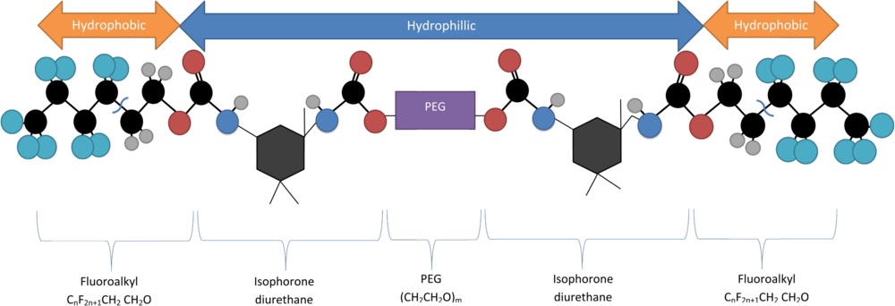
In chemistry, hydrophobicity is the physical property of a molecule that is seemingly repelled from a mass of water. In contrast, hydrophiles are attracted to water.

<span class="mw-page-title-main">Gel</span> Highly viscous liquid exhibiting a kind of semi-solid behavior

A gel is a semi-solid that can have properties ranging from soft and weak to hard and tough. Gels are defined as a substantially dilute cross-linked system, which exhibits no flow when in the steady state, although the liquid phase may still diffuse through this system. A gel has been defined phenomenologically as a soft, solid or solid-like material consisting of two or more components, one of which is a liquid, present in substantial quantity.

A monolayer is a single, closely packed layer of entities, commonly atoms or molecules. Monolayers can also be made out of cells. Self-assembled monolayers form spontaneously on surfaces. Monolayers of layered crystals like graphene and molybdenum disulfide are generally called 2D materials.

<span class="mw-page-title-main">Micelle</span> Group of fatty molecules suspended in liquid by soaps and/or detergents

A micelle or micella is an aggregate of surfactant amphipathic lipid molecules dispersed in a liquid, forming a colloidal suspension. A typical micelle in water forms an aggregate with the hydrophilic "head" regions in contact with surrounding solvent, sequestering the hydrophobic single-tail regions in the micelle centre.

<span class="mw-page-title-main">Quantum dot</span> Zero-dimensional, nano-scale semiconductor particles with novel optical and electronic properties

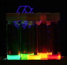
Quantum dots (QDs) or semiconductor nanocrystals are semiconductor particles a few nanometres in size with optical and electronic properties that differ from those of larger particles via quantum mechanical effects. They are a central topic in nanotechnology and materials science. When a quantum dot is illuminated by UV light, an electron in the quantum dot can be excited to a state of higher energy. In the case of a semiconducting quantum dot, this process corresponds to the transition of an electron from the valence band to the conductance band. The excited electron can drop back into the valence band releasing its energy as light. This light emission (photoluminescence) is illustrated in the figure on the right. The color of that light depends on the energy difference between the conductance band and the valence band, or the transition between discrete energy states when the band structure is no longer well-defined in QDs.

<span class="mw-page-title-main">Hydrogel</span> Soft water-rich polymer gel

A hydrogel is a biphasic material, a mixture of porous, permeable solids and at least 10% by weight or volume of interstitial fluid composed completely or mainly by water. In hydrogels the porous permeable solid is a water insoluble three dimensional network of natural or synthetic polymers and a fluid, having absorbed a large amount of water or biological fluids. These properties underpin several applications, especially in the biomedical area. Many hydrogels are synthetic, but some are derived from nature. The term 'hydrogel' was coined in 1894.

<span class="mw-page-title-main">Dendrimer</span> Highly ordered, branched polymeric molecule

Dendrimers are highly ordered, branched polymeric molecules. Synonymous terms for dendrimer include arborols and cascade molecules. Typically, dendrimers are symmetric about the core, and often adopt a spherical three-dimensional morphology. The word dendron is also encountered frequently. A dendron usually contains a single chemically addressable group called the focal point or core. The difference between dendrons and dendrimers is illustrated in the top figure, but the terms are typically encountered interchangeably.

A drug carrier or drug vehicle is a substrate used in the process of drug delivery which serves to improve the selectivity, effectiveness, and/or safety of drug administration. Drug carriers are primarily used to control the release of drugs into systemic circulation. This can be accomplished either by slow release of a particular drug over a long period of time or by triggered release at the drug's target by some stimulus, such as changes in pH, application of heat, and activation by light. Drug carriers are also used to improve the pharmacokinetic properties, specifically the bioavailability, of many drugs with poor water solubility and/or membrane permeability.

<span class="mw-page-title-main">Janus particles</span> Type of nanoparticle or microparticle

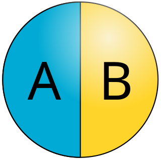
Janus particles are special types of nanoparticles or microparticles whose surfaces have two or more distinct physical properties. This unique surface of Janus particles allows two different types of chemistry to occur on the same particle. The simplest case of a Janus particle is achieved by dividing the particle into two distinct parts, each of them either made of a different material, or bearing different functional groups. For example, a Janus particle may have one half of its surface composed of hydrophilic groups and the other half hydrophobic groups, the particles might have two surfaces of different color, fluorescence, or magnetic properties. This gives these particles unique properties related to their asymmetric structure and/or functionalization.

A nanogel is a polymer-based, crosslinked hydrogel particle on the sub-micron scale. These complex networks of polymers present a unique opportunity in the field of drug delivery at the intersection of nanoparticles and hydrogel synthesis. Nanogels can be natural, synthetic, or a combination of the two and have a high degree of tunability in terms of their size, shape, surface functionalization, and degradation mechanisms. Given these inherent characteristics in addition to their biocompatibility and capacity to encapsulate small drugs and molecules, nanogels are a promising strategy to treat disease and dysfunction by serving as delivery vehicles capable of navigating across challenging physiological barriers within the body. 

<span class="mw-page-title-main">Nanocarrier</span>

A nanocarrier is nanomaterial being used as a transport module for another substance, such as a drug. Commonly used nanocarriers include micelles, polymers, carbon-based materials, liposomes and other substances. Nanocarriers are currently being studied for their use in drug delivery and their unique characteristics demonstrate potential use in chemotherapy. This class of materials was first reported by a team of researchers of University of Évora, Alentejo in early 1960's, and grew exponentially in relevance since then.

<span class="mw-page-title-main">Self-assembly of nanoparticles</span> Physical phenomenon

Nanoparticles are classified as having at least one of its dimensions in the range of 1-100 nanometers (nm). The small size of nanoparticles allows them to have unique characteristics which may not be possible on the macro-scale. Self-assembly is the spontaneous organization of smaller subunits to form larger, well-organized patterns. For nanoparticles, this spontaneous assembly is a consequence of interactions between the particles aimed at achieving a thermodynamic equilibrium and reducing the system’s free energy. The thermodynamics definition of self-assembly was introduced by Professor Nicholas A. Kotov. He describes self-assembly as a process where components of the system acquire non-random spatial distribution with respect to each other and the boundaries of the system. This definition allows one to account for mass and energy fluxes taking place in the self-assembly processes.

<span class="mw-page-title-main">Surface chemistry of neural implants</span>

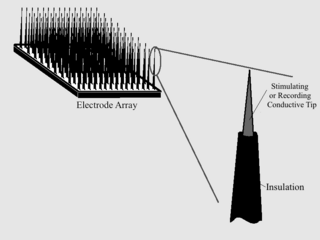
As with any material implanted in the body, it is important to minimize or eliminate foreign body response and maximize effectual integration. Neural implants have the potential to increase the quality of life for patients with such disabilities as Alzheimer's, Parkinson's, epilepsy, depression, and migraines. With the complexity of interfaces between a neural implant and brain tissue, adverse reactions such as fibrous tissue encapsulation that hinder the functionality, occur. Surface modifications to these implants can help improve the tissue-implant interface, increasing the lifetime and effectiveness of the implant.

<span class="mw-page-title-main">Self-healing hydrogels</span> Type of hydrogel

Self-healing hydrogels are a specialized type of polymer hydrogel. A hydrogel is a macromolecular polymer gel constructed of a network of crosslinked polymer chains. Hydrogels are synthesized from hydrophilic monomers by either chain or step growth, along with a functional crosslinker to promote network formation. A net-like structure along with void imperfections enhance the hydrogel's ability to absorb large amounts of water via hydrogen bonding. As a result, hydrogels, self-healing alike, develop characteristic firm yet elastic mechanical properties. Self-healing refers to the spontaneous formation of new bonds when old bonds are broken within a material. The structure of the hydrogel along with electrostatic attraction forces drive new bond formation through reconstructive covalent dangling side chain or non-covalent hydrogen bonding. These flesh-like properties have motivated the research and development of self-healing hydrogels in fields such as reconstructive tissue engineering as scaffolding, as well as use in passive and preventive applications.

<span class="mw-page-title-main">DNA-functionalized quantum dots</span>

DNA-functionalization of quantum dots is the attachment of strands of DNA to the surface of a quantum dot. Although quantum dots with cadmium (Cd) have some cytotoxic release, researchers have functionalized quantum dots for biocompatibility and bound them to DNA in order to combine the advantages of both materials. Quantum dots are commonly used for imaging biological systems in vitro and in vivo in animal studies due to their excellent optical properties when excited by light, while DNA has numerous bioengineering applications, including: genetic engineering, self-assembling nanostructures, protein binding, and biomarkers. The ability to visualize the chemical and biological processes of DNA allows feedback to optimize and learn about these small scale behaviors.

Quantum dots (QDs) are semiconductor nanoparticles with a size less than 10 nm. They exhibited size-dependent properties especially in the optical absorption and the photoluminescence (PL). Typically, the fluorescence emission peak of the QDs can be tuned by changing their diameters. So far, QDs were consisted of different group elements such as CdTe, CdSe, CdS in the II-VI category, InP or InAs in the III-V category, CuInS2 or AgInS2 in the I–III–VI2 category, and PbSe/PbS in the IV-VI category. These QDs are promising candidates as fluorescent labels in various biological applications such as bioimaging, biosensing and drug delivery.

Nanoparticle drug delivery systems are engineered technologies that use nanoparticles for the targeted delivery and controlled release of therapeutic agents. The modern form of a drug delivery system should minimize side-effects and reduce both dosage and dosage frequency. Recently, nanoparticles have aroused attention due to their potential application for effective drug delivery.

<span class="mw-page-title-main">Polymer-protein hybrid</span> Nanostructures of protein-polymer conjugates

Polymer-protein hybrids are a class of nanostructure composed of protein-polymer conjugates. The protein component generally gives the advantages of biocompatibility and biodegradability, as many proteins are produced naturally by the body and are therefore well tolerated and metabolized. Although proteins are used as targeted therapy drugs, the main limitations—the lack of stability and insufficient circulation times still remain. Therefore, protein-polymer conjugates have been investigated to further enhance pharmacologic behavior and stability. By adjusting the chemical structure of the protein-polymer conjugates, polymer-protein particles with unique structures and functions, such as stimulus responsiveness, enrichment in specific tissue types, and enzyme activity, can be synthesized. Polymer-protein particles have been the focus of much research recently because they possess potential uses including bioseparations, imaging, biosensing, gene and drug delivery.

<span class="mw-page-title-main">Nanoparticle interfacial layer</span> Interfacial layer of nanoparticles

A nanoparticle interfacial layer is a well structured layer of typically organic molecules around a nanoparticle. These molecules are known as stabilizers, capping and surface ligands or passivating agents. The interfacial layer has a significant effect on the properties of the nanoparticle and is therefore often considered as an integral part of a nanoparticle. The interfacial layer has an typical thickness between 0.1 and 4 nm, which is dependent on the type of the molecules the layer is made of. The organic molecules that make up the interfacial layer are often amphiphilic molecules, meaning that they have a polar head group combined with a non-polar tail.

<span class="mw-page-title-main">Dextran drug delivery systems</span> Polymeric drug carrier

Dextran drug delivery systems involve the use of the natural glucose polymer dextran in applications as a prodrug, nanoparticle, microsphere, micelle, and hydrogel drug carrier in the field of targeted and controlled drug delivery. According to several in vitro and animal research studies, dextran carriers reduce off-site toxicity and improve local drug concentration at the target tissue site. This technology has significant implications as a potential strategy for delivering therapeutics to treat cancer, cardiovascular diseases, pulmonary diseases, bone diseases, liver diseases, colonic diseases, infections, and HIV.

References

  1. Glazer, ES; SA Curley (July 2010). "Radiofrequency field-induced thermal cytotoxicity in cancer cells treated with fluorescent nanoparticles". Cancer. 116 (13): 3285–93. doi:10.1002/cncr.25135. PMC   2928886 . PMID   20564640.
  2. Resch-Genger, Ute; Grabolle; Cavaliere-Jaricot; Nitschke; Nann (August 2008). "Quantum dots versus organic dyes as fluorescent labels". Nature Methods. 5 (9): 763–775. doi:10.1038/nmeth.1248. PMID   18756197. S2CID   9007994.
  3. Angell, Joshua. "Synthesis and Characterization of CdSe-ZnS Core-Shell Quantum Dots".{{cite journal}}: Cite journal requires |journal= (help)
  4. Jin, Shan; Yanxi Hu; Zhanjun Gu; Lei Liu; Hai-Chen Wu (July 2011). "Application of Quantum Dots in Biological Imaging". Journal of Nanomaterials. 2011: 1–13. doi: 10.1155/2011/834139 .
  5. Lundberg, D.J.; R.G. Brown; J.E. Glass; R.R. Eley (1994). "Synthesis, Characterization, and Solution Rheology of Model Hydrophobically-Modified, Water-Soluble Ethoxylated Urethanes". Langmuir. 10 (9): 3027–3034. doi:10.1021/la00021a028.
  6. 1 2 3 Mathias, Errol V.; Julia Aponte; Julia A. Kornfield; Yong Ba (October 2010). "Properties of Small Molecular Drug Loading and Diffusion in a Fluorinated PEG Hydrogel Studied by 1H Molecular Diffusion NMR and 19F Spin Diffusion NMR". Colloid and Polymer Science. 288 (18): 1655–1663. doi:10.1007/s00396-010-2304-9. PMC   2982959 . PMID   21170115.
  7. Horie, K, et al., 890.
  8. 1 2 Tae, Giyoong; Julia A. Kornfield; Jeffry A. Hubbell; Diethelm Johannsmann; Thieo E. Hogen-Esch (May 2001). "Hydrogels with Controlled, Surface Erosion Characteristics from Self-Assembly of Fluoroalkyl-Ended Poly(ethylene glycol)". Macromolecules. 34 (18): 6409–6419. Bibcode:2001MaMol..34.6409T. doi:10.1021/ma0107511.
  9. Wyss, Hans; Ryan J. Larson; David A. Weitz (2007). "Oscillatory Rheology: Measuring the Viscoelastic Behaviour of Soft Materials" (PDF). G.I.T. Laboratory. 3 (4): 68–70.
  10. Rubinstein, Michael; Dobrynin, Andrey V. (1997). "Solutions of Associative Polymers". Trends in Polymer Science. 5 (6): 181.
  11. Aust EF, Ito S, Sawodny M, Knoll W (1994). "Surface plasmon and guided optical wave microscopies". Scanning. 16 (3): 353–362.
  12. Tae, G.; J.A. Kornfield; J.A. Hubbell; Diethelm Johannsmann (17 September 2002). "Anomalous Sorption in Thin Films of Fluoroalkyl-Ended Poly(Ethylene Glycol)s". Langmuir. 18 (21): 8241–8245. doi:10.1021/la020255l.
  13. Yuan, Jipei; Dan Wen; Nikolai Gaponik; Alexander Eychmuller (22 November 2012). "Enzyme-Encapsulating Quantum Dot Hydrogels and Xerogels as Biosensors: Multifunctional Platforms for Both Biocatalysis and Fluorescent Probing". Angewandte Chemie International Edition. 52 (3): 976–979. doi:10.1002/anie.201205791. PMID   23172829.
  14. Roullier, Victor; Fabien Grasset; Fouzia Boulmedais; Franck Artzner; Olivier Cador; Vale´rie Marchi-Artzner (15 October 2008). "Small Bioactivated Magnetic Quantum Dot Micelles" (PDF). Chemistry of Materials. 20 (21): 6657–6665. doi:10.1021/cm801423r . Retrieved 8 June 2013.
  15. Huff, Terry; Ling Tong; Yan Zhao; Matthew Hansen; Jin-Xin Cheng; Alexander Wei (2007). "Hyperthermic effect of gold nanorods on tumor cells" (PDF). Nanomedicine. 2 (1): 125–132. doi:10.2217/17435889.2.1.125. PMC   2597406 . PMID   17716198.