Hypnogram

Last updated
Example hypnogram of a normal, healthy adult
Here, both stage 3 and stage 4 are shown; these are often combined as stage 3. HYPNOGRAM created by Natasha k.jpg
Example hypnogram of a normal, healthy adult
Here, both stage 3 and stage 4 are shown; these are often combined as stage 3.

A hypnogram is a form of polysomnography; it is a graph that represents the stages of sleep as a function of time. It was developed as an easy way to present the recordings of the brain wave activity from an electroencephalogram (EEG) during a period of sleep. It allows the different stages of sleep: rapid eye movement sleep (REM) and non-rapid eye movement sleep (NREM) to be identified during the sleep cycle. NREM sleep can be further classified into NREM stage 1, 2 and 3. The previously considered 4th stage of NREM sleep has been included within stage 3; this stage is also called slow wave sleep (SWS) and is the deepest stage of sleep. [1]

Contents

Method

A hypnogram (top) with other readings below Interactive dreaming (Dutch group).jpg
A hypnogram (top) with other readings below

Hypnograms are usually obtained by visually scoring the recordings from electroencephalogram (EEGs), electrooculography (EOGs) and electromyography (EMGs). [2]

The output from these three sources is recorded simultaneously on a graph by a monitor or computer as a hypnogram. Certain frequencies displayed by EEGs, EOGs and EMGs are characteristic and determine what stage of sleep or wake the subject is in. There is a protocol defined by the American Academy of Sleep Medicine (AASM) for sleep scoring, whereby the sleep or wake state is recorded in 30-second epochs. [3] Prior to this the Rechtschaffen and Kales (RK) rules were used to classify sleep stages. [4]

Output

Normal sleep

Cycles of REM and non-REM stages make up sleep. A normal healthy adult requires 7–9 hours of sleep per night. The number of hours of sleep is variable, however the proportion of sleep spent in a particular stage remains mostly consistent; healthy adults normally spend 20–25% of their sleep in REM sleep. [5] During rest following a sleep-deprived state, there is a period of rebound sleep which has longer and deeper episodes of SWS to make up for the lack of sleep. [6]

On a hypnogram, a sleep cycle is usually around 90 minutes and there are four to six cycles of REM/NREM stages that occur during a major period of sleep. Most SWS occurs in the first one or two cycles; this is the deepest period of sleep. The second half of the sleeping period contains most REM sleep and little or no SWS and may contain brief periods of wakefulness which can be recorded but are not usually perceived. [7] The stage that occurs before waking is normally REM sleep. [8]

Hypnograms for healthy persons vary slightly according to age, emotional state, and environmental factors. [9]

Disrupted sleep

Sleep architecture can be evaluated using hypnograms, demonstrating irregular sleeping patterns associated with sleep disorders. Disruptions or irregularities to the normal sleep cycle or sleep stage transitions can be detected; for example a hypnogram can show that in obstructive sleep apnea (OSA) the stability of transition between REM and NREM stages is disrupted. [10]

The effects of certain medications on sleep architecture can be visualised on a hypnogram. For example, the anticonvulsant Phenytoin (PHT) can be seen to disrupt sleep by increasing the duration of NREM stage 1 and decreasing the duration of SWS; whereas the drug Gabapentin is seen to revive sleep by increasing the duration of SWS.[ unreliable medical source? ] [11]

Analysis

The main use of a hypnogram is as a qualitative method to visualise the time period of each stage of sleep, as well as the number of transitions between stages. Hypnograms are rarely used to provide quantitative data, however it has been suggested that statistical evaluation can be carried out using multistate survival analysis and log-linear models to provide numerical significance. [12]

Limitations

The restrictions of measuring sleep at short 30-second epochs limits the ability to record events shorter than 30 seconds; hence, the macrostructure of sleep can be evaluated while the microstructure is not. The sleep process is smoothened out in hypnogram results unlike it occurs naturally. Also some specific features of sleep such as sleep spindles and K complexes may not be defined in the hypnogram; this is particularly true for sleep scoring that is automated. [13]

The method of obtaining the data used in a hypnogram is restricted to the input from an EEG, EOG or EMG. The interval of recording may include features from several stages, in which case it is recorded as the stage whose features occupy the recording for the longest duration. For this reason, the stage of sleep may be misrepresented on the hypnogram.[ citation needed ]

Research directions

Suggestions to improve the automated output of hypnograms to provide more reliable and accurate results include increasing the measures of sleep, for example by additionally measuring sleep with an electrocardiogram (ECG). [14] Another advancement involves combining hypnograms with color density spectral arrays to improve the quality of sleep analysis. [15]

Related Research Articles

<span class="mw-page-title-main">Sleep</span> Naturally recurring resting state of mind and body

Sleep is a state of reduced mental and physical activity in which consciousness is altered and sensory activity is inhibited to a certain extent. During sleep, there is a decrease in muscle activity, and interactions with the surrounding environment. While sleep differs from wakefulness in terms of the ability to react to stimuli, it still involves active brain patterns, making it more reactive than a coma or disorders of consciousness.

<span class="mw-page-title-main">Sleep disorder</span> Medical disorder of the sleep patterns of a person

A sleep disorder, or somnipathy, is a medical disorder of an individual's sleep patterns. Some sleep disorders are severe enough to interfere with normal physical, mental, social and emotional functioning. Polysomnography and actigraphy are tests commonly ordered for diagnosing sleep disorders.

<span class="mw-page-title-main">Rapid eye movement sleep</span> Phase of sleep characterized by random & rapid eye movements

Rapid eye movement sleep is a unique phase of sleep in humans, mammals and birds, characterized by random rapid movement of the eyes, accompanied by low muscle tone throughout the body, and the propensity of the sleeper to dream vividly.

<span class="mw-page-title-main">Sleep cycle</span> Oscillation between the slow-wave and REM phases of sleep

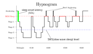
The sleep cycle is an oscillation between the slow-wave and REM (paradoxical) phases of sleep. It is sometimes called the ultradian sleep cycle, sleep–dream cycle, or REM-NREM cycle, to distinguish it from the circadian alternation between sleep and wakefulness. In humans, this cycle takes 70 to 110 minutes.

<span class="mw-page-title-main">Delta wave</span> High amplitude low frequency brain wave

{

<span class="mw-page-title-main">Nightmare disorder</span> Medical condition

Nightmare disorder is a sleep disorder characterized by repeated intense nightmares that most often center on threats to physical safety and security. The nightmares usually occur during the REM stage of sleep, and the person who experiences the nightmares typically remembers them well upon waking. More specifically, nightmare disorder is a type of parasomnia, a subset of sleep disorders categorized by abnormal movement or behavior or verbal actions during sleep or shortly before or after. Other parasomnias include sleepwalking, sleep terrors, bedwetting, and sleep paralysis.

Non-rapid eye movement sleep (NREM), also known as quiescent sleep, is, collectively, sleep stages 1–3, previously known as stages 1–4. Rapid eye movement sleep (REM) is not included. There are distinct electroencephalographic and other characteristics seen in each stage. Unlike REM sleep, there is usually little or no eye movement during these stages. Dreaming occurs during both sleep states, and muscles are not paralyzed as in REM sleep. People who do not go through the sleeping stages properly get stuck in NREM sleep, and because muscles are not paralyzed a person may be able to sleepwalk. According to studies, the mental activity that takes place during NREM sleep is believed to be thought-like, whereas REM sleep includes hallucinatory and bizarre content. NREM sleep is characteristic of dreamer-initiated friendliness, compared to REM sleep where it's more aggressive, implying that NREM is in charge of simulating friendly interactions. The mental activity that occurs in NREM and REM sleep is a result of two different mind generators, which also explains the difference in mental activity. In addition, there is a parasympathetic dominance during NREM. The reported differences between the REM and NREM activity are believed to arise from differences in the memory stages that occur during the two types of sleep.

Sexsomnia, also known as sleep sex, is a distinct form of parasomnia, or an abnormal activity that occurs while an individual is asleep. Sexsomnia is characterized by an individual engaging in sexual acts while in non-rapid eye movement (NREM) sleep. Sexual behaviors that result from sexsomnia are not to be mistaken with normal nocturnal sexual behaviors, which do not occur during NREM sleep. Sexual behaviors that are viewed as normal during sleep and are accompanied by extensive research and documentation include nocturnal emissions, nocturnal erections, and sleep orgasms.

<span class="mw-page-title-main">Polysomnography</span> Multi-parameter study of sleep and sleep disorders

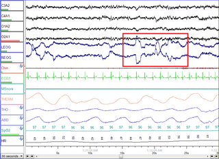
Polysomnography (PSG), a type of sleep study, is a multi-parameter study of sleep and a diagnostic tool in sleep medicine. The test result is called a polysomnogram, also abbreviated PSG. The name is derived from Greek and Latin roots: the Greek πολύς, the Latin somnus ("sleep"), and the Greek γράφειν.

The cyclic alternating pattern is a pattern of two, long-lasting alternate electroencephalogram (EEG) patterns that occur in sleep, as described by Terzano, et al., in 1985. It is a pattern of spontaneous cortical activity, which is ongoing and in the absence of sensory stimulation. It is the reorganization of the sleeping brain challenged by the modification of environmental conditions and it is characterized by periodic abnormal electrocortical activity that recurs with a frequency of up to one minute. It is considered "the EEG marker of unstable sleep". CAP does not occur during REM. In Lennox-Gastaut syndrome, CAP modulates the occurrence of clinical seizures and generalized epileptic discharges by means of a gate-control mechanism.

<span class="mw-page-title-main">K-complex</span>

A K-complex is a waveform that may be seen on an electroencephalogram (EEG). It occurs during stage 2 NREM sleep. It is the "largest event in healthy human EEG". They are more frequent in the first sleep cycles.

Sleep spindles are bursts of neural oscillatory activity that are generated by interplay of the thalamic reticular nucleus (TRN) and other thalamic nuclei during stage 2 NREM sleep in a frequency range of ~11 to 16 Hz with a duration of 0.5 seconds or greater. After generation as an interaction of the TRN neurons and thalamocortical cells, spindles are sustained and relayed to the cortex by thalamo-thalamic and thalamo-cortical feedback loops regulated by both GABAergic and NMDA-receptor mediated glutamatergic neurotransmission. Sleep spindles have been reported for all tested mammalian species. Considering animals in which sleep-spindles were studied extensively, they appear to have a conserved main frequency of roughly 9–16 Hz. Only in humans, rats and dogs is a difference in the intrinsic frequency of frontal and posterior spindles confirmed, however.

<span class="mw-page-title-main">Slow-wave sleep</span> Period of sleep in humans and other animals

Slow-wave sleep (SWS), often referred to as deep sleep, consists of stage three of non-rapid eye movement sleep. It usually lasts between 70 and 90 minutes and takes place during the first hours of the night. Initially, SWS consisted of both Stage 3, which has 20–50 percent delta wave activity, and Stage 4, which has more than 50 percent delta wave activity.

Parasomnias are a category of sleep disorders that involve abnormal movements, behaviors, emotions, perceptions, and dreams that occur while falling asleep, sleeping, between sleep stages, or during arousal from sleep. Parasomnias are dissociated sleep states which are partial arousals during the transitions between wakefulness, NREM sleep, and REM sleep, and their combinations.

Allan Rechtschaffen was a noted pioneer in the field of sleep research whose work includes some of the first laboratory studies of insomnia, narcolepsy, sleep apnea, and napping. He received his PhD from Northwestern University in 1956.

<span class="mw-page-title-main">Sleep in animals</span>

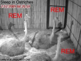
Sleep in animals refers to a behavioral and physiological state characterized by altered consciousness, reduced responsiveness to external stimuli, and homeostatic regulation observed in various animals. Sleep has been observed in mammals, birds, reptiles, amphibians, and some fish, and, in some form, in insects and even in simpler animals such as nematodes. The internal circadian clock promotes sleep at night for diurnal organisms and in the day for nocturnal organisms. Sleep patterns vary widely among species. It appears to be a requirement for all mammals and most other animals.

Sleep onset is the transition from wakefulness into sleep. Sleep onset usually transmits into non-rapid eye movement sleep but under certain circumstances it is possible to transit from wakefulness directly into rapid eye movement sleep.

<span class="mw-page-title-main">Sleep and memory</span> Relationship between sleep and memory

The relationship between sleep and memory has been studied since at least the early 19th century. Memory, the cognitive process of storing and retrieving past experiences, learning and recognition, is a product of brain plasticity, the structural changes within synapses that create associations between stimuli. Stimuli are encoded within milliseconds; however, the long-term maintenance of memories can take additional minutes, days, or even years to fully consolidate and become a stable memory that is accessible. Therefore, the formation of a specific memory occurs rapidly, but the evolution of a memory is often an ongoing process.

<span class="mw-page-title-main">Neuroscience of sleep</span> Study of the neuroscientific and physiological basis of the nature of sleep

The neuroscience of sleep is the study of the neuroscientific and physiological basis of the nature of sleep and its functions. Traditionally, sleep has been studied as part of psychology and medicine. The study of sleep from a neuroscience perspective grew to prominence with advances in technology and the proliferation of neuroscience research from the second half of the twentieth century.

An electrogram (EGM) is a recording of electrical activity of organs such as the brain and heart, measured by monitoring changes in electric potential.

References

  1. Silber MH, Ancoli-Israel S, Bonnet MH, Chokroverty S, Grigg-Damberger MM, et al. (2007). "The visual scoring of sleep in adults". Journal of Clinical Sleep Medicine. 3 (2): 121–31. doi: 10.5664/jcsm.26814 . PMID   17557422.
  2. Cabiddu R, Cerutti S, Viardot G, Werner S, Bianchi AM (2012). "Modulation of the Sympatho-Vagal Balance during Sleep: Frequency Domain Study of Heart Rate Variability and Respiration". Front Physiol . 3: 45. doi: 10.3389/fphys.2012.00045 . PMC   3299415 . PMID   22416233.
  3. McGrogan N, Braithwaite E, Tarassenko L (2001). "BioSleep: A comprehensive sleep analysis system". 2001 Conference Proceedings of the 23rd Annual International Conference of the IEEE Engineering in Medicine and Biology Society. Vol. 2. pp. 1608–11. doi:10.1109/IEMBS.2001.1020520. ISBN   978-0-7803-7211-5. S2CID   2966203.
  4. Danker-Hopfe H, Anderer P, Zeitlhofer J, et al. (March 2009). "Interrater reliability for sleep scoring according to the Rechtschaffen & Kales and the new AASM standard". Journal of Sleep Research. 18 (1): 74–84. doi:10.1111/j.1365-2869.2008.00700.x. PMID   19250176. S2CID   38993280.
  5. Lee-Chiong TL (2009). Sleep Medicine Essentials. Wiley-Blackwell. p. 2. ISBN   978-0470195666.
  6. Ferrara M, De Gennaro L, Casagrande M, Bertini M (July 2000). "Selective slow-wave sleep deprivation and time-of-night effects on cognitive performance upon awakening". Psychophysiology. 37 (4): 440–6. doi:10.1111/1469-8986.3740440. PMID   10934902.
  7. Lee-Chiong TL (2009). Sleep Medicine Essentials. Wiley-Blackwell. pp. 2–3. ISBN   978-0470195666.
  8. Merica H, Fortune RD (December 2004). "State transitions between wake and sleep, and within the ultradian cycle, with focus on the link to neuronal activity". Sleep Med Rev. 8 (6): 473–85. doi:10.1016/j.smrv.2004.06.006. PMID   15556379.
  9. Wilson S, Nutt D (1999). "Treatment of sleep disorders in adults". Advances in Psychiatric Treatment. 5: 11–18. doi: 10.1192/apt.5.1.11 .
  10. Bianchi MT, Cash SS, Mietus J, Peng CK, Thomas R (2010). "Obstructive sleep apnea alters sleep stage transition dynamics". PLOS ONE. 5 (6): e11356. Bibcode:2010PLoSO...511356B. doi: 10.1371/journal.pone.0011356 . PMC   2893208 . PMID   20596541.
  11. [ unreliable medical source? ]Legros B, Bazil CW (January 2003). "Effects of antiepileptic drugs on sleep architecture: a pilot study". Sleep Med. 4 (1): 51–5. doi:10.1016/s1389-9457(02)00217-4. PMID   14592360.
  12. Swihart BJ, Caffo B, Bandeen-Roche K, Punjabi NM (August 2008). "Characterizing sleep structure using the hypnogram". J Clin Sleep Med. 4 (4): 349–55. doi:10.5664/jcsm.27236. PMC   2542492 . PMID   18763427.
  13. Barcaro U, Navona C, Belloli S, Bonanni E, Gneri C, Murri L (May 1998). "A simple method for the quantitative description of sleep microstructure". Electroencephalogr Clin Neurophysiol. 106 (5): 429–32. doi:10.1016/S0013-4694(98)00008-X. PMID   9680156.
  14. Krakovská A, Mezeiová K (September 2011). "Automatic sleep scoring: a search for an optimal combination of measures". Artif Intell Med. 53 (1): 25–33. doi:10.1016/j.artmed.2011.06.004. PMID   21742473.
  15. Pracki T, Pracka D, Ziółkowska-Kochan M, Tafll-Klawe M, Szota A, Wiłkość M (2008). "The modified Color Density Spectral Array--an alternative method for sleep presentation". Acta Neurobiol Exp . 68 (4): 516–8. PMID   19112475.