Identification of cell death

Last updated

Standards for the identification of cell death have changed. Cell death used to be defined and described based on morphology. Now there is a switch in classifying it basing on molecular and genetic definitions. This description is more functional and applies to both in vitro and in vivo, so cell death subroutines are now described by a series of precise, measurable, biochemical features. A set of recommendations for describing the terminology of cell death was proposed by the Nomenclature Committee on Cell Death (NCCD) in 2009, because misusing words and concepts may slow down progress in the area of cell death research. [1]

Contents

The classic definition of death defines it as a state characterized by the cessation of signs of life. It is when a cell has lost the integrity of its plasma membrane and/or has undergone complete disintegration, including its nucleus, and/or its fragments have been engulfed by a neighboring cell in vivo. It is caused by an irreversible functional imbalance and collapse of the internal organization of a system. The role of cell death is the maintenance of tissue and organ homeostasis, for example, the regular loss of skin cells or a more active role seen in involuting tissues like the thymus.

Cells die either by accident or design. In fact there are two mechanisms of cell death; necrosis and apoptosis (apoptosis in invertebrates is called cell deletion). Dying cells are engaged in a process that is reversible until a first irreversible phase or "point-of-no-return" is trespassed.

Necrosis is an unprogrammed death of cells, which involves early plasma membrane changes leading to loss of calcium and sodium imbalance. This causes acidosis, osmotic shock, clumping of chromatin and nuclear pyknosis. These changes are accompanied by a loss of oxidative phosphorylation, a drop in ATP production, and a loss of homeostatic capability. There are also mitochondrial changes which include calcium overload and activation of phospholipases leading to membrane diffusion signals, a stage of irreversible damage. The secondary stage involves swelling of the lysosome, dilation of the endoplasmic reticulum, a leakage of enzymes and proteins and a loss of compartmentalization.

Apoptosis, or programmed cell death, is generally characterized by distinct morphological characteristics and energy-dependent biochemical mechanisms. It is considered a vital component of various processes of life including normal cell turnover, proper development and functioning of the immune system, hormone dependent atrophy, embryonic development and chemical-induced cell death. For example, the differentiation of fingers and toes in a developing human embryo occurs because cells between the fingers apoptose, resulting in separate digits.

Cell death methodology

DefinitionNotesMethods of detection3-5
Molecular or morphological criteria to define dead cells
Loss of plasma membrane integrityPlasma membrane was broken down, leading to the loss of cell's identity(IF) Microscopy and/or FACS to assess the elimination of the vital dyes, in vitro
Cell fragmentationThe whole cell has undergone complete fragmentation into discrete bodies (apoptotic bodies)(IF) Microscopy

FACS quantification of hypodiploid events

(sub-G1 peak)

Engulfment by adjacent cellsNeighboring cells phagocytosed the corpse or its fragments(IF) Microscopy

FACS colocalization studies

Proposed points-of-no return to define dying cells
Massive activation of caspasesThe classic apoptotic program is initiated by Caspases, yet in several cases, caspase-independent death occurs. Moreover, caspases are involved in non-lethal other processes including differentiation and activation of cellsImmunoblotting

FACS quantification by means of fluorogenic substrates or specific antibodies

dissipationtransient dissipation is not always a lethal event, but protracted loss usually precedes MMP and cell death;FACS quantification with ΔΨm-sensitive probes Calcein-cobalt technique
MMPComplete MMP results in the release of lethal catabolic enzymes or activators of such enzymes. Nonetheless, partial permeabilization may not always lead to cell deathIF colocalization studies

Immunoblotting after subcellular fractionation

PS exposureAn early event of apoptosis is the PS exposure on the outer leaflet of the plasma membrane, but it may be reversible. PS exposure occurs also in T-cell activation, without cell death.FACS quantification of Annexin V binding
Operative definition of cell death, in particular in cancer research
Loss of clonogenic survivalLong-lasting or irreversible cell cycle arrest that leads to cell death can't be distinguished by this method.Clonogenic assays

[1]

The morphometric method

The morphometric method is a way to demonstrate cell death in the laboratory. Morphometric measurement provides the result of cell death as a volume, size, weight and length of tissue, organ and the whole organism that compares with before and after the occurrence of cell death. [2] This method was observed by Attalah and Johnson who used electronic particle analyses to determine cell viability.

Histochemical and autoradiographical techniques

Another indicator of cell death is acid hydrolysis, which is released from digestion during phagocytosis of dead cells by macrophages or neighboring cells, and the intravital dye is a marker of secondary phagocytosis.

Histological and cytochemical techniques

To demonstrate cell death in some cases a vital dye is used to detect when cellular function is disrupted. This procedure uses living tissue that is immersed in diluted 1:0000 solution of Nile blue sulphate in saline. The measurement of cell death by using this dye is observing a change of color or the formation of fluorescence. When the cell died the nucleus went through destruction stages, one of them pyknosis, which lead to the release of a basic histone group and this happened when the irreversible condensation of chromatins occurred. The phagocytosis process took place in secondary lysosomes and the autophagy and heterophagy controlled the dead cell by acid hydrolysis activity. The techniques used to explained this is by the detection of (6-3H)-thymidine and acid phosphates' activity in cryostat.

Procedure

Specimen was injected with (6-3H)-thymidine, and then the tissue was sacrificed, removed after 1 hour and quenched in liquid nitrogen. Then 4 μm cryostat sections were cut and mounted on clean cover slips, the cover slips were held with sections in cryostat, fixed in cold analar acetone for 10 minutes and the cover slips were rinsed in buffer-incubated in acid phosphate medium (15 minutes). Naphthol AS TR phosphate [3] was used as the substrate and hexazonium paraosaniline as coupler. Again the sections were rinsed thoroughly in distilled water and the sections were dipped in autoradiography emulsion (I1ford L4 diluted 1:5). Preparations were exposed to be (0-4 °C) 2 to 3 weeks in dark room. The photographical slide was processed, counterstained in haematoxylin and mounted for microscopy.

Results

The result of this experiment is the red color which is caused by azo-dye technique done above and this is an indicator that cell autolysis occurred. This was the major aim in the morphometric method. Another thing is the production of silver grains in the photographic emulsion.

Discussion

This change in color is due to fine homogeneous red reaction of acid phosphatase activity. In the lysosome there's a lot of indication of cell death like the free hydrolase. Incorporate tritiated thymidine gives silver grains in the photographic emulsion which happened in the cell nuclei. The ideal tissue for this procedure is thymus tissue. This discussion focuses on two changes that occur in the thymus of mouse as an example study. The first change is the ratio of cells that are dying (diffusing acid phosphatase) and the second is the thymidine incorporating cells (cells synthesizing DNA). The results are compared according to the age of the mouse. After measuring the ratios and numbers, the conclusion is that the level of cell death in involuting thymus doubled in comparison to the young thymus and the thymidine decreased in the older thymus compared to the young thymus. To further show these results it was observed that some thymocytes contained lysosomal sites of acid phosphatase activity. When the macrophages engulfed the dying cells the levels of acid phosphatase increased.

Autoradiography technique with histological staining

Lewis employed autoradiographic incorporation of 3H-thymidine to calculate mitotic indices and estimate pyknotic indices. This technique can be used to study the tissue kinetics of tumors and has applications in the scanning electron microscope. [4]

Scanning electron microscopy

The scanning electron microscope was employed by Hodges and Muir (1975) to study autoradiograph. This approach was combined with the cytochemical method for demonstrating free acid phosphate and cell lysis. Dying cells which are rich in free acid phosphate will contain a brominated reaction product and will give a characteristic signal for bromine when subjected to x-ray microanalysis. Fine structural studies There are common fine-structural changes occurring in dying cells. This was concluded after some attempts from the scientists like Kerr (1972) who proposed the general concept of apoptosis in vertebrates, While Scheweichel and Merker (1973) described induced and physiological cell death in prenatal mouse tissues. Using fine structural distinctions, it is possible to recognize and differentiate between the types of cell death, Acid phosphate and cell deletion:

Acid phosphate is an enzyme or group of iso-enzymes which can be used in demonstrating cell death and this can be easily observed by the electron microscope. P-nitrophoenyl phosphate activity can be used as a good marker for cell death. This marker has been used to localize the cell death in cells during embryological development, and the result noticed was the release of exoplasmic nonlysosomal acid phosphate. This appears as a sign of cell death. There are many experiments showing that the ectoplasmic p-nitrophoenyl phosphate which was released is related to ribosomes not to lysosomes.

DNA, RNA and protein synthesis in cell death

Programmed cell death means genetic control of the process, thus genes specifying cell death in a developmental sequence must be present. Many authors show that there may be a premonitory increase in protein synthesis as a primer for programmed cell death.

necrosis Structural changes of cells undergoing necrosis or apoptosis.png
necrosis

Related Research Articles

<span class="mw-page-title-main">Endocytosis</span> Cellular process

Endocytosis is a cellular process in which substances are brought into the cell. The material to be internalized is surrounded by an area of cell membrane, which then buds off inside the cell to form a vesicle containing the ingested materials. Endocytosis includes pinocytosis and phagocytosis. It is a form of active transport.

<span class="mw-page-title-main">Glycolysis</span> Series of interconnected biochemical reactions

Glycolysis is the metabolic pathway that converts glucose into pyruvate and, in most organisms, occurs in the liquid part of cells. The free energy released in this process is used to form the high-energy molecules adenosine triphosphate (ATP) and reduced nicotinamide adenine dinucleotide (NADH). Glycolysis is a sequence of ten reactions catalyzed by enzymes.

<span class="mw-page-title-main">Histology</span> Study of the microscopic anatomy of cells and tissues of plants and animals

Histology, also known as microscopic anatomy or microanatomy, is the branch of biology that studies the microscopic anatomy of biological tissues. Histology is the microscopic counterpart to gross anatomy, which looks at larger structures visible without a microscope. Although one may divide microscopic anatomy into organology, the study of organs, histology, the study of tissues, and cytology, the study of cells, modern usage places all of these topics under the field of histology. In medicine, histopathology is the branch of histology that includes the microscopic identification and study of diseased tissue. In the field of paleontology, the term paleohistology refers to the histology of fossil organisms.

<span class="mw-page-title-main">Lysosome</span> Cell membrane organelle

A lysosome is a single membrane-bound organelle found in many animal cells. They are spherical vesicles that contain hydrolytic enzymes that digest many kinds of biomolecules. A lysosome has a specific composition, of both its membrane proteins and its lumenal proteins. The lumen's pH (~4.5–5.0) is optimal for the enzymes involved in hydrolysis, analogous to the activity of the stomach. Besides degradation of polymers, the lysosome is involved in cell processes of secretion, plasma membrane repair, apoptosis, cell signaling, and energy metabolism.

<span class="mw-page-title-main">Vesicle (biology and chemistry)</span> Any small, fluid-filled, spherical organelle enclosed by a membrane

In cell biology, a vesicle is a structure within or outside a cell, consisting of liquid or cytoplasm enclosed by a lipid bilayer. Vesicles form naturally during the processes of secretion (exocytosis), uptake (endocytosis), and the transport of materials within the plasma membrane. Alternatively, they may be prepared artificially, in which case they are called liposomes. If there is only one phospholipid bilayer, the vesicles are called unilamellar liposomes; otherwise they are called multilamellar liposomes. The membrane enclosing the vesicle is also a lamellar phase, similar to that of the plasma membrane, and intracellular vesicles can fuse with the plasma membrane to release their contents outside the cell. Vesicles can also fuse with other organelles within the cell. A vesicle released from the cell is known as an extracellular vesicle.

<span class="mw-page-title-main">Necrosis</span> Unprogrammed cell death caused by external cell injury

Necrosis is a form of cell injury which results in the premature death of cells in living tissue by autolysis. The term "necrosis" came about in the mid-19th century and is commonly attributed to German pathologist Rudolf Virchow, who is often regarded as one of the founders of modern pathology. Necrosis is caused by factors external to the cell or tissue, such as infection, or trauma which result in the unregulated digestion of cell components. In contrast, apoptosis is a naturally occurring programmed and targeted cause of cellular death. While apoptosis often provides beneficial effects to the organism, necrosis is almost always detrimental and can be fatal.

<span class="mw-page-title-main">Kinase</span> Enzyme catalyzing transfer of phosphate groups onto specific substrates

In biochemistry, a kinase is an enzyme that catalyzes the transfer of phosphate groups from high-energy, phosphate-donating molecules to specific substrates. This process is known as phosphorylation, where the high-energy ATP molecule donates a phosphate group to the substrate molecule. As a result, kinase produces a phosphorylated substrate and ADP. Conversely, it is referred to as dephosphorylation when the phosphorylated substrate donates a phosphate group and ADP gains a phosphate group. These two processes, phosphorylation and dephosphorylation, occur four times during glycolysis.

<span class="mw-page-title-main">Phagocytosis</span> Process by which a cell uses its plasma membrane to engulf a large particle

Phagocytosis is the process by which a cell uses its plasma membrane to engulf a large particle, giving rise to an internal compartment called the phagosome. It is one type of endocytosis. A cell that performs phagocytosis is called a phagocyte.

Biology is the study of life and its processes. Biologists study all aspects of living things, including all of the many life forms on earth and the processes in them that enable life. These basic processes include the harnessing of energy, the synthesis and duplication of the materials that make up the body, the reproduction of the organism and many other functions. Biology, along with chemistry and physics is one of the major disciplines of natural science.

<span class="mw-page-title-main">Autoradiograph</span> Radiograph made by recording radiation emitted by samples on photographic plates

An autoradiograph is an image on an X-ray film or nuclear emulsion produced by the pattern of decay emissions from a distribution of a radioactive substance. Alternatively, the autoradiograph is also available as a digital image, due to the recent development of scintillation gas detectors or rare-earth phosphorimaging systems. The film or emulsion is apposed to the labeled tissue section to obtain the autoradiograph. The auto- prefix indicates that the radioactive substance is within the sample, as distinguished from the case of historadiography or microradiography, in which the sample is marked using an external source. Some autoradiographs can be examined microscopically for localization of silver grains in which the process is termed micro-autoradiography. For example, micro-autoradiography was used to examine whether atrazine was being metabolized by the hornwort plant or by epiphytic microorganisms in the biofilm layer surrounding the plant.

A histiocyte is a vertebrate cell that is part of the mononuclear phagocyte system. The mononuclear phagocytic system is part of the organism's immune system. The histiocyte is a tissue macrophage or a dendritic cell. Part of their job is to clear out neutrophils once they've reached the end of their lifespan.

Cardiolipin is an important component of the inner mitochondrial membrane, where it constitutes about 20% of the total lipid composition. It can also be found in the membranes of most bacteria. The name "cardiolipin" is derived from the fact that it was first found in animal hearts. It was first isolated from the beef heart in the early 1940s by Mary C. Pangborn. In mammalian cells, but also in plant cells, cardiolipin (CL) is found almost exclusively in the inner mitochondrial membrane, where it is essential for the optimal function of numerous enzymes that are involved in mitochondrial energy metabolism.

<span class="mw-page-title-main">Charles Philippe Leblond</span> Canadian cell biology researcher, professor

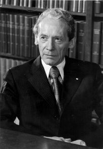
Charles Philippe Leblond was a pioneer of cell biology and stem cell research and a Canadian former professor of anatomy. Leblond is notable for developing autoradiography and his work showing how cells continuously renew themselves, regardless of age.

<span class="mw-page-title-main">Ceramide</span> Family of waxy lipid molecules

Ceramides are a family of waxy lipid molecules. A ceramide is composed of sphingosine and a fatty acid joined by an amide bond. Ceramides are found in high concentrations within the cell membrane of eukaryotic cells, since they are component lipids that make up sphingomyelin, one of the major lipids in the lipid bilayer. Contrary to previous assumptions that ceramides and other sphingolipids found in cell membrane were purely supporting structural elements, ceramide can participate in a variety of cellular signaling: examples include regulating differentiation, proliferation, and programmed cell death (PCD) of cells.

<span class="mw-page-title-main">Glucocerebrosidase</span> Mammalian protein found in humans

β-Glucocerebrosidase is an enzyme with glucosylceramidase activity that cleaves by hydrolysis the β-glycosidic linkage of the chemical glucocerebroside, an intermediate in glycolipid metabolism that is abundant in cell membranes. It is localized in the lysosome, where it remains associated with the lysosomal membrane. β-Glucocerebrosidase is 497 amino acids in length and has a molecular mass of 59,700 Da.

<span class="mw-page-title-main">Acridine orange</span> Organic dye used in biochemistry

Acridine orange is an organic compound that serves as a nucleic acid-selective fluorescent dye with cationic properties useful for cell cycle determination. Acridine orange is cell-permeable, which allows the dye to interact with DNA by intercalation, or RNA via electrostatic attractions. When bound to DNA, acridine orange is very similar spectrally to an organic compound known as fluorescein. Acridine orange and fluorescein have a maximum excitation at 502nm and 525 nm (green). When acridine orange associates with RNA, the fluorescent dye experiences a maximum excitation shift from 525 nm (green) to 460 nm (blue). The shift in maximum excitation also produces a maximum emission of 650 nm (red). Acridine orange is able to withstand low pH environments, allowing the fluorescent dye to penetrate acidic organelles such as lysosomes and phagolysosomes that are membrane-bound organelles essential for acid hydrolysis or for producing products of phagocytosis of apoptotic cells. Acridine orange is used in epifluorescence microscopy and flow cytometry. The ability to penetrate the cell membranes of acidic organelles and cationic properties of acridine orange allows the dye to differentiate between various types of cells. The shift in maximum excitation and emission wavelengths provides a foundation to predict the wavelength at which the cells will stain.

<span class="mw-page-title-main">Lipid signaling</span> Biological signaling using lipid molecules

Lipid signaling, broadly defined, refers to any biological cell signaling event involving a lipid messenger that binds a protein target, such as a receptor, kinase or phosphatase, which in turn mediate the effects of these lipids on specific cellular responses. Lipid signaling is thought to be qualitatively different from other classical signaling paradigms because lipids can freely diffuse through membranes. One consequence of this is that lipid messengers cannot be stored in vesicles prior to release and so are often biosynthesized "on demand" at their intended site of action. As such, many lipid signaling molecules cannot circulate freely in solution but, rather, exist bound to special carrier proteins in serum.

<span class="mw-page-title-main">Alveolar macrophage</span>

An alveolar macrophage, pulmonary macrophage, is a type of macrophage, a professional phagocyte, found in the airways and at the level of the alveoli in the lungs, but separated from their walls.

<span class="mw-page-title-main">Outline of cell biology</span> Overview of and topical guide to cell biology

The following outline is provided as an overview of and topical guide to cell biology:

Crystallopathy is a harmful state or disease associated with the formation and aggregation of crystals in tissues or cavities, or in other words, a heterogeneous group of diseases caused by intrinsic or environmental microparticles or crystals, promoting tissue inflammation and scarring.

References

  1. 1 2 Kroemer, G., Galluzzi, L., Vandenabeele, P., Abrams, J., Alnemri, E., Baehrecke, E., … Melino, G. (2009). "Classification of cell death: recommendations of the Nomenclature Committee on Cell Death 2009". Cell Death and Differentiation, 16(1), 3–11. (http://doi.org/10.1038/cdd.2008.150.)
  2. Encyclopædia Britannica . (2015). "Homeostasis". Accessed 22 December 2016
  3. 2016. "Naphthol AS-TR phosphate". Accessed 29 December 2016.
  4. I. Davies and D.C. Sigee (1984) Cell ageing and cell death. Pages 5-32. Cambridge University Press, New York NY.