Industrial computed tomography

Last updated
Animated set of computed tomography transmission images of a Logitech C500 webcam

Industrial computed tomography (CT) scanning is any computer-aided tomographic process, usually X-ray computed tomography, that uses irradiation to produce three-dimensional internal and external representations of a scanned object. Industrial CT scanning has been used in many areas of industry for internal inspection of components. Some of the key uses for industrial CT scanning have been flaw detection, failure analysis, metrology, assembly analysis and reverse engineering applications. [1] [2] Just as in medical imaging, industrial imaging includes both nontomographic radiography (industrial radiography) and computed tomographic radiography (computed tomography).

Contents

Types of scanners

Line beam scanner CT Scanner Line Beam.jpg
Line beam scanner

Line beam scanning is the traditional process of industrial CT scanning. [3] X-rays are produced and the beam is collimated to create a line. The X-ray line beam is then translated across the part and data is collected by the detector. The data is then reconstructed to create a 3-D volume rendering of the part.

In cone beam scanning, the part to be scanned is placed on a rotary table. [3] As the part rotates, the cone of X-rays produce a large number of 2D images that are collected by the detector. The 2D images are then processed to create a 3D volume rendering of the external and internal geometries of the part.

Cone beam scanner Cone Beam CT Scanner.png
Cone beam scanner

History

Industrial CT scanning technology was introduced in 1972 with the invention of the CT scanner for medical imaging by Godfrey Hounsfield. The invention earned him a Nobel Prize in medicine, which he shared with Allan McLeod Cormack. [4] [5] Many advances in CT scanning have allowed for its use in the industrial field for metrology in addition to the visual inspection primarily used in the medical field (medical CT scan).

Analysis and inspection techniques

Various inspection uses and techniques include part-to-CAD comparisons, part-to-part comparisons, assembly and defect analysis, void analysis, wall thickness analysis, and generation of CAD data. The CAD data can be used for reverse engineering, geometric dimensioning and tolerance analysis, and production part approval. [6]

Assembly

One of the most recognized forms of analysis using CT is for assembly, or visual analysis. CT scanning provides views inside components in their functioning position, without disassembly. Some software programs for industrial CT scanning allow for measurements to be taken from the CT dataset volume rendering. These measurements are useful for determining the clearances between assembled parts or the dimension of an individual feature.

An industrial computed tomography (CT) scan conducted on an aluminum casting to identify internal failures such as voids. All color coordinated particles within casting are voids/porosity/air pockets, which can additionally be measured and are color coordinated according to size. Industrial Computed Tomography - Failures - Void.jpg
An industrial computed tomography (CT) scan conducted on an aluminum casting to identify internal failures such as voids. All color coordinated particles within casting are voids/porosity/air pockets, which can additionally be measured and are color coordinated according to size.

Void, crack and defect detection

Flight through a 3D reconstruction of a disposable pepper grinder. Glass in blue.

Traditionally, determining defects, voids and cracks within an object would require destructive testing. CT scanning can detect internal features and flaws displaying this information in 3D without destroying the part. Industrial CT scanning (3D X-ray) is used to detect flaws inside a part such as porosity, [7] an inclusion, or a crack. [8] It has been also used to detect the origin and propagation of damages in concrete. [9]

Metal casting and moulded plastic components are typically prone to porosity because of cooling processes, transitions between thick and thin walls, and material properties. Void analysis can be used to locate, measure, and analyze voids inside plastic or metal components.

Geometric dimensioning and tolerancing analysis

Traditionally, without destructive testing, full metrology has only been performed on the exterior dimensions of components, such as with a coordinate-measuring machine (CMM) or with a vision system to map exterior surfaces. Internal inspection methods would require using a 2D X-ray of the component or the use of destructive testing. Industrial CT scanning allows for full non-destructive metrology. With unlimited geometrical complexity, 3D printing allows for complex internal features to be created with no impact on cost, such features are not accessible using traditional CMM. The first 3D printed artefact that is optimised for characterisation of form using computed tomography CT [10]

Image-based finite element methods

Image-based finite element method converts the 3D image data from X-ray computed tomography directly into meshes for finite element analysis. Benefits of this method include modelling complex geometries (e.g. composite materials) or accurately modelling "as manufactured" components at the micro-scale. [11]

The industrial computed tomography market is forecast to reach a size of USD 773.45 million to USD 1,116.5 million between 2029 and 2030. Regional trends show that strong market growth is expected, particularly in the Asia-Pacific region, but also in North America and Europe, due to strict safety regulations and preventive maintenance of industrial equipment. [12] [13]  Growth is being driven primarily by the ongoing development of CT devices and services that enable precise and non-destructive testing of components. Innovations such as the use of artificial intelligence for automated fault analyses and the development of mobile CT systems are expanding the possibilities. [14]

See also

Related Research Articles

<span class="mw-page-title-main">CT scan</span> Medical imaging procedure using X-rays to produce cross-sectional images

A computed tomography scan is a medical imaging technique used to obtain detailed internal images of the body. The personnel that perform CT scans are called radiographers or radiology technologists.

<span class="mw-page-title-main">Radiography</span> Imaging technique using ionizing and non-ionizing radiation

Radiography is an imaging technique using X-rays, gamma rays, or similar ionizing radiation and non-ionizing radiation to view the internal form of an object. Applications of radiography include medical and industrial radiography. Similar techniques are used in airport security,. To create an image in conventional radiography, a beam of X-rays is produced by an X-ray generator and it is projected towards the object. A certain amount of the X-rays or other radiation are absorbed by the object, dependent on the object's density and structural composition. The X-rays that pass through the object are captured behind the object by a detector. The generation of flat two-dimensional images by this technique is called projectional radiography. In computed tomography, an X-ray source and its associated detectors rotate around the subject, which itself moves through the conical X-ray beam produced. Any given point within the subject is crossed from many directions by many different beams at different times. Information regarding the attenuation of these beams is collated and subjected to computation to generate two-dimensional images on three planes which can be further processed to produce a three-dimensional image.

<span class="mw-page-title-main">Medical imaging</span> Technique and process of creating visual representations of the interior of a body

Medical imaging is the technique and process of imaging the interior of a body for clinical analysis and medical intervention, as well as visual representation of the function of some organs or tissues (physiology). Medical imaging seeks to reveal internal structures hidden by the skin and bones, as well as to diagnose and treat disease. Medical imaging also establishes a database of normal anatomy and physiology to make it possible to identify abnormalities. Although imaging of removed organs and tissues can be performed for medical reasons, such procedures are usually considered part of pathology instead of medical imaging.

<span class="mw-page-title-main">Nondestructive testing</span> Evaluating the properties of a material, component, or system without causing damage

Nondestructive testing (NDT) is any of a wide group of analysis techniques used in science and technology industry to evaluate the properties of a material, component or system without causing damage. The terms nondestructive examination (NDE), nondestructive inspection (NDI), and nondestructive evaluation (NDE) are also commonly used to describe this technology. Because NDT does not permanently alter the article being inspected, it is a highly valuable technique that can save both money and time in product evaluation, troubleshooting, and research. The six most frequently used NDT methods are eddy-current, magnetic-particle, liquid penetrant, radiographic, ultrasonic, and visual testing. NDT is commonly used in forensic engineering, mechanical engineering, petroleum engineering, electrical engineering, civil engineering, systems engineering, aeronautical engineering, medicine, and art. Innovations in the field of nondestructive testing have had a profound impact on medical imaging, including on echocardiography, medical ultrasonography, and digital radiography.

<span class="mw-page-title-main">Tomography</span> Imaging by sections or sectioning using a penetrative wave

Tomography is imaging by sections or sectioning that uses any kind of penetrating wave. The method is used in radiology, archaeology, biology, atmospheric science, geophysics, oceanography, plasma physics, materials science, cosmochemistry, astrophysics, quantum information, and other areas of science. The word tomography is derived from Ancient Greek τόμος tomos, "slice, section" and γράφω graphō, "to write" or, in this context as well, "to describe." A device used in tomography is called a tomograph, while the image produced is a tomogram.

<span class="mw-page-title-main">Tomographic reconstruction</span> Estimate object properties from a finite number of projections

Tomographic reconstruction is a type of multidimensional inverse problem where the challenge is to yield an estimate of a specific system from a finite number of projections. The mathematical basis for tomographic imaging was laid down by Johann Radon. A notable example of applications is the reconstruction of computed tomography (CT) where cross-sectional images of patients are obtained in non-invasive manner. Recent developments have seen the Radon transform and its inverse used for tasks related to realistic object insertion required for testing and evaluating computed tomography use in airport security.

<span class="mw-page-title-main">X-ray microtomography</span> X-ray 3D imaging method

In radiography, X-ray microtomography uses X-rays to create cross-sections of a physical object that can be used to recreate a virtual model without destroying the original object. It is similar to tomography and X-ray computed tomography. The prefix micro- is used to indicate that the pixel sizes of the cross-sections are in the micrometre range. These pixel sizes have also resulted in creation of its synonyms high-resolution X-ray tomography, micro-computed tomography, and similar terms. Sometimes the terms high-resolution computed tomography (HRCT) and micro-CT are differentiated, but in other cases the term high-resolution micro-CT is used. Virtually all tomography today is computed tomography.

Digital radiography is a form of radiography that uses x-ray–sensitive plates to directly capture data during the patient examination, immediately transferring it to a computer system without the use of an intermediate cassette. Advantages include time efficiency through bypassing chemical processing and the ability to digitally transfer and enhance images. Also, less radiation can be used to produce an image of similar contrast to conventional radiography.

<span class="mw-page-title-main">Industrial radiography</span> Type of non-destructive testing

Industrial radiography is a modality of non-destructive testing that uses ionizing radiation to inspect materials and components with the objective of locating and quantifying defects and degradation in material properties that would lead to the failure of engineering structures. It plays an important role in the science and technology needed to ensure product quality and reliability. In Australia, industrial radiographic non-destructive testing is colloquially referred to as "bombing" a component with a "bomb".

Terahertz tomography is a class of tomography where sectional imaging is done by terahertz radiation. Terahertz radiation is electromagnetic radiation with a frequency between 0.1 and 10 THz; it falls between radio waves and light waves on the spectrum; it encompasses portions of the millimeter waves and infrared wavelengths. Because of its high frequency and short wavelength, terahertz wave has a high signal-to-noise ratio in the time domain spectrum. Tomography using terahertz radiation can image samples that are opaque in the visible and near-infrared regions of the spectrum. Terahertz wave three-dimensional (3D) imaging technology has developed rapidly since its first successful application in 1997, and a series of new 3D imaging technologies have been proposed successively.

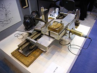
The 5DX was an automated X-ray inspection robot, which belonged to the set of automated test equipment robots and industrial robots utilizing machine vision. The 5DX was manufactured by Hewlett Packard, then later Agilent Technologies when HP was split into Hewlett Packard and Agilent Technologies in 1999. The 5DX performed a non-destructive structural test using X-ray laminography (tomography) to take 3D images of an assembled printed circuit board using 8-bit grayscale to indicate solder thickness. It was used in the assembled printed circuit board (PCB) electronics manufacturing industry to provide process feedback to a surface mount technology assembly line, as well as defect capture.

<span class="mw-page-title-main">Tomosynthesis</span> Medical imaging technique

Tomosynthesis, also digital tomosynthesis (DTS), is a method for performing high-resolution limited-angle tomography at radiation dose levels comparable with projectional radiography. It has been studied for a variety of clinical applications, including vascular imaging, dental imaging, orthopedic imaging, mammographic imaging, musculoskeletal imaging, and chest imaging.

Flat-panel Volume CT is a technique under development to make computed tomography images with improved performance. The key difference between volume CT and traditional CT is that volume CT uses a two-dimensional x-ray detector orientation, to take multiple two-dimensional images. On the other hand, the conventional CT uses a one-dimensional x-ray detector orientation to take one-dimensional x-ray images.

John Fitzallen Moore was an American physicist, the son of authors Virginia Moore and Louis Untermeyer. His last name was legally changed after his parents' divorce. His work in military electronics, communications, and spectroscopy culminated in medical electronics and x-ray products with the founding of the company Bio-Imaging Research.

<span class="mw-page-title-main">Avizo (software)</span> Software for scientific and industrial data visualization and analysis

Avizo is a general-purpose commercial software application for scientific and industrial data visualization and analysis.

<span class="mw-page-title-main">Cone beam computed tomography</span> Medical imaging technique

Cone beam computed tomography is a medical imaging technique consisting of X-ray computed tomography where the X-rays are divergent, forming a cone.

<span class="mw-page-title-main">Operation of computed tomography</span>

X-ray computed tomography operates by using an X-ray generator that rotates around the object; X-ray detectors are positioned on the opposite side of the circle from the X-ray source.

Jesse Garant Metrology Center is a part inspection company, providing NDT and metrology services using advanced imaging equipment.

<span class="mw-page-title-main">History of computed tomography</span> History of CT scanning technology

The history of X-ray computed tomography dates back to at least 1917 with the mathematical theory of the Radon transform In the early 1900s an Italian radiologist named Alessandro Vallebona invented tomography which used radiographic film to see a single slice of the body. It was not widely used until the 1930s, when Dr Bernard George Ziedses des Plantes developed a practical method for implementing the technique.

X-ray diffraction computed tomography is an experimental technique that combines X-ray diffraction with the computed tomography data acquisition approach. X-ray diffraction (XRD) computed tomography (CT) was first introduced in 1987 by Harding et al. using a laboratory diffractometer and a monochromatic X-ray pencil beam. The first implementation of the technique at synchrotron facilities was performed in 1998 by Kleuker et al.

References

  1. Flisch, A., et al. Industrial Computer Tomography in Reverse Engineering Applications. DGZfP-Proceedings BB 67-CD Paper 8, Computerized Tomography for Industrial Applications and Image Processing in Radiology, March 15–17, 1999, Berlin, Germany.
  2. Woods, Susan. "3-D CT inspection offers a full view of microparts", November 1, 2010.
  3. 1 2 Hofmann, J., Flisch, A., Obrist, A., Adaptive CT scanning-mesh based optimisation methods for industrial X-ray computer tomography applications. NDT&E International (37), 2004, pp. 271–278.
  4. Zoofan, Bahman. "3D Micro-Tomography – A Powerful Engineering Tool" Archived 2011-07-07 at the Wayback Machine . 3D Scanning Technologies. July 5, 2010.
  5. Noel, Julien. "Advantages of CT in 3D Scanning of Industrial Parts Archived 2011-07-07 at the Wayback Machine . August 18, 2010.
  6. "Reducing Preproduction Inspection Costs with Industrial (CT) Computed Tomography." Micro Manufacturing Magazine for the global micro manufacturing technology industry, August 2010.
  7. Lambert, J.; Chambers, A. R.; Sinclair, I.; Spearing, S. M. (2012). "3D damage characterisation and the role of voids in the fatigue of wind turbine blade materials". Composites Science and Technology. 72 (2): 337. doi:10.1016/j.compscitech.2011.11.023.
  8. Bull, D. J.; Helfen, L.; Sinclair, I.; Spearing, S. M.; Baumbach, T. (2013). "A comparison of multi-scale 3D X-ray tomographic inspection techniques for assessing carbon fibre composite impact damage" (PDF). Composites Science and Technology. 75: 55–61. doi:10.1016/j.compscitech.2012.12.006.
  9. Joshi, Nirmal Raj; Matsumoto, Ayumu; Asamoto, Shingo; Miura, Taito; Kawabata, Yuichiro (2022-04-01). "Investigation of the mechanical behaviour of concrete with severe delayed ettringite formation expansion focusing on internal damage propagation under various compressive loading patterns". Cement and Concrete Composites. 128: 104433. doi: 10.1016/j.cemconcomp.2022.104433 . ISSN   0958-9465. S2CID   246514058.
  10. Shah, Paras; Racasan, Radu; Bills, Paul (2016-11-01). "Comparison of different additive manufacturing methods using computed tomography". Case Studies in Nondestructive Testing and Evaluation. 6: 69–78. doi: 10.1016/j.csndt.2016.05.008 . ISSN   2214-6571.
  11. Evans, Ll. M.; Margetts, L.; Casalegno, V.; Lever, L. M.; Bushell, J.; Lowe, T.; Wallwork, A.; Young, P.; Lindemann, A. (2015-05-28). "Transient thermal finite element analysis of CFC–Cu ITER monoblock using X-ray tomography data". Fusion Engineering and Design. 100: 100–111. Bibcode:2015FusED.100..100E. doi: 10.1016/j.fusengdes.2015.04.048 . hdl: 10871/17772 .
  12. "Industrial CT Scanning Market - Computed Tomography - Size, Share & Industry Analysis". www.mordorintelligence.com. Mordor Intelligence. Retrieved 2024-04-11.
  13. "Industrial Computed Tomography Market Size Report, 2030". www.grandviewresearch.com. Grand View Research, Inc. Retrieved 2024-04-11.
  14. "Insights into trends, market development and technological innovations". www.microvista.de. Microvista GmbH. 2024-04-10. Retrieved 2024-04-11.