The tensor veli palatini muscle is a thin, triangular muscle of the head that tenses the soft palate and opens the Eustachian tube to equalise pressure in the middle ear.

A jugular foramen is one of the two large foramina (openings) in the base of the skull, located behind the carotid canal. It is formed by the temporal bone and the occipital bone. It allows many structures to pass, including the inferior petrosal sinus, three cranial nerves, the sigmoid sinus, and meningeal arteries.

The internal auditory meatus is a canal within the petrous part of the temporal bone of the skull between the posterior cranial fossa and the inner ear.

The inferior thyroid artery is an artery in the neck. It arises from the thyrocervical trunk and passes upward, in front of the vertebral artery and longus colli muscle. It then turns medially behind the carotid sheath and its contents, and also behind the sympathetic trunk, the middle cervical ganglion resting upon the vessel.

The posterior auricular artery is a small artery that arises from the external carotid artery. It ascends along the side of the head. It supplies several muscles of the neck and several structures of the head.

The inferior alveolar artery is an artery of the head. It is a branch of the maxillary artery. It descends through the infratemporal fossa as part of a neurovascular bundle with the inferior alveolar nerve and vein to the mandibular foramen where it enters and passes anteriorly inside the mandible, suplying the body of mandible and the dental pulp of the lower molar and premolar teeth. Its terminal incisor branch supplies the rest of the lower teeth. Its mental branch exits the mandibula anteriorly through the mental foramen to supply adjacent lip and skin.

The ascending pharyngeal artery is an artery of the neck that supplies the pharynx.

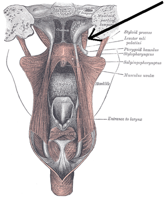
The temporal styloid process is a slender bony process of the temporal bone extending downward and forward from the undersurface of the temporal bone just below the ear. The styloid process gives attachments to several muscles, and ligaments.

The facial canal is a Z-shaped canal in the temporal bone of the skull. It extends between the internal acoustic meatus and stylomastoid foramen. It transmits the facial nerve.
The deep auricular artery is a branch of the maxillary artery. The deep auricular artery pierces the external acoustic meatus. It provides arterial supply to the skin of the external acoustic meatus, and contributes arterial supply to the tympanic membrane, and the temporomandibular joint.
The anterior tympanic artery is a branch of the maxillary artery. It passes through the petrotympanic fissure to entre the middle ear where it contributes to the formation of the circular anastomosis around the tympanic membrane. It provides arterial supply to part of the lining of the middle ear. It is accompanied by the chorda tympani nerve.
The infraorbital artery is a small artery in the head that arises from the maxillary artery and passes through the inferior orbital fissure to enter the orbit, then passes forward along the floor of the orbit, finally exiting the orbit through the infraorbital foramen to reach the face.

The posterior superior alveolar artery is a branch of the maxillary artery. It is one of two or three superior alveolar arteries. It provides arterial suply to the molar and premolar teeth, maxillary sinus and adjacent bone, and the gingiva.

The lesser petrosal nerve is the general visceral efferent (GVE) nerve conveying pre-ganglionic parasympathetic secretomotor fibers for the parotid gland from the tympanic plexus to the otic ganglion. It passes out of the tympanic cavity through the petrous part of the temporal bone into the middle cranial fossa of the cranial cavity, then exits the cranial cavity through its own canaliculus to reach the infratemporal fossa.

The tympanic canaliculus is a minute canal in the bony wedge/ridge that separates the carotid canal and jugular foramen/jugular fossa. The proximal opening of the canal is situated upon the inferior surface of the petrous part of the temporal bone; its distal opening is situated upon the floor of the tympanic cavity. The canal gives passage to the tympanic nerve i.e. tympanic branch of glossopharyngeal nerve ), and inferior tympanic artery.

The infraorbital nerve is a branch of the maxillary nerve. It arises in the pterygopalatine fossa. It passes through the inferior orbital fissure to enter the orbit. It travels through the orbit, then enters and traverses the infraorbital canal, exiting the canal at the infraorbital foramen to reach the face. It provides sensory innervation to the skin and mucous membranes around the middle of the face.
The artery of the pterygoid canal is an artery in the pterygoid canal, in the head.

The submental artery is the largest branch of the facial artery in the neck. It first runs forward under the mouth, then turns upward upon reaching the chin.

The artery to the ductus deferens is an artery in males that provides blood to the ductus deferens.
The caroticotympanic nerves are post-ganglionic sympathetic branches from the internal carotid plexus which leave the carotid canal through the wall of this canal to enter the tympanic cavity and participate in the formation of the tympanic plexus upon the promontory of tympanic cavity. They travel with the caroticotympanic artery.
