Related Research Articles
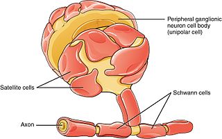
Schwann cells or neurolemmocytes are the principal glia of the peripheral nervous system (PNS). Glial cells function to support neurons and in the PNS, also include satellite cells, olfactory ensheathing cells, enteric glia and glia that reside at sensory nerve endings, such as the Pacinian corpuscle. The two types of Schwann cells are myelinating and nonmyelinating. Myelinating Schwann cells wrap around axons of motor and sensory neurons to form the myelin sheath. The Schwann cell promoter is present in the downstream region of the human dystrophin gene that gives shortened transcript that are again synthesized in a tissue-specific manner.
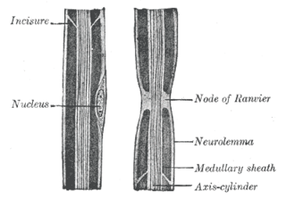
Neurilemma is the outermost nucleated cytoplasmic layer of Schwann cells that surrounds the axon of the neuron. It forms the outermost layer of the nerve fiber in the peripheral nervous system.

The nephron is the minute or microscopic structural and functional unit of the kidney. It is composed of a renal corpuscle and a renal tubule. The renal corpuscle consists of a tuft of capillaries called a glomerulus and a cup-shaped structure called Bowman's capsule. The renal tubule extends from the capsule. The capsule and tubule are connected and are composed of epithelial cells with a lumen. A healthy adult has 1 to 1.5 million nephrons in each kidney. Blood is filtered as it passes through three layers: the endothelial cells of the capillary wall, its basement membrane, and between the foot processes of the podocytes of the lining of the capsule. The tubule has adjacent peritubular capillaries that run between the descending and ascending portions of the tubule. As the fluid from the capsule flows down into the tubule, it is processed by the epithelial cells lining the tubule: water is reabsorbed and substances are exchanged ; first with the interstitial fluid outside the tubules, and then into the plasma in the adjacent peritubular capillaries through the endothelial cells lining that capillary. This process regulates the volume of body fluid as well as levels of many body substances. At the end of the tubule, the remaining fluid—urine—exits: it is composed of water, metabolic waste, and toxins.

Friedrich Gustav Jakob Henle was a German physician, pathologist, and anatomist. He is credited with the discovery of the loop of Henle in the kidney. His essay, "On Miasma and Contagia," was an early argument for the germ theory of disease. He was an important figure in the development of modern medicine.

The hair follicle is an organ found in mammalian skin. It resides in the dermal layer of the skin and is made up of 20 different cell types, each with distinct functions. The hair follicle regulates hair growth via a complex interaction between hormones, neuropeptides, and immune cells. This complex interaction induces the hair follicle to produce different types of hair as seen on different parts of the body. For example, terminal hairs grow on the scalp and lanugo hairs are seen covering the bodies of fetuses in the uterus and in some newborn babies. The process of hair growth occurs in distinct sequential stages: anagen is the active growth phase, catagen is the regression of the hair follicle phase, telogen is the resting stage, exogen is the active shedding of hair phase and kenogen is the phase between the empty hair follicle and the growth of new hair.
Artificial hair integrations, more commonly known as hair extensions, hair weaves, and fake hair add length and fullness to human hair. Hair extensions are usually clipped, glued, or sewn on natural hair by incorporating additional human or synthetic hair. These methods include tape-in extensions, clip-in or clip-on extensions, micro/nano rings, fusion method, weaving method, and wigs.

The epidermis is a single layer of cells that covers the leaves, flowers, roots and stems of plants. It forms a boundary between the plant and the external environment. The epidermis serves several functions: it protects against water loss, regulates gas exchange, secretes metabolic compounds, and absorbs water and mineral nutrients. The epidermis of most leaves shows dorsoventral anatomy: the upper (adaxial) and lower (abaxial) surfaces have somewhat different construction and may serve different functions. Woody stems and some other stem structures such as potato tubers produce a secondary covering called the periderm that replaces the epidermis as the protective covering.

The cervical loop is the location on an enamel organ in a developing tooth where the outer enamel epithelium and the inner enamel epithelium join. The cervical loop is a histologic term indicating a specific epithelial structure at the apical side of the tooth germ, consisting of loosely aggregated stellate reticulum in the center surrounded by stratum intermedium. These tissues are enveloped by a basal layer of epithelium known on the outside of the tooth as outer enamel epithelium and on the inside as inner enamel epithelium. During root formation the inner layers of epithelium disappear and only the basal layers are left creating Hertwig's epithelial root sheath (HERS). At this point it is usually referred to as HERS instead of the cervical loop to indicate the structural difference.

The pileipellis is the uppermost layer of hyphae in the pileus of a fungal fruit body. It covers the trama, the fleshy tissue of the fruit body. The pileipellis is more or less synonymous with the cuticle, but the cuticle generally describes this layer as a macroscopic feature, while pileipellis refers to this structure as a microscopic layer. Pileipellis type is an important character in the identification of fungi. Pileipellis types include the cutis, trichoderm, epithelium, and hymeniderm types.
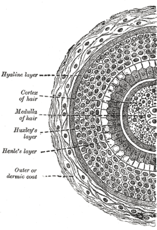
Huxley's layer is the second layer of the inner root sheath of the hair and consists of one or two layers of horny, flattened, nucleated cells. It lies between Henle's layer and the cuticle.
Henle's layer is the third and the outermost layer of the inner root sheath of the hair follicle, consisting of a single layer of cubical cells with clear flattened nuclei. It is named after German physician, pathologist and anatomist Friedrich Gustav Jakob Henle.
In mammals, trichocytes are the specialized epithelial cells from which the highly mechanically resilient tissues hair and nails are formed. They can be identified by the fact that they express "hard", "trichocyte" or "hair" keratin proteins. These are modified keratins containing large amounts of the amino acid cysteine, which facilitates chemical cross-linking of these proteins to form the tough material from which hair and nail is composed. These cells give rise to non-hair non-keratinized IRSC as well.
This page provides a glossary of plant morphology. Botanists and other biologists who study plant morphology use a number of different terms to classify and identify plant organs and parts that can be observed using no more than a handheld magnifying lens. This page provides help in understanding the numerous other pages describing plants by their various taxa. The accompanying page—Plant morphology—provides an overview of the science of the external form of plants. There is also an alphabetical list: Glossary of botanical terms. In contrast, this page deals with botanical terms in a systematic manner, with some illustrations, and organized by plant anatomy and function in plant physiology.
Rhizodermis is the root epidermis, the outermost primary cell layer of the root.
The inner or epidermic coat of the hair follicle is closely adherent to the root of the hair, and consists of two strata named respectively the outer and inner root sheaths.
The outer root sheath or external root sheath of the hair follicle encloses the inner root sheath and hair shaft. It is continuous with the basal layer of the interfollicular epidermis (skin).
A cuticle, or cuticula, is any of a variety of tough but flexible, non-mineral outer coverings of an organism, or parts of an organism, that provide protection. Various types of "cuticle" are non-homologous, differing in their origin, structure, function, and chemical composition.

The hair cuticle is the outermost part of the hair shaft. It is formed from dead cells, overlapping in layers, which form scales that strengthen and protect the hair shaft.

Loose anagen syndrome, also known as loose anagen hair syndrome, is a hair disorder related to dermatology. It is characterised by the easy and pain free detachment of anagen staged hairs from the scalp. This hair condition can be spontaneous or genetically inherited.
Root sheath may refer to any of these biological structures:
References
- ↑ James, William; Berger, Timothy; Elston, Dirk (2005) Andrews' Diseases of the Skin: Clinical Dermatology (10th ed.). Saunders. Page 8. ISBN 0-7216-2921-0.
- 1 2 Bailey, Frederick Randolph (1910), A text-book of histology, W. Wood, University of California, pp. 359–560
- ↑ Rajiv S Joshi (2011). "The Inner Root Sheath and the Men Associated with it Eponymically". Int J Trichology. 3 (1): 57–62. doi: 10.4103/0974-7753.82119 . PMC 3129131 . PMID 21769243.