Interstitial nephritis

Last updated
Interstitial nephritis
Acute Interstitial Nephritis.jpg
Acute interstitial nephritis on light microscopy
Specialty Nephrology   OOjs UI icon edit-ltr-progressive.svg

Interstitial nephritis, also known as tubulointerstitial nephritis, is inflammation of the area of the kidney known as the renal interstitium, which consists of a collection of cells, extracellular matrix, and fluid surrounding the renal tubules. It is also known as intestinal nephritis because the clinical picture may include mesenteric lymphadenitis in some cases of acute pyelonephritis (mostly due to use of NSAIDs). More specifically, in case of recurrent urinary tract infection, secondary infection can spread to adjacent intestine. [1] In addition to providing a scaffolding support for the tubular architecture, the interstitium has been shown to participate in the fluid and electrolyte exchange as well as endocrine functions of the kidney. [1]

Contents

There are a variety of known factors that can provoke the inflammatory process within the renal interstitium, including pharmacologic, environmental, infectious and systemic disease contributors. The spectrum of disease presentation can range from an acute process to a chronic condition with progressive tubular cell damage and renal dysfunction.

Signs and symptoms

Interstitial nephritis may present with a variety of signs and symptoms, many of these nonspecific. Fever is the most common, occurring in 30-50% of patients, particularly those with drug-induced interstitial nephritis. [2] Other general symptoms that occur with variable frequency include nausea, vomiting, fatigue, lack of appetite, and weight loss. More specific symptoms, such as flank pain, pain with urination, and visible blood in the urine, as well as signs like hypertension can be helpful in increasing suspicion for the diagnosis. [3] The "classic" triad of symptoms reported in early documented cases consisted of rash, joint pain, and increased eosinophils in the blood; however, more recent epidemiology suggests that this grouping of symptoms only occurs in a small minority (5-10%) of patients. [4] With modern drugs causing between 70 and 90% of current cases, [5] [6] the possibility of a change in presentation exists.

Causes

Common causes include infection, or reaction to medication such as an analgesic, anti-inflammatory, [7] or antibiotics such as methicillin (meticillin). Reaction to medications causes 71% [5] to 92% [6] of cases.

Drugs


Examples of Drugs Associated With Interstitial Nephritis [2] [8]
ClassExamples
Antibioticβ-lactams (e.g. penicillins, cephalosporins), sulfonamides (e.g. trimethoprim-sulfamethoxazole), fluoroquinolones (e.g. ciprofloxacin), macrolides (e.g. erythromycin), anti-tuberculins (e.g. rifampin, ethambutol), chloramphenicol
Anti-inflammatoryNonsteroidal antiinflammatory drugs (e.g. ibuprofen, naproxen), selective COX-2 inhibitors (e.g. celecoxib)
AntiviralAcyclovir, atazanavir, abacavir, indinavir
AnalgesicAspirin, acetaminophen
GastrointestinalProton pump inhibitors (e.g. omeprazole, lansoprazole), H2-receptor blockers (e.g. cimetidine), 5-aminosalicylates (e.g. mesalamine)
AntiseizurePhenytoin, carbamazepine, phenobarbital
DiureticHydrochlorothiazide, furosemide, triamterene, chlorthalidone
ChemotherapyTyrosine kinase inhibitors (e.g. sunitinib), checkpoint inhibitors (e.g. ipilimumab, nivolumab, pembrolizumab, atezolizumab)
Contrast agentGadolininum based contrast agent [9] [10]
OtherAllopurinol, Chinese herbs

This disease is also caused by other diseases and toxins that damage the kidney. Both acute and chronic tubulointerstitial nephritis can be caused by a bacterial infection in the kidneys known as pyelonephritis, but the most common cause is by an adverse reaction to a medication. The medications that are known to cause this sort of reaction are β-lactam antibiotics such as penicillin [11] and cephalexin, and nonsteroidal anti-inflammatory drugs (aspirin less frequently than others), as well as proton-pump inhibitors, rifampicin, sulfa medications, fluoroquinolones, diuretics, allopurinol, mesalamine, and phenytoin. The time between exposure to the drug and the development of acute tubulointerstitial nephritis can be anywhere from 5 days to 5 months (fenoprofen-induced).[ citation needed ]

Diagnosis

The condition can appear without symptoms. When present they may appear widely varied and can occur rapidly or gradually. [5] [12] [13] [14] [15]

When caused by an allergic reaction, the symptoms of acute tubulointerstitial nephritis are fever (27% of patients), [5] rash (15% of patients), [5] and enlarged kidneys. Some people experience dysuria, and lower back pain.

In chronic tubulointerstitial nephritis the patient can experience symptoms such as nausea, vomiting, fatigue, and weight loss. Other conditions that may develop include a high concentration of potassium in the blood, metabolic acidosis, and kidney failure.[ citation needed ]

Blood tests

About 23% of patients have a high level of eosinophils in the blood. [5]

Urinary findings

Urinary findings include:

Pathology

While non-invasive patient evaluation (physical examination, blood and urine testing, imaging studies) can be suggestive, the only way to definitively diagnosis interstitial nephritis is with a tissue diagnosis obtained by kidney biopsy. Pathologic examination will reveal the presence of interstitial edema and inflammatory infiltration with various white blood cells, including neutrophils, eosinophils, and lymphocytes. Generally, blood vessels and glomeruli are not affected. Electron microscopy shows mitochondrial damage in the tubular epithelial cells, vacuoles in the cytoplasm, and enlarged endoplasmic reticulum. [23]

Gallium scan

The sensitivity of an abnormal gallium scan has been reported to range from 60% [24] to 100%. [25] A study of Gallium scan in 76 patients [23 with AIN, 8 with biopsy-proven AIN] showed an AUC of 0.75. [26]

Novel biomarkers

Given the challenges with clinical diagnosis of AIN due to lack of clinical features and lack of accuracy of existing tests, there has been significant interest in identifying non-invasive biomarkers for this disease. One study showed that monocyte chemotactic protein-1 (chemokine CCL-2) and neutrophil gelatinase associated lipocalin (NGAL) were higher in patients with AIN than in controls (in this case healthy participants). [27] A more recent study, showed that urine cytokines interleukin-9 and tumor necrosis factor-α were higher in patients with AIN than in controls without AIN who underwent a biopsy for evaluation of acute kidney injury and showed an AUC of 0.79. [28] This study also showed that the biomarkers had higher AUC than the clinician's pre-biopsy impression of AIN and, when added to a model of clinical variables, showed an AUC of 0.84. In a subsequent study, interleukin-9 was also shown to identify patients most likely to respond to corticosteroid therapy. [29]

Treatment

Treatment consists of addressing the cause, such as by removing an offending drug. There is no clear evidence that corticosteroids help. [6] Nutrition therapy consists of adequate fluid intake, which can require several liters of extra fluid. [30]

Prognosis

The kidneys are the only body system that are directly affected by tubulointerstitial nephritis. Kidney function is usually reduced; the kidneys can be just slightly dysfunctional, or fail completely.[ citation needed ]

In chronic tubulointerstitial nephritis, the most serious long-term effect is kidney failure. When the proximal tubule is injured, sodium, potassium, bicarbonate, uric acid, and phosphate reabsorption may be reduced or changed, resulting in low bicarbonate, known as metabolic acidosis, low potassium, low uric acid known as hypouricemia, and low phosphate known as hypophosphatemia. Damage to the distal tubule may cause loss of urine-concentrating ability and polyuria.[ citation needed ]

In most cases of acute tubulointerstitial nephritis, the function of the kidneys will return after the harmful drug is discontinued, or when the underlying disease is cured by treatment.

If the illness is caused by an allergic reaction, a corticosteroid may speed the recovery kidney function; however, this is often not the case.

Chronic tubulointerstitial nephritis has no cure. Some patients may require dialysis. Eventually, a kidney transplant may be needed.[ citation needed ]

Epidemiology

Interstitial nephritis is uncommon (<1% incidence) in patients without any symptoms but occurs in about 10-15% of hospitalized patients with acute kidney injury of unknown cause. [2] While it can occur in patients of all ages, it is more common in elderly patients, perhaps due to increased exposure to drugs and other triggering causes. [2]

Related Research Articles

<span class="mw-page-title-main">Nephrology</span> Medical study concerned with the kidneys

Nephrology is a specialty for both adult internal medicine and pediatric medicine that concerns the study of the kidneys, specifically normal kidney function and kidney disease, the preservation of kidney health, and the treatment of kidney disease, from diet and medication to renal replacement therapy. The word "renal" is an adjective meaning "relating to the kidneys", and its roots are French or late Latin. Whereas according to some opinions, "renal" and "nephro" should be replaced with "kidney" in scientific writings such as "kidney medicine" or "kidney replacement therapy", other experts have advocated preserving the use of renal and nephro as appropriate including in "nephrology" and "renal replacement therapy", respectively.

<span class="mw-page-title-main">Proteinuria</span> Presence of an excess of serum proteins in the urine

Proteinuria is the presence of excess proteins in the urine. In healthy persons, urine contains very little protein, less than 150 mg/day; an excess is suggestive of illness. Excess protein in the urine often causes the urine to become foamy. Severe proteinuria can cause nephrotic syndrome in which there is worsening swelling of the body.

<span class="mw-page-title-main">Kidney failure</span> Disease where the kidneys fail to adequately filter waste products from the blood

Kidney failure, also known as end-stage renal disease (ESRD), is a medical condition in which the kidneys can no longer adequately filter waste products from the blood, functioning at less than 15% of normal levels. Kidney failure is classified as either acute kidney failure, which develops rapidly and may resolve; and chronic kidney failure, which develops slowly and can often be irreversible. Symptoms may include leg swelling, feeling tired, vomiting, loss of appetite, and confusion. Complications of acute and chronic failure include uremia, hyperkalemia, and volume overload. Complications of chronic failure also include heart disease, high blood pressure, and anaemia.

<span class="mw-page-title-main">Nephritis</span> Inflammation of the kidneys

Nephritis is inflammation of the kidneys and may involve the glomeruli, tubules, or interstitial tissue surrounding the glomeruli and tubules. It is one of several different types of nephropathy.

<span class="mw-page-title-main">Kidney disease</span> Damage to or disease of a kidney

Kidney disease, or renal disease, technically referred to as nephropathy, is damage to or disease of a kidney. Nephritis is an inflammatory kidney disease and has several types according to the location of the inflammation. Inflammation can be diagnosed by blood tests. Nephrosis is non-inflammatory kidney disease. Nephritis and nephrosis can give rise to nephritic syndrome and nephrotic syndrome respectively. Kidney disease usually causes a loss of kidney function to some degree and can result in kidney failure, the complete loss of kidney function. Kidney failure is known as the end-stage of kidney disease, where dialysis or a kidney transplant is the only treatment option.

<span class="mw-page-title-main">Acute kidney injury</span> Medical condition

Acute kidney injury (AKI), previously called acute renal failure (ARF), is a sudden decrease in kidney function that develops within 7 days, as shown by an increase in serum creatinine or a decrease in urine output, or both.

<span class="mw-page-title-main">IgA nephropathy</span> Disease of the kidney

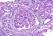
IgA nephropathy (IgAN), also known as Berger's disease, or synpharyngitic glomerulonephritis, is a disease of the kidney and the immune system; specifically it is a form of glomerulonephritis or an inflammation of the glomeruli of the kidney. Aggressive Berger's disease can attack other major organs, such as the liver, skin and heart.

<span class="mw-page-title-main">Cystinuria</span> Amino acid metabolic disorder involving cystine stones forming in the kidneys, ureter, and bladder

Cystinuria is an inherited autosomal recessive disease characterized by high concentrations of the amino acid cystine in the urine, leading to the formation of cystine stones in the kidneys, ureters, and bladder. It is a type of aminoaciduria. "Cystine", not "cysteine," is implicated in this disease; the former is a dimer of the latter.

<span class="mw-page-title-main">Pyelonephritis</span> Medical condition

Pyelonephritis is inflammation of the kidney, typically due to a bacterial infection. Symptoms most often include fever and flank tenderness. Other symptoms may include nausea, burning with urination, and frequent urination. Complications may include pus around the kidney, sepsis, or kidney failure.

<span class="mw-page-title-main">Bartter syndrome</span> Medical condition

Bartter syndrome (BS) is a rare inherited disease characterised by a defect in the thick ascending limb of the loop of Henle, which results in low potassium levels (hypokalemia), increased blood pH (alkalosis), and normal to low blood pressure. There are two types of Bartter syndrome: neonatal and classic. A closely associated disorder, Gitelman syndrome, is milder than both subtypes of Bartter syndrome.

Phosphate nephropathy or nephrocalcinosis is an adverse renal condition that arises with a formation of phosphate crystals within the kidney's tubules. This renal insufficiency is associated with the use of oral sodium phosphate (OSP) such as C.B. Fleet's Phospho soda and Salix's Visocol, for bowel cleansing prior to a colonoscopy.   

<span class="mw-page-title-main">Analgesic nephropathy</span> Medical condition

Analgesic nephropathy is injury to the kidneys caused by analgesic medications such as aspirin, bucetin, phenacetin, and paracetamol. The term usually refers to damage induced by excessive use of combinations of these medications, especially combinations that include phenacetin. It may also be used to describe kidney injury from any single analgesic medication.

<span class="mw-page-title-main">Urine specific gravity</span> Topic related to human medicine

Specific gravity, in the context of clinical pathology, is a urinalysis parameter commonly used in the evaluation of kidney function and can aid in the diagnosis of various renal diseases.

<span class="mw-page-title-main">Nephrocalcinosis</span> Medical condition caused by the deposition of calcium salts in the kidneys

Nephrocalcinosis, once known as Albright's calcinosis after Fuller Albright, is a term originally used to describe the deposition of poorly soluble calcium salts in the renal parenchyma due to hyperparathyroidism. The term nephrocalcinosis is used to describe the deposition of both calcium oxalate and calcium phosphate. It may cause acute kidney injury. It is now more commonly used to describe diffuse, fine, renal parenchymal calcification in radiology. It is caused by multiple different conditions and is determined by progressive kidney dysfunction. These outlines eventually come together to form a dense mass. During its early stages, nephrocalcinosis is visible on x-ray, and appears as a fine granular mottling over the renal outlines. It is most commonly seen as an incidental finding with medullary sponge kidney on an abdominal x-ray. It may be severe enough to cause renal tubular acidosis or even end stage kidney disease, due to disruption of the kidney tissue by the deposited calcium salts.

Primary hyperoxaluria is a rare condition, resulting in increased excretion of oxalate, with oxalate stones being common.

<span class="mw-page-title-main">Cholesterol embolism</span> Medical condition

Cholesterol embolism occurs when cholesterol is released, usually from an atherosclerotic plaque, and travels as an embolus in the bloodstream to lodge causing an obstruction in blood vessels further away. Most commonly this causes skin symptoms, gangrene of the extremities and sometimes kidney failure; problems with other organs may arise, depending on the site at which the cholesterol crystals enter the bloodstream. When the kidneys are involved, the disease is referred to as atheroembolic renal disease. The diagnosis usually involves biopsy from an affected organ. Cholesterol embolism is treated by removing the cause and giving supportive therapy; statin drugs have been found to improve the prognosis.

Urologic diseases or conditions include urinary tract infections, kidney stones, bladder control problems, and prostate problems, among others. Some urologic conditions do not affect a person for that long and some are lifetime conditions. Kidney diseases are normally investigated and treated by nephrologists, while the specialty of urology deals with problems in the other organs. Gynecologists may deal with problems of incontinence in women.

Glomerulonephrosis is a non-inflammatory disease of the kidney (nephrosis) presenting primarily in the glomerulus as nephrotic syndrome. The nephron is the functional unit of the kidney and it contains the glomerulus, which acts as a filter for blood to retain proteins and blood lipids. Damage to these filtration units results in important blood contents being released as waste in urine. This disease can be characterized by symptoms such as fatigue, swelling, and foamy urine, and can lead to chronic kidney disease and ultimately end-stage renal disease, as well as cardiovascular diseases. Glomerulonephrosis can present as either primary glomerulonephrosis or secondary glomerulonephrosis.

<span class="mw-page-title-main">Distal renal tubular acidosis</span> Medical condition

Distal renal tubular acidosis (dRTA) is the classical form of RTA, being the first described. Distal RTA is characterized by a failure of acid secretion by the alpha intercalated cells of the distal tubule and cortical collecting duct of the distal nephron. This failure of acid secretion may be due to a number of causes. It leads to relatively alkaline urine, due to the kidney's inability to acidify the urine to a pH of less than 5.3.

Malarial nephropathy is kidney failure attributed to malarial infection. Among various complications due to infection, renal-related disorders are often the most life-threatening. Including malaria-induced renal lesions, infection may lead to both tubulointerstitial damage and glomerulonephritis. In addition, malarial acute kidney failure has emerged as a serious problem due to its high mortality rate in non-immune adult patients.

References

  1. 1 2 Zeisberg, Michael; Kalluri, Reghu (October 2015). "Physiology of the Renal Interstitium". Clinical Journal of the American Society of Nephrology. 10 (10): 1831–1840. doi:10.2215/CJN.00640114. PMC   4594057 . PMID   25813241.
  2. 1 2 3 4 Brewster, Ursula C.; Rastegar, Asghar (2014), "Acute Interstitial Nephritis", National Kidney Foundation Primer on Kidney Diseases (6th ed.), Elsevier, pp. 312–317, doi:10.1016/b978-1-4557-4617-0.00035-2, ISBN   9781455746170
  3. Meyers, Catherine M. (2014), "Chronic Tubulointerstitial Disease", National Kidney Foundation Primer on Kidney Diseases, Elsevier, pp. 390–396, doi:10.1016/b978-1-4557-4617-0.00045-5, ISBN   9781455746170
  4. Brewster, Ursula C.; Rastegar, Asghar (2014), "Acute Interstitial Nephritis", National Kidney Foundation Primer on Kidney Diseases, Elsevier, pp. 312–317, doi:10.1016/b978-1-4557-4617-0.00035-2, ISBN   9781455746170
  5. 1 2 3 4 5 6 Baker R, Pusey C (2004). "The changing profile of acute tubulointerstitial nephritis". Nephrol Dial Transplant. 19 (1): 8–11. doi: 10.1093/ndt/gfg464 . PMID   14671029.
  6. 1 2 3 Clarkson M, Giblin L, O'Connell F, O'Kelly P, Walshe J, Conlon P, O'Meara Y, Dormon A, Campbell E, Donohoe J (2004). "Acute interstitial nephritis: clinical features and response to corticosteroid therapy". Nephrol Dial Transplant. 19 (11): 2778–83. doi:10.1093/ndt/gfh485. PMID   15340098.
  7. National Kidney Organization. “Heavy or long-term use of some of these medicines, such as ibuprofen, naproxen, and higher dose aspirin, can cause chronic kidney disease known as chronic interstitial nephritis.”
  8. Praga, Manuel; Appel, Gerald (2018-11-01). "Clinical manifestations and diagnosis of acute interstitial nephritis". UpToDate. Retrieved 2018-12-10.
  9. Elmholdt, T. R.; Olesen, A. B.; Jørgensen, B.; Kvist, S.; Skov, L.; Thomsen, H. S.; Marckmann, P.; Pedersen, M. (2013). "Nephrogenic Systemic Fibrosis in Denmark– A Nationwide Investigation". PLOS ONE. 8 (12): e82037. Bibcode:2013PLoSO...882037E. doi: 10.1371/journal.pone.0082037 . PMC   3857209 . PMID   24349178.
  10. Akgun, Hulya; Gonlusen, Gulfiliz; Cartwright, Jr, Joiner; Suki, Wadi N. (2006-09-01). "Are Gadolinium-Based Contrast Media Nephrotoxic?: A Renal Biopsy Study". Archives of Pathology & Laboratory Medicine. 130 (9): 1354–1357. doi:10.5858/2006-130-1354-AGCMNA. ISSN   0003-9985. PMID   16948524 . Retrieved 2022-07-25.{{cite journal}}: CS1 maint: multiple names: authors list (link)
  11. Ohlsson, Arne; Shah, Vibhuti S; Ohlsson, Arne (2014). "Intrapartum antibiotics for known maternal Group B streptococcal colonization". Cochrane Database of Systematic Reviews (6): CD007467. doi:10.1002/14651858.CD007467.pub4. PMID   24915629.
  12. 1 2 Rossert J (2001). "Drug-induced acute interstitial nephritis". Kidney Int. 60 (2): 804–17. doi: 10.1046/j.1523-1755.2001.060002804.x . PMID   11473672.
  13. Pusey C, Saltissi D, Bloodworth L, Rainford D, Christie J (1983). "Drug associated acute interstitial nephritis: clinical and pathological features and the response to high dose steroid therapy". Q J Med. 52 (206): 194–211. PMID   6604293.
  14. Handa S (1986). "Drug-induced acute interstitial nephritis: report of 10 cases". CMAJ. 135 (11): 1278–81. PMC   1491384 . PMID   3779558.
  15. Buysen J, Houthoff H, Krediet R, Arisz L (1990). "Acute interstitial nephritis: a clinical and morphological study in 27 patients". Nephrol Dial Transplant. 5 (2): 94–9. doi:10.1093/ndt/5.2.94. PMID   2113219.
  16. Schwarz A, Krause P, Kunzendorf U, Keller F, Distler A (2000). "The outcome of acute interstitial nephritis risk factors for the transition from acute to chronic interstitial nephritis". Clin Nephrol. 54 (3): 179–90. PMID   11020015.
  17. Muriithi, A. K.; Nasr, S. H.; Leung, N. (2013). "Utility of urine eosinophils in the diagnosis of acute interstitial nephritis". Clinical Journal of the American Society of Nephrology. 8 (11): 1857–62. doi:10.2215/CJN.01330213. PMC   3817898 . PMID   24052222.
  18. Perazella, M. A.; Bomback, A. S. (2013). "Urinary eosinophils in AIN: Farewell to an old biomarker?". Clinical Journal of the American Society of Nephrology. 8 (11): 1841–3. doi:10.2215/CJN.08620813. PMC   3817912 . PMID   24052220.
  19. Lins R, Verpooten G, De Clerck D, De Broe M (1986). "Urinary indices in acute interstitial nephritis". Clin Nephrol. 26 (3): 131–3. PMID   3769228.
  20. Fogazzi, G. B.; Ferrari, B.; Garigali, G.; Simonini, P.; Consonni, D. (2012). "Urinary sediment findings in acute interstitial nephritis". American Journal of Kidney Diseases. 60 (2): 330–2. doi:10.1053/j.ajkd.2012.05.002. PMID   22677261.
  21. 1 2 Frank B. Cortazar, Zoe A. Kibbelaar, Ilya G. Glezerman, Ala Abudayyeh, Omar Mamlouk, Shveta S. Motwani, Naoka Murakami, Sandra M. Herrmann, Sandhya Manohar, Anushree C. Shirali, Abhijat Kitchlu, Shayan Shirazian, Amer Assal, Anitha Vijayan, Amanda DeMauro Renaghan, David I. Ortiz-Melo, Sunil Rangarajan, A. Bilal Malik, Jonathan J. Hogan, Alex R. Dinh, Daniel Sanghoon Shin, Kristen A. Marrone, Zain Mithani, Douglas B. Johnson, Afrooz Hosseini, Deekchha Uprety, Shreyak Sharma, Shruti Gupta, Kerry L. Reynolds, Meghan E. Sise, David E. Leaf JASN Feb 2020, 31 (2) 435–446; DOI: 10.1681/ASN.2019070676
  22. Urinary Sediment Findings in Acute Interstitial Nephritis Fogazzi, Giovanni B. et al. American Journal of Kidney Diseases, Volume 60, Issue 2, 330 - 332
  23. Alon, Uri S. (2015-11-12), "Pediatric Tubulointerstitial Nephritis", Pediatric Nephrology, Springer Berlin Heidelberg, pp. 1407–1428, doi:10.1007/978-3-662-43596-0_40, ISBN   9783662435953
  24. Graham G, Lundy M, Moreno A (1983). "Failure of Gallium-67 scintigraphy to identify reliably noninfectious interstitial nephritis: concise communication". J Nucl Med. 24 (7): 568–70. PMID   6864309.
  25. Linton A, Richmond J, Clark W, Lindsay R, Driedger A, Lamki L (1985). "Gallium67 scintigraphy in the diagnosis of acute renal disease". Clin Nephrol. 24 (2): 84–7. PMID   3862487.
  26. Graham F, Lord M, Froment D, Cardinal H, Bollée G. The use of gallium-67 scintigraphy in the diagnosis of acute interstitial nephritis. Clin Kidney J. 2016;9(1):76-81. doi:10.1093/ckj/sfv129
  27. Wu Y, Yang L, Su T, Wang C, Liu G, Li XM. Pathological significance of a panel of urinary biomarkers in patients with drug-induced tubulointerstitial nephritis. Clin J Am Soc Nephrol. 2010;5(11):1954-1959. doi:10.2215/CJN.02370310
  28. Moledina DG, Wilson FP, Pober JS, et al. Urine TNF-α and IL-9 for clinical diagnosis of acute interstitial nephritis. JCI Insight. 2019;4(10):e127456. Published 2019 May 16. doi:10.1172/jci.insight.127456
  29. Dennis G Moledina, F Perry Wilson, Lidiya Kukova, Wassim Obeid, Randy Luciano, Michael Kuperman, Gilbert W Moeckel, Michael Kashgarian, Mark A Perazella, Lloyd G Cantley, Chirag R Parikh, Urine interleukin-9 and tumor necrosis factor-α for prognosis of human acute interstitial nephritis, Nephrology Dialysis Transplantation, 2020;, gfaa169, https://doi.org/10.1093/ndt/gfaa169
  30. Mahan KL, Escott-Stump S (2003). "39" . In Alexopolos Y (ed.). Krause's Food, Nutrition, & Diet Therapy (11th ed.). Philadelphia Pennsylvania: Saunders. p.  968. ISBN   978-0-7216-9784-0.