Kasugamycin

Last updated
Kasugamycin
Kasugamycin structure.svg
Names
IUPAC name
2-amino-2-[(2R,3S,5S,6R)-5-amino-2-methyl-6-[(2R,3S,5S,6S)-2,3,4,5,6-pentahydroxycyclohexyl]oxyoxan-3-yl]iminoacetic acid
Other names
Kasumin; 3-O-[2-Amino-4-[(carboxyiminomethyl)amino]-2,3,4,6-tetradeoxy-D-arabino-hexopyranosyl]-D-chiro-inositol
Identifiers
3D model (JSmol)
ChEBI
ChemSpider
ECHA InfoCard 100.116.563 OOjs UI icon edit-ltr-progressive.svg
KEGG
PubChem CID
UNII
  • InChI=1S/C14H25N3O9/c1-3-5(17-12(16)13(23)24)2-4(15)14(25-3)26-11-9(21)7(19)6(18)8(20)10(11)22/h3-11,14,18-22H,2,15H2,1H3,(H2,16,17)(H,23,24)/t3-,4+,5+,6-,7+,8+,9-,10+,11+,14-/m1/s1 Yes check.svgY
    Key: PVTHJAPFENJVNC-MHRBZPPQSA-N Yes check.svgY
  • InChI=1/C14H25N3O9/c1-3-5(17-12(16)13(23)24)2-4(15)14(25-3)26-11-9(21)7(19)6(18)8(20)10(11)22/h3-11,14,18-22H,2,15H2,1H3,(H2,16,17)(H,23,24)/t3-,4+,5+,6-,7+,8+,9-,10+,11+,14-/m1/s1
    Key: PVTHJAPFENJVNC-MHRBZPPQBX
  • OC(=O)C(=N)N[C@H]2C[C@H](N)[C@@H](O[C@H]1[C@H](O)[C@@H](O)[C@@H](O)[C@H](O)[C@@H]1O)O[C@@H]2C
Properties
C14H25N3O9
Molar mass 379.366 g·mol−1
Except where otherwise noted, data are given for materials in their standard state (at 25 °C [77 °F], 100 kPa).
X mark.svgN  verify  (what is  Yes check.svgYX mark.svgN ?)

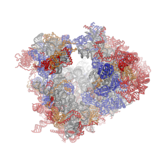
Kasugamycin (Ksg) is an aminoglycoside antibiotic that was originally isolated in 1965, from Streptomyces kasugaensis , a Streptomyces strain found near the Kasuga shrine in Nara, Japan. Kasugamycin was discovered by Hamao Umezawa, who also discovered kanamycin and bleomycin, as a drug that prevent growth of a fungus causing rice blast disease. It was later found to inhibit bacterial growth also. It exists as a white, crystalline substance with the chemical formula C14H28ClN3O10 (kasugamycin hydrochloride). It is also known as kasumin. [1]

Contents

Mechanism of action

Like many of the known natural antibiotics, kasugamycin inhibits proliferation of bacteria by tampering with their ability to make new proteins, the ribosome being the major target. Kasugamycin inhibits protein synthesis at the step of translation initiation. Kasugamycin inhibition is thought to occur by direct competition with initiator transfer RNA. Recent experiments suggest that kasugamycin indirectly induces dissociation of P-site-bound fMet-tRNAfMet from 30S subunits through perturbation of the mRNA, thereby interfering with translation initiation.[ citation needed ]

Kasugamycin specifically inhibits translation initiation of canonical but not of leaderless mRNA. For initiation on leaderless mRNA, the overlap between mRNA and kasugamycin is reduced and the binding of tRNA is further stabilized by the presence of the 50S subunit, minimizing Ksg efficacy. Kasugamycin also induces the formation of unusual 61S ribosomes in vivo, which are proficient in selectively translating leaderless mRNA. 61S particles are stable and are devoid of more than six proteins of the small subunit, including the functionally important proteins S1 and S12.[ citation needed ]

Structural basis for kasugamycin action

The structure of the kasugamycin-70S ribosome complex from Escherichia coli has been determined by X-ray crystallography at 3.5-A resolution. The drug binds within the messenger RNA channel of the 30S subunit between the universally conserved G926 and A794 nucleotides in 16S ribosomal RNA, which are sites of kasugamycin resistance. The kasugamycin binding sites are present on top of helix 44 (h44), spanning the region between h24 and h28, which contacts the conserved nucleotides A794 and G926. Neither binding position overlaps with P-site tRNA. Instead, kasugamycin mimics the codon nucleotides at the P and E sites by binding within the path of the mRNA, thus perturbing the mRNA-tRNA codon-anticodon interaction.[ citation needed ]

Resistance

Low level resistance to kasugamycin is acquired by mutations in the 16S rRNA methyltransferase KsgA which methylates the nucleotides A1518 and A1519 in 16S rRNA. Spontaneous ksgA mutations conferring a modest level of resistance to kasugamycin occur at a high frequency of 10−6. Once cells acquire the ksgA mutations, they produce high-level kasugamycin resistance at an extraordinarily high frequency (100-fold greater frequency than that observed in the ksgA+ strain).[ citation needed ]

Surprisingly, kasugamycin resistance mutations do not inhibit binding of the drug to the ribosome. Present structural and biochemical results indicate that inhibition by kasugamycin and kasugamycin resistance are closely linked to the structure of the mRNA at the junction of the peptidyl-tRNA and exit-tRNA sites (P and E sites)[ citation needed ]

Related Research Articles

<span class="mw-page-title-main">Protein biosynthesis</span> Assembly of proteins inside biological cells

Protein biosynthesis is a core biological process, occurring inside cells, balancing the loss of cellular proteins through the production of new proteins. Proteins perform a number of critical functions as enzymes, structural proteins or hormones. Protein synthesis is a very similar process for both prokaryotes and eukaryotes but there are some distinct differences.

<span class="mw-page-title-main">Ribosome</span> Intracellular organelle consisting of RNA and protein functioning to synthesize proteins

Ribosomes are macromolecular machines, found within all cells, that perform biological protein synthesis. Ribosomes link amino acids together in the order specified by the codons of messenger RNA (mRNA) molecules to form polypeptide chains. Ribosomes consist of two major components: the small and large ribosomal subunits. Each subunit consists of one or more ribosomal RNA (rRNA) molecules and many ribosomal proteins. The ribosomes and associated molecules are also known as the translational apparatus.

<span class="mw-page-title-main">Translation (biology)</span> Cellular process of protein synthesis

In biology, translation is the process in living cells in which proteins are produced using RNA molecules as templates. The generated protein is a sequence of amino acids. This sequence is determined by the sequence of nucleotides in the RNA. The nucleotides are considered three at a time. Each such triple results in addition of one specific amino acid to the protein being generated. The matching from nucleotide triple to amino acid is called the genetic code. The translation is performed by a large complex of functional RNA and proteins called ribosomes. The entire process is called gene expression.

The 5′ untranslated region is the region of a messenger RNA (mRNA) that is directly upstream from the initiation codon. This region is important for the regulation of translation of a transcript by differing mechanisms in viruses, prokaryotes and eukaryotes. While called untranslated, the 5′ UTR or a portion of it is sometimes translated into a protein product. This product can then regulate the translation of the main coding sequence of the mRNA. In many organisms, however, the 5′ UTR is completely untranslated, instead forming a complex secondary structure to regulate translation.

<span class="mw-page-title-main">Ribosomal RNA</span> RNA component of the ribosome, essential for protein synthesis in all living organisms

Ribosomal ribonucleic acid (rRNA) is a type of non-coding RNA which is the primary component of ribosomes, essential to all cells. rRNA is a ribozyme which carries out protein synthesis in ribosomes. Ribosomal RNA is transcribed from ribosomal DNA (rDNA) and then bound to ribosomal proteins to form small and large ribosome subunits. rRNA is the physical and mechanical factor of the ribosome that forces transfer RNA (tRNA) and messenger RNA (mRNA) to process and translate the latter into proteins. Ribosomal RNA is the predominant form of RNA found in most cells; it makes up about 80% of cellular RNA despite never being translated into proteins itself. Ribosomes are composed of approximately 60% rRNA and 40% ribosomal proteins by mass.

Bacterial translation is the process by which messenger RNA is translated into proteins in bacteria.

<span class="mw-page-title-main">EF-Tu</span> Prokaryotic elongation factor

EF-Tu is a prokaryotic elongation factor responsible for catalyzing the binding of an aminoacyl-tRNA (aa-tRNA) to the ribosome. It is a G-protein, and facilitates the selection and binding of an aa-tRNA to the A-site of the ribosome. As a reflection of its crucial role in translation, EF-Tu is one of the most abundant and highly conserved proteins in prokaryotes. It is found in eukaryotic mitochondria as TUFM.

Translation initiation is essential to protein synthesis and regulates mRNA translation fidelity and efficiency in bacteria. The 30S ribosomal subunit, initiator tRNA, and mRNA form an initiation complex for elongation. This complex process requires three essential protein factors in bacteria – IF1, IF2, and IF3. These factors bind to the 30S subunit and promote correct initiation codon selection on the mRNA.

<span class="mw-page-title-main">4EGI-1</span> Chemical compound

4EGI-1 is a synthetic chemical compound which has been found to interfere with the growth of certain types of cancer cells in vitro. Its mechanism of action involves interruption of the binding of cellular initiation factor proteins involved in the translation of transcribed mRNA at the ribosome. The inhibition of these initiation factors prevents the initiation and translation of many proteins whose functions are essential to the rapid growth and proliferation of cancer cells.

A ribosome binding site, or ribosomal binding site (RBS), is a sequence of nucleotides upstream of the start codon of an mRNA transcript that is responsible for the recruitment of a ribosome during the initiation of translation. Mostly, RBS refers to bacterial sequences, although internal ribosome entry sites (IRES) have been described in mRNAs of eukaryotic cells or viruses that infect eukaryotes. Ribosome recruitment in eukaryotes is generally mediated by the 5' cap present on eukaryotic mRNAs.

<span class="mw-page-title-main">Prokaryotic small ribosomal subunit</span> Smaller subunit of the 70S ribosome found in prokaryote cells

The prokaryotic small ribosomal subunit, or 30S subunit, is the smaller subunit of the 70S ribosome found in prokaryotes. It is a complex of the 16S ribosomal RNA (rRNA) and 19 proteins. This complex is implicated in the binding of transfer RNA to messenger RNA (mRNA). The small subunit is responsible for the binding and the reading of the mRNA during translation. The small subunit, both the rRNA and its proteins, complexes with the large 50S subunit to form the 70S prokaryotic ribosome in prokaryotic cells. This 70S ribosome is then used to translate mRNA into proteins.

<span class="mw-page-title-main">23S ribosomal RNA</span> A component of the large subunit of the prokaryotic ribosome

The 23S rRNA is a 2,904 nucleotide long component of the large subunit (50S) of the bacterial/archean ribosome and makes up the peptidyl transferase center (PTC). The 23S rRNA is divided into six secondary structural domains titled I-VI, with the corresponding 5S rRNA being considered domain VII. The ribosomal peptidyl transferase activity resides in domain V of this rRNA, which is also the most common binding site for antibiotics that inhibit translation, making it a target for ribosomal engineering. A well-known member of this antibiotic class, chloramphenicol, acts by inhibiting peptide bond formation, with recent 3D-structural studies showing two different binding sites depending on the species of ribosome. Numerous mutations in domains of the 23S rRNA with Peptidyl transferase activity have resulted in antibiotic resistance. 23S rRNA genes typically have higher sequence variations, including insertions and/or deletions, compared to other rRNAs.

<span class="mw-page-title-main">EF-G</span> Prokaryotic elongation factor

EF-G is a prokaryotic elongation factor involved in protein translation. As a GTPase, EF-G catalyzes the movement (translocation) of transfer RNA (tRNA) and messenger RNA (mRNA) through the ribosome.

<span class="mw-page-title-main">Protein synthesis inhibitor</span> Inhibitors of translation

A protein synthesis inhibitor is a compound that stops or slows the growth or proliferation of cells by disrupting the processes that lead directly to the generation of new proteins.

Eukaryotic translation initiation factor 4 G (eIF4G) is a protein involved in eukaryotic translation initiation and is a component of the eIF4F cap-binding complex. Orthologs of eIF4G have been studied in multiple species, including humans, yeast, and wheat. However, eIF4G is exclusively found in domain Eukarya, and not in domains Bacteria or Archaea, which do not have capped mRNA. As such, eIF4G structure and function may vary between species, although the human EIF4G1 has been the focus of extensive studies.

Eukaryotic Initiation Factor 2 (eIF2) is an eukaryotic initiation factor. It is required for most forms of eukaryotic translation initiation. eIF2 mediates the binding of tRNAiMet to the ribosome in a GTP-dependent manner. eIF2 is a heterotrimer consisting of an alpha, a beta, and a gamma subunit.

<span class="mw-page-title-main">Eukaryotic ribosome</span> Large and complex molecular machine

Ribosomes are a large and complex molecular machine that catalyzes the synthesis of proteins, referred to as translation. The ribosome selects aminoacylated transfer RNAs (tRNAs) based on the sequence of a protein-encoding messenger RNA (mRNA) and covalently links the amino acids into a polypeptide chain. Ribosomes from all organisms share a highly conserved catalytic center. However, the ribosomes of eukaryotes are much larger than prokaryotic ribosomes and subject to more complex regulation and biogenesis pathways. Eukaryotic ribosomes are also known as 80S ribosomes, referring to their sedimentation coefficients in Svedberg units, because they sediment faster than the prokaryotic (70S) ribosomes. Eukaryotic ribosomes have two unequal subunits, designated small subunit (40S) and large subunit (60S) according to their sedimentation coefficients. Both subunits contain dozens of ribosomal proteins arranged on a scaffold composed of ribosomal RNA (rRNA). The small subunit monitors the complementarity between tRNA anticodon and mRNA, while the large subunit catalyzes peptide bond formation.

Translational regulation refers to the control of the levels of protein synthesized from its mRNA. This regulation is vastly important to the cellular response to stressors, growth cues, and differentiation. In comparison to transcriptional regulation, it results in much more immediate cellular adjustment through direct regulation of protein concentration. The corresponding mechanisms are primarily targeted on the control of ribosome recruitment on the initiation codon, but can also involve modulation of peptide elongation, termination of protein synthesis, or ribosome biogenesis. While these general concepts are widely conserved, some of the finer details in this sort of regulation have been proven to differ between prokaryotic and eukaryotic organisms.

The P-site is the second binding site for tRNA in the ribosome. The other two sites are the A-site (aminoacyl), which is the first binding site in the ribosome, and the E-site (exit), the third. During protein translation, the P-site holds the tRNA which is linked to the growing polypeptide chain. When a stop codon is reached, the peptidyl-tRNA bond of the tRNA located in the P-site is cleaved releasing the newly synthesized protein. During the translocation step of the elongation phase, the mRNA is advanced by one codon, coupled to movement of the tRNAs from the ribosomal A to P and P to E sites, catalyzed by elongation factor EF-G.

16S rRNA (adenine1518-N6/adenine1519-N6)-dimethyltransferase (EC 2.1.1.182, S-adenosylmethionine-6-N',N'-adenosyl (rRNA) dimethyltransferase, KsgA, ksgA methyltransferase) is an enzyme with systematic name S-adenosyl-L-methionine:16S rRNA (adenine1518-N6/adenine1519-N6)-dimethyltransferase. This enzyme catalyses the following chemical reaction

References

  1. Franz Müller; Peter Ackermann; Paul Margot (2012). "Fungicides, Agricultural, 2. Individual Fungicides". Ullmann's Encyclopedia of Industrial Chemistry. Weinheim: Wiley-VCH. doi:10.1002/14356007.o12_o06. ISBN   978-3-527-30673-2.

Further reading

1. Okuyama, A., Machiyama, N., Kinoshita, T., and Tanaka, N. (1971). Inhibition by kasugamycin of initiation complex formation on 30S ribosomes. Biochem. Biophys. Res. Commun. 43, 196–199.

2. Schluenzen, F., Takemoto, C., Wilson, D.N., Kaminishi, T., Harms, J.M., Hanawa-Suetsugu, K., Szaflarski, W., Kawazoe, M., Shirouzu, M., Nierhaus, K.H., et al. (2006). The antibiotic kasugamycin mimics mRNA nucleotides to destabilize tRNA binding and inhibit canonical translation initiation. Nat. Struct. Mol. Biol. 13, 871–878.

3. Schuwirth, B.S., Day, J.M., Hau, C.W., Janssen, G.R., Dahlberg, A.E., Cate, J.H., and Vila-Sanjurjo, A. (2006). Structural analysis of kasugamycin inhibition of translation. Nat. Struct. Mol. Biol. 13, 879–886.

4. Kaberdina A.C., Szaflarski W., Nierhaus K.H., and Moll I. (2009). An Unexpected Type of Ribosomes Induced by Kasugamycin: A Look into Ancestral Times of Protein Synthesis?. Mol. Cell. 33(2):141-2.

5. Ochi K., Kim J., Tanaka Y., Wang G., Masuda K., Nanamiya H., Okamoto S., Tokuyama S., Adachi Y. and Kawamura F. (2009). Inactivation of KsgA, a 16S rRNA Methyltransferase, Causes Vigorous Emergence of Mutants with High-Level Kasugamycin Resistance. Antimicrobial Agents and Chemotherapy, 53,1 (193-201).

6. Mankin A. (2006). Antibiotic blocks mRNA path on the ribosome. Nature Structural & Molecular Biology - 13, 858 – 860.