Knee dislocation

Last updated

Knee dislocation
PosteriorKneeDIsclocation.jpg
Plain lateral X-ray of the left knee showing a posterior knee dislocation [1]
Specialty Orthopedic surgery  OOjs UI icon edit-ltr-progressive.svg
Symptoms Knee pain, knee deformity [2]
Complications Injury to the artery behind the knee, compartment syndrome [3] [4]
TypesAnterior, posterior, lateral, medial, rotatory [4]
CausesTrauma [3]
Diagnostic method Based on history of the injury and physical examination, supported by medical imaging [5] [2]
Differential diagnosis Femur fracture, tibial fracture, patellar dislocation, ACL tear [6]
Treatment Reduction, splinting, surgery [4]
Prognosis 10% risk of amputation [4]
Frequency1 per 100,000 per year [3]

A knee dislocation is an injury in which there is disruption of the knee joint between the tibia and the femur. [3] [4] Symptoms include pain and instability of the knee. [2] Complications may include injury to an artery, most commonly the popliteal artery behind the knee, or compartment syndrome. [3] [4] [7]

Contents

About half of cases are the result of major trauma and about half as a result of minor trauma. [3] About 50% of the time, the joint spontaneously reduces before arrival at hospital. [3] Typically there is a tear of the anterior cruciate ligament, posterior cruciate ligament, and either the medial collateral ligament or lateral collateral ligament. [3] If the ankle–brachial pressure index is less than 0.9, CT angiography is recommended to detect blood vessel injury. [3] Otherwise repeated physical exams may be sufficient. [2] More recently, the FAST-D protocol, assessing the posterior tibial and dorsalis pedis arteries for a ‘tri-phasic wave pattern’ with ultrasound, has been shown to be reliable in ruling out significant arterial injury. [8]

If the joint remains dislocated, reduction and splinting is indicated; [4] this is typically carried out under procedural sedation. [2] If signs of arterial injury are present, immediate surgery is generally recommended. [3] Multiple surgeries may be required. [4] In just over 10% of cases, an amputation of part of the leg is required. [4]

Knee dislocations are rare, occurring in about 1 per 100,000 people per year. [3] Males are more often affected than females. [2] Younger adults are most often affected. [2] Descriptions of this injury date back to at least 20 BC by Meges of Sidon. [9]

Signs and symptoms

CT angiogram 3D reconstruction, posterior view showing a normal artery on the left, and occlusion to right popliteal artery as a result of a knee dislocation CTAngioOcclusionRtPop.jpg
CT angiogram 3D reconstruction, posterior view showing a normal artery on the left, and occlusion to right popliteal artery as a result of a knee dislocation

Symptoms include knee pain. [2] The joint may also have lost its normal shape and contour. [2] A joint effusion may, or may not, be present. [2]

Complications

Complications may include injury to the artery behind the knee (popliteal artery) in about 20% of cases or compartment syndrome. [3] [4] Damage to the common peroneal nerve or tibial nerve may also occur. [2] Nerve problems, if they occur, often persist to a variable degree. [11]

Cause

About half are the result of major trauma, the other half as a result of minor trauma. [3] Major trauma may include mechanisms such as falls from a significant height, motor vehicle collisions, or a pedestrian being hit by a motor vehicle. [2] Cases due to major trauma often have other injuries. [5]

Minor trauma may include tripping while walking or while playing sports. [2] Risk factors include obesity. [2]

The condition may also occur in a number of genetic disorders such as Ellis–van Creveld syndrome, Larsen syndrome, and Ehlers–Danlos syndrome. [12]

Diagnosis

A Segond fracture seen on X-ray SegondFracture.JPG
A Segond fracture seen on X-ray

As the injury may have self-reduced before arrival at hospital, the diagnosis may not be readily apparent. [2] Diagnosis may be suspected based on the history of the injury and physical examination [5] which may include anterior drawer test, valgus stress test, varus stress test, and posterior sag test. [5] An accurate physical exam can be difficult due to pain. [5]

Plain X-rays, CT scan, ultrasonography, or MRI may help with the diagnosis. [2] [11] Findings on X-ray that may be useful among those who have already reduced include a variable joint space, subluxation of the joint, or a Segond fracture. [5]

If the ankle–brachial pressure index (ABI) is less than 0.9, CT angiography is recommended. [3] Standard angiography may also be used. [2] If the ABI is greater than 0.9 repeated physical exams over the next 24 hours to verify good blood flow may be sufficient. [2] [11] The ABI is calculated by taking the systolic blood pressure at the ankle and dividing it by the systolic blood pressure in the arm. [2] More recently, the FAST-D protocol, using ultrasound to assess the posterior tibial and dorsalis pedis arteries for a ‘tri-phasic wave pattern’, has been shown to be reliable in ruling out significant arterial injury. [8]

Classification

A lateral dislocation of the knee Lateral-knee-dislocation-1.jpg
A lateral dislocation of the knee

They may be divided into five types: anterior, posterior, lateral, medial, and rotatory. [4] This classification is based on the movement of the tibia with respect to the femur. [11] Anterior dislocations, followed by posterior, are the most common. [2] They may also be classified on the basis of which ligaments are injured. [2]

Treatment

Initial management is often based on Advanced Trauma Life Support. [5] If the joint remains dislocated reduction and splinting is indicated. [4] Reduction can often be done with simple traction after the person has received procedural sedation. [11] If the joint cannot be reduced in the emergency department, then emergency surgery is recommended. [2]

In those with signs of arterial injury, immediate surgery is generally carried out. [3] If the joint does not stay reduced external fixation may be needed. [2] If the nerves and artery are intact the ligaments may be repaired after a few days. [11] Multiple surgeries may be required. [4] In just over 10% of cases an amputation of part of the leg is required. [4]

Epidemiology

Knee dislocations are rare: they represent about 1 in 5,000 orthopedic injuries, [5] and about 1 knee dislocation occurs annually per 100,000 people. [3] Males are more often affected than females, and young adults the most often. [2]

Related Research Articles

<span class="mw-page-title-main">Human leg</span> Lower extremity or limb of the human body (foot, lower leg, thigh and hip)

The human leg is the entire lower limb of the human body, including the foot, thigh or sometimes even the hip or buttock region. The major bones of the leg are the femur, tibia, and adjacent fibula. The thigh is between the hip and knee, while the calf (rear) and shin (front) are between the knee and foot.

<span class="mw-page-title-main">Knee</span> Leg joint in primates

In humans and other primates, the knee joins the thigh with the leg and consists of two joints: one between the femur and tibia, and one between the femur and patella. It is the largest joint in the human body. The knee is a modified hinge joint, which permits flexion and extension as well as slight internal and external rotation. The knee is vulnerable to injury and to the development of osteoarthritis.

<span class="mw-page-title-main">Femoral artery</span> Large artery in the thigh

The femoral artery is a large artery in the thigh and the main arterial supply to the thigh and leg. The femoral artery gives off the deep femoral artery and descends along the anteromedial part of the thigh in the femoral triangle. It enters and passes through the adductor canal, and becomes the popliteal artery as it passes through the adductor hiatus in the adductor magnus near the junction of the middle and distal thirds of the thigh.

<span class="mw-page-title-main">Vascular surgery</span> Medical specialty, operative procedures for the treatment of vascular disorders

Vascular surgery is a surgical subspecialty in which vascular diseases involving the arteries, veins, or lymphatic vessels, are managed by medical therapy, minimally-invasive catheter procedures and surgical reconstruction. The specialty evolved from general and cardiovascular surgery where it refined the management of just the vessels, no longer treating the heart or other organs. Modern vascular surgery includes open surgery techniques, endovascular techniques and medical management of vascular diseases - unlike the parent specialities. The vascular surgeon is trained in the diagnosis and management of diseases affecting all parts of the vascular system excluding the coronaries and intracranial vasculature. Vascular surgeons also are called to assist other physicians to carry out surgery near vessels, or to salvage vascular injuries that include hemorrhage control, dissection, occlusion or simply for safe exposure of vascular structures.

<span class="mw-page-title-main">Popliteal artery</span> Continuation of the femoral artery that supplies the lower leg

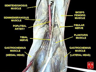
The popliteal artery is a deeply placed continuation of the femoral artery opening in the distal portion of the adductor magnus muscle. It courses through the popliteal fossa and ends at the lower border of the popliteus muscle, where it branches into the anterior and posterior tibial arteries.

<span class="mw-page-title-main">Joint dislocation</span> Medical injury

A joint dislocation, also called luxation, occurs when there is an abnormal separation in the joint, where two or more bones meet. A partial dislocation is referred to as a subluxation. Dislocations are often caused by sudden trauma on the joint like an impact or fall. A joint dislocation can cause damage to the surrounding ligaments, tendons, muscles, and nerves. Dislocations can occur in any major joint or minor joint. The most common joint dislocation is a shoulder dislocation.

<span class="mw-page-title-main">Tibialis posterior muscle</span> Muscle in the most central of all the leg muscles

The tibialis posterior muscle is the most central of all the leg muscles, and is located in the deep posterior compartment of the leg. It is the key stabilizing muscle of the lower leg.

<span class="mw-page-title-main">Plantaris muscle</span> One of the superficial muscles of the superficial posterior compartment of the leg,

The plantaris is one of the superficial muscles of the superficial posterior compartment of the leg, one of the fascial compartments of the leg.

The common fibular nerve is a nerve in the lower leg that provides sensation over the posterolateral part of the leg and the knee joint. It divides at the knee into two terminal branches: the superficial fibular nerve and deep fibular nerve, which innervate the muscles of the lateral and anterior compartments of the leg respectively. When the common fibular nerve is damaged or compressed, foot drop can ensue.

<span class="mw-page-title-main">Hip dislocation</span> Orthopedic injury

A hip dislocation is when the thighbone (femur) separates from the hip bone (pelvis). Specifically it is when the ball–shaped head of the femur separates from its cup–shaped socket in the hip bone, known as the acetabulum. The joint of the femur and pelvis is very stable, secured by both bony and soft-tissue constraints. With that, dislocation would require significant force which typically results from significant trauma such as from a motor vehicle collision or from a fall from elevation. Hip dislocations can also occur following a hip replacement or from a developmental abnormality known as hip dysplasia.

<span class="mw-page-title-main">Popliteal fossa</span> Depression at back of knee joint

The popliteal fossa is a shallow depression located at the back of the knee joint. The bones of the popliteal fossa are the femur and the tibia. Like other flexion surfaces of large joints, it is an area where blood vessels and nerves pass relatively superficially, and with an increased number of lymph nodes.

The knee examination, in medicine and physiotherapy, is performed as part of a physical examination, or when a patient presents with knee pain or a history that suggests a pathology of the knee joint.

<span class="mw-page-title-main">Sternoclavicular joint</span> Joint between the manubrium of the sternum and the clavicle bone.

The sternoclavicular joint or sternoclavicular articulation is a synovial saddle joint between the manubrium of the sternum, and the clavicle, and the first costal cartilage. The joint possesses a joint capsule, and an articular disc, and is reinforced by multiple ligaments.

<span class="mw-page-title-main">Popliteal artery entrapment syndrome</span> Abnormal popliteal artery compression by surrounding structures

The popliteal artery entrapment syndrome (PAES) is an uncommon pathology that occurs when the popliteal artery is compressed by the surrounding popliteal fossa myofascial structures. This results in claudication and chronic leg ischemia. This condition mainly occurs more in young athletes than in the elderlies. Elderlies, who present with similar symptoms, are more likely to be diagnosed with peripheral artery disease with associated atherosclerosis. Patients with PAES mainly present with intermittent feet and calf pain associated with exercises and relieved with rest. PAES can be diagnosed with a combination of medical history, physical examination, and advanced imaging modalities such as duplex ultrasound, computer tomography, or magnetic resonance angiography. Management can range from non-intervention to open surgical decompression with a generally good prognosis. Complications of untreated PAES can include stenotic artery degeneration, complete popliteal artery occlusion, distal arterial thromboembolism, or even formation of an aneurysm.

<span class="mw-page-title-main">Dislocated shoulder</span> Injury

A dislocated shoulder is a condition in which the head of the humerus is detached from the shoulder joint. Symptoms include shoulder pain and instability. Complications may include a Bankart lesion, Hill-Sachs lesion, rotator cuff tear, or injury to the axillary nerve.

<span class="mw-page-title-main">Malleolus</span> Ankle bone protrusion

A malleolus is the bony prominence on each side of the human ankle.

<span class="mw-page-title-main">Posterior compartment of thigh</span> One of the fascial compartments that contains the knee flexors and hip extensors

The posterior compartment of the thigh is one of the fascial compartments that contains the knee flexors and hip extensors known as the hamstring muscles, as well as vascular and nervous elements, particularly the sciatic nerve.

<span class="mw-page-title-main">Elbow</span> Joint between the upper and lower parts of the arm

The elbow is the region between the upper arm and the forearm that surrounds the elbow joint. The elbow includes prominent landmarks such as the olecranon, the cubital fossa, and the lateral and the medial epicondyles of the humerus. The elbow joint is a hinge joint between the arm and the forearm; more specifically between the humerus in the upper arm and the radius and ulna in the forearm which allows the forearm and hand to be moved towards and away from the body. The term elbow is specifically used for humans and other primates, and in other vertebrates forelimb plus joint is used.

<span class="mw-page-title-main">Tibial plateau fracture</span> Medical condition

A tibial plateau fracture is a break of the upper part of the tibia (shinbone) that involves the knee joint. Symptoms include pain, swelling, and a decreased ability to move the knee. People are generally unable to walk. Complication may include injury to the artery or nerve, arthritis, and compartment syndrome.

<span class="mw-page-title-main">Medial knee injuries</span> Medical condition

Medial knee injuries are the most common type of knee injury. The medial ligament complex of the knee consists of:

References

  1. Duprey K, Lin M (February 2010). "Posterior knee dislocation". The Western Journal of Emergency Medicine. 11 (1): 103–4. PMC   2850837 . PMID   20411095.
  2. 1 2 3 4 5 6 7 8 9 10 11 12 13 14 15 16 17 18 19 20 21 22 23 24 Boyce RH, Singh K, Obremskey WT (December 2015). "Acute Management of Traumatic Knee Dislocations for the Generalist". The Journal of the American Academy of Orthopaedic Surgeons. 23 (12): 761–8. doi:10.5435/JAAOS-D-14-00349. PMID   26493970. S2CID   10713473.
  3. 1 2 3 4 5 6 7 8 9 10 11 12 13 14 15 16 Maslaris A, Brinkmann O, Bungartz M, Krettek C, Jagodzinski M, Liodakis E (August 2018). "Management of knee dislocation prior to ligament reconstruction: What is the current evidence? Update of a universal treatment algorithm". European Journal of Orthopaedic Surgery & Traumatology. 28 (6): 1001–1015. doi:10.1007/s00590-018-2148-4. PMID   29470650. S2CID   3482099.
  4. 1 2 3 4 5 6 7 8 9 10 11 12 13 14 Bryant B, Musahl V, Harner CD (2011). "59. The Dislocated Knee". In W. Norman Scott (ed.). Insall & Scott Surgery of the Knee E-Book (5th ed.). Elsevier Churchill Livingstone. p. 565. ISBN   978-1-4377-1503-3.
  5. 1 2 3 4 5 6 7 8 Lachman JR, Rehman S, Pipitone PS (October 2015). "Traumatic Knee Dislocations: Evaluation, Management, and Surgical Treatment". The Orthopedic Clinics of North America. 46 (4): 479–93. doi:10.1016/j.ocl.2015.06.004. PMID   26410637.
  6. Eiff MP, Hatch RL (2011). Fracture Management for Primary Care E-Book. Elsevier Health Sciences. p. ix. ISBN   978-1455725021.
  7. Medina O, Arom GA, Yeranosian MG, Petrigliano FA, McAllister DR (September 2014). "Vascular and nerve injury after knee dislocation: a systematic review". Clinical Orthopaedics and Related Research. 472 (9): 2621–9. doi:10.1007/s11999-014-3511-3. PMC   4117866 . PMID   24554457.
  8. 1 2 Montorfano, Miguel Angel; Montorfano, Lisandro Miguel; Perez Quirante, Federico; Rodríguez, Federico; Vera, Leonardo; Neri, Luca (December 2017). "The FAST D protocol: a simple method to rule out traumatic vascular injuries of the lower extremities". Critical Ultrasound Journal. 9 (1): 8. doi: 10.1186/s13089-017-0063-2 . PMC   5360748 . PMID   28324353.
  9. Elliott JS (1914). Outlines of Greek and Roman Medicine. Creatikron Company. p. 76. ISBN   9781449985219.
  10. Godfrey AD, Hindi F, Ettles C, Pemberton M, Grewal P (2017). "Acute Thrombotic Occlusion of the Popliteal Artery following Knee Dislocation: A Case Report of Management, Local Unit Practice, and a Review of the Literature". Case Reports in Surgery. 2017: 5346457. doi: 10.1155/2017/5346457 . PMC   5299179 . PMID   28246569.
  11. 1 2 3 4 5 6 Pallin DJ, Hockberger R, Gausche-Hill M (2018). "50. Knee and lower leg". In Walls RM (ed.). Rosen's Emergency Medicine – Concepts and Clinical Practice E-Book (9th ed.). Philadelphia: Elsevier Health Sciences. p. 618. ISBN   978-0-323-35479-0.
  12. Graham JM, Sanchez-Lara PA (2016). "12. Knee dislocation (Genu Recurvatum)". Smith's Recognizable Patterns of Human Deformation E-Book (4th ed.). Philadelphia: Elsevier. p. 81. ISBN   978-0-323-29494-2.