
X-ray crystallography is the experimental science determining the atomic and molecular structure of a crystal, in which the crystalline structure causes a beam of incident X-rays to diffract into many specific directions. By measuring the angles and intensities of these diffracted beams, a crystallographer can produce a three-dimensional picture of the density of electrons within the crystal. From this electron density, the positions of the atoms in the crystal can be determined, as well as their chemical bonds, crystallographic disorder, and various other information.
Solid-state chemistry, also sometimes referred as materials chemistry, is the study of the synthesis, structure, and properties of solid phase materials. It therefore has a strong overlap with solid-state physics, mineralogy, crystallography, ceramics, metallurgy, thermodynamics, materials science and electronics with a focus on the synthesis of novel materials and their characterization. A diverse range of synthetic techniques, such as the ceramic method and chemical vapour depostion, make solid-state materials. Solids can be classified as crystalline or amorphous on basis of the nature of order present in the arrangement of their constituent particles. Their elemental compositions, microstructures, and physical properties can be characterized through a variety of analytical methods.

A synchrotron light source is a source of electromagnetic radiation (EM) usually produced by a storage ring, for scientific and technical purposes. First observed in synchrotrons, synchrotron light is now produced by storage rings and other specialized particle accelerators, typically accelerating electrons. Once the high-energy electron beam has been generated, it is directed into auxiliary components such as bending magnets and insertion devices in storage rings and free electron lasers. These supply the strong magnetic fields perpendicular to the beam that are needed to stimulate the high energy electrons to emit photons.
Gas electron diffraction (GED) is one of the applications of electron diffraction techniques. The target of this method is the determination of the structure of gaseous molecules, i.e., the geometrical arrangement of the atoms from which a molecule is built up. GED is one of two experimental methods to determine the structure of free molecules, undistorted by intermolecular forces, which are omnipresent in the solid and liquid state. The determination of accurate molecular structures by GED studies is fundamental for an understanding of structural chemistry.
Seismic analysis is a subset of structural analysis and is the calculation of the response of a building structure to earthquakes. It is part of the process of structural design, earthquake engineering or structural assessment and retrofit in regions where earthquakes are prevalent.
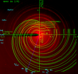
Electron crystallography is a method to determine the arrangement of atoms in solids using a transmission electron microscope (TEM). It can involve the use of high-resolution transmission electron microscopy images, electron diffraction patterns including convergent-beam electron diffraction or combinations of these. It has been successful in determining some bulk structures, and also surface structures. Two related methods are low-energy electron diffraction which has solved the structure of many surfaces, and reflection high-energy electron diffraction which is used to monitor surfaces often during growth.
In X-ray crystallography, the Flack parameter is a factor used to estimate the absolute configuration of a structural model determined by single-crystal structure analysis.
Rietveld refinement is a technique described by Hugo Rietveld for use in the characterisation of crystalline materials. The neutron and X-ray diffraction of powder samples results in a pattern characterised by reflections at certain positions. The height, width and position of these reflections can be used to determine many aspects of the material's structure.
Powder diffraction is a scientific technique using X-ray, neutron, or electron diffraction on powder or microcrystalline samples for structural characterization of materials. An instrument dedicated to performing such powder measurements is called a powder diffractometer.
Hugo M. Rietveld was a Dutch crystallographer who is famous for his publication on the full profile refinement method in powder diffraction, which became later known as the Rietveld refinement method. The method is used for the characterisation of crystalline materials from X-ray powder diffraction data. The Rietveld refinement uses a least squares approach to refine a theoretical line profile until it matches the measured profile. The introduction of this technique which used the full profile instead of individual reflections was a significant step forward in the diffraction analysis of powder samples.
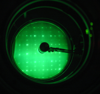
Low-energy electron diffraction (LEED) is a technique for the determination of the surface structure of single-crystalline materials by bombardment with a collimated beam of low-energy electrons (30–200 eV) and observation of diffracted electrons as spots on a fluorescent screen.
In crystallography, the R-factor is a measure of the agreement between the crystallographic model and the experimental X-ray diffraction data. In other words, it is a measure of how well the refined structure predicts the observed data. The value is also sometimes called the discrepancy index, as it mathematically describes the difference between the experimental observations and the ideal calculated values. It is defined by the following equation:
Molecular replacement (MR) is a method of solving the phase problem in X-ray crystallography. MR relies upon the existence of a previously solved protein structure which is similar to our unknown structure from which the diffraction data is derived. This could come from a homologous protein, or from the lower-resolution protein NMR structure of the same protein.
In physics, strain scanning is the general name for various techniques that aim to measure the strain in a crystalline material through its effect on the diffraction of X-rays and neutrons. In these methods the material itself is used as a form of strain gauge.
A crystallographic database is a database specifically designed to store information about the structure of molecules and crystals. Crystals are solids having, in all three dimensions of space, a regularly repeating arrangement of atoms, ions, or molecules. They are characterized by symmetry, morphology, and directionally dependent physical properties. A crystal structure describes the arrangement of atoms, ions, or molecules in a crystal.
Meyrowitzite, Ca(UO2)(CO3)2·5H2O, is a carbonate mineral verified in May of 2018 by the Commission of New Minerals, Nomenclature and Classification of the International Mineralogical Association. It is an extremely rare mineral, discovered in the Markey mine Utah, U.S.A. The mineral is a transparent yellow and has blades up to approximately 0.2 mm in length. It is soluble in water or aqueous solutions. Meyrowitzite is named in honor of Robert Meyrowitz (1916–2013), an American analytical chemist. After serving in WW II, he joined the United States Geological Survey (USGS). He was known for developing innovative new methods for analyzing small and difficult to study mineralogical samples along with his formulation of the high-index immersion liquids.
Vanarsite, NaCa12(As3+V4+3.5V5+8.5As5+6O51)2・78H2O, is a mineral that forms as very dark blue blades that are flattened on {100} and elongated on [010]. The crystals form in sub-parallel intergrowths in aggregates up to 5 mm long and are part of the cubic crystal system. The mineral is found in the Packrat mine in Colorado, USA in association with three mineral of similar composition, packratite, Ca11(As3+V4+2V5+10As5+6O51)2・83H2O, morrisonite, Ca6(As3+V4+2V5+10As5+6O51)2・78H2O, and gatewayite, Ca11(As3+V4+3V5+9As5+6O51)・31H2O. Vanarsite has been determined to be part of the vanadate, arsenite, and arsenate groups. Analytical methods were used to determine its chemical composition, physical properties, and structure.
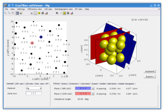
CrysTBox is a suite of computer tools designed to accelerate material research based on transmission electron microscope images via highly accurate automated analysis and interactive visualization. Relying on artificial intelligence and computer vision, CrysTBox makes routine crystallographic analyses simpler, faster and more accurate compared to human evaluators. The high level of automation together with sub-pixel precision and interactive visualization makes the quantitative crystallographic analysis accessible even for non-crystallographers allowing for an interdisciplinary research. Simultaneously, experienced material scientists can take advantage of advanced functionalities for comprehensive analyses.
X-ray diffraction computed tomography is an experimental technique that combines X-ray diffraction with the computed tomography data acquisition approach. X-ray diffraction (XRD) computed tomography (CT) was first introduced in 1987 by Harding et al. using a laboratory diffractometer and a monochromatic X-ray pencil beam. The first implementation of the technique at synchrotron facilities was performed in 1998 by Kleuker et al.
