Leucocytozoon

Last updated

Leucocytozoon
Leucocytozoon smithi.jpg
Leucocytozoon smithi in a stained blood smear from a turkey
Scientific classification OOjs UI icon edit-ltr.svg
Domain: Eukaryota
Clade: Diaphoretickes
Clade: SAR
Clade: Alveolata
Phylum: Apicomplexa
Class: Aconoidasida
Order: Haemosporida
Family: Leucocytozoidae
Genus: Leucocytozoon
Species

Leucocytozoon acridotheris
Leucocytozoon alcedinis
Leucocytozoon alcedis
Leucocytozoon anatis
Leucocytozoon andrewsi
Leucocytozoon anellobiae
Leucocytozoon apiaster
Leucocytozoon ardeolae
Leucocytozoon artamidis
Leucocytozoon anseris
Leucocytozoon atkinsoni
Leucocytozoon balmorali
Leucocytozoon beaurepairei
Leucocytozoon bennetti
Leucocytozoon berestneffi
Leucocytozoon bishopae
Leucocytozoon bishopi
Leucocytozoon bisi
Leucocytozoon bonasae
Leucocytozoon bouffardi
Leucocytozoon brimonti
Leucocytozoon bucerotis
Leucocytozoon cambournaci
Leucocytozoon capitonis
Leucocytozoon caprimulgi
Leucocytozoon caulleryi
Leucocytozoon centropi
Leucocytozoon chloropsidis
Leucocytozoon coccyzus
Leucocytozoon colius
Leucocytozoon communis
Leucocytozoon coracinae
Leucocytozoon costai
Leucocytozoon dacelo
Leucocytozoon danilewskyi Ziemann 1898
Leucocytozoon deswardti
Leucocytozoon dinizi
Leucocytozoon dubreuili Mathis & Leger 1911
Leucocytozoon dutoiti
Leucocytozoon enriquesi
Leucocytozoon eurystomi
Leucocytozoon francae
Leucocytozoon francolini
Leucocytozoon frascai
Leucocytozoon fringillinarum Woodcock 1910
Leucocytozoon galli
Leucocytozoon gallinarum
Leucocytozoon gentili
Leucocytozoon giovannolai
Leucocytozoon grallariae
Leucocytozoon greineri
Leucocytozoon grusi
Leucocytozoon hamiltoni
Leucocytozoon huchzermeyeri
Leucocytozoon ibisi
Leucocytozoon icteris
Leucocytozoon iowense
Leucocytozoon irenae
Leucocytozoon jakamowi
Leucocytozoon kerandeli
Leucocytozoon lairdi
Leucocytozoon lanium
Leucocytozoon laverani
Leucocytozoon leboeufi
Leucocytozoon leitaoi
Leucocytozoon liothricis
Leucocytozoon lovati
Leucocytozoon maccluri
Leucocytozoon macleani
Leucocytozoon major
Leucocytozoon majoris Laveran 1902
Leucocytozoon mansoni
Leucocytozoon marchouxi
Leucocytozoon martini
Leucocytozoon mathisi
Leucocytozoon mcclurei
Leucocytozoon melloi
Leucocytozoon mesnili
Leucocytozoon molpastis
Leucocytozoon muscicapa
Leucocytozoon mutus
Leucocytozoon neavei
Leucocytozoon nectariniae
Leucocytozoon neotropicalis
Leucocytozoon numidae
Leucocytozoon oriolis
Leucocytozoon otidis
Leucocytozoon pittae
Leucocytozoon pogoniuli
Leucocytozoon pycnonoti
Leucocytozoon rimondi
Leucocytozoon roubaudi
Leucocytozoon sabrezi
Leucocytozoon sakharoffi
Leucocytozoon schoutedeni
Leucocytozoon schuffneri
Leucocytozoon simondi
Leucocytozoon smithi
Leucocytozoon squamatus
Leucocytozoon struthionis
Leucocytozoon sturni
Leucocytozoon sunibdu
Leucocytozoon tawaki
Leucocytozoon timaliae
Leucocytozoon toddi Sambon 1908
Leucocytozoon trachyphoni
Leucocytozoon whitworthi
Leucocytozoon ziemanni
Leucocytozoon zosteropis

Contents

Leucocytozoon (or Leukocytozoon) is a genus of parasitic alveolates belonging to the phylum Apicomplexa (which also includes the malaria parasites).

The species of this genus use either blackflies ( Simulium species) or a biting midge as their definitive host and birds as their intermediate host. [1] There are over 100 species in this genus. Over 100 species of birds have been recorded as hosts to these parasites.

Life cycle

Blood from a chicken infected with a male gametocyte (left) and a female gametocyte (right). Leucocytozoon sabrazesi gametocytes from blood of domestic chicken.tif
Blood from a chicken infected with a male gametocyte (left) and a female gametocyte (right).

Parasites in the genus Leucocytozoon have a life cycle that involves both a bird host, and a black fly (with the exception of Leucocytozoon caulleryi which cycles between a bird host and a biting midge). [1] Parasites enter the bird host in a form called a sporozoite through the bite of the blood-sucking black fly. [1] The sporozoites invade host cells in the liver where they undergo asexual replication, forming numerous daughter cells called merozoites within 4–5 days. [1] The duration of this stage depends in part upon the species. In some species this stage may occur in the endothelial cells instead of the liver.

The newly released merozoites infect either erythrocytes, leukocytes, macrophages or endothelial cells. Those infecting the macrophages or endothelial cells develop into megaloschizonts. The megaloschizonts divide into primary cytomeres, which in turn multiply into smaller cytomeres, which mature into schizonts, which in turn divide into merozoites. In erythrocytes or leukocytes, merozoites develop into gametocytes.

The gametocytes are taken up by a blood-sucking fly as it bites the bird host. Gametocytes then mature in the insect midgut into macrogametocytes (female) with red-staining nuclei and microgametocytes (male) with pale-staining diffuse nuclei: these fuse to form an ookinete. The male gametocytes normally give rise to eight microgametes. The ookinete penetrates an intestinal cell of the fly and matures into an oocyst. After several days the oocyst produces ~100 sporozoites that leave and migrate to the salivary glands of the fly.

Description

The nuclei of the schizonts is enclosed in a trilaminar membrane with peripherally arranged chromatin. The schizonts also contain numerous cytomers also with trilaminar membranes and containing multiple ribosomes. Repeated invagination of the cytomeres gives rise to the merozoites which also have a trilaminar membrane.

The merozoites have rhoptries, micronemes and three apical rings. The mitochondrion contains vesicular cristae. There may be one or two paranuclear bodies in some species. Their function is unknown.

Two gametocyte forms are recognised: an elongated (sail-like) form and a compact spherical form. These are usually 12–14 micrometres long. The large gametocytes tend to grossly distort the infected cells and make cell identification difficult. A pseudopigment known as volutin may be present.

Evolution

It has been suggested that this genus arose after the two others genera Plasmodium and Haemoproteus, the latter originating from the late Oligocene or early Eocene at about the same time as Piciformes and Coraciiformes. [2]

Taxonomy

The genus is divided into two subgenera—Akiba and Leucocytozoon—based on the vector species.

The only known member of the subgenus Akiba is Leucocytozoon (Akiba) caulleryi which uses members of the genus Culicoides as its vectors.

The remaining species in the genus use members of the genus Simulium as their vectors.

In 1977, Greiner and Kocan in an extensive examination of species in the order Falconiformes declared that the only valid species infecting this order was L. toddi. [3]

L. dubreuili is considered to be restricted to the family Turdidae ; L. fringillinarum is considered to be restricted to several passiform families; and L. majoris is considered restricted to the family Paridae .[ citation needed ]

L. ziemanni infects owls. [4]

Diagnostic criteria

Form gamonts in white blood cells and/or erythrocytes. Gametocytes cause marked enlargement and distortion of the infected cell producing a football-like appearance.

No merogony occurs in either leucocytes or erythrocytes.

Merogony occurs in the parenchyma of liver, heart, kidney, or other organs. Meronts may form large bodies divided into cytomeres.

Hemozoin deposits (pigment) are not formed—a useful distinguishing feature for Leucocytozoon from Haemoproteus and Plasmodium .

Oocysts develop rapidly in 3–5 days. The oocysts are small and nonexpanding, reaching 13 micrometres in diameter and typically have less than 100 short, thick sporozoites.

The vectors are Simulium or Culicoides species.

The vertebrate hosts are birds.

Type species: Leucocytozoon ziemanni

Pathology

The typical pathology of infection with these parasites includes anaemia and enlargement of the liver and spleen. Gross lesions also include pulmonary congestion and pericardial effusion.

Megaloschizonts appear as grey-white nodules found in the heart, liver, lung or spleen. Microscopically there is ischemic necrosis and associated inflammation in the heart, brain, spleen and liver due to occlusion of blood vessels by megaloschizonts in endothelial cells. Ruptured schizonts may induce granulomatous reactions in the surrounding tissues.

Clinically the majority of birds affected with leucocytozoonosis exhibit no signs. Among those that do the signs include mild to severe signs of anorexia, ataxia, weakness, anemia, emaciation and difficulty breathing.

The excess mortality due to Leucocytozoon in adult birds seems to occur as a result of debilitation and increased susceptibility to secondary infection.

Epidemiology

L. simondi is suspected to be a major parasite of Canada geese in some areas, including the upper Midwestern United States and Canada. L. smithi affects turkey farms in the southeastern United States.

Host range

Bird hosts

Vectors

Notes

Like many protist species and genera this genus is subject to ongoing revision especially in the light of DNA based taxonomy. Described species may be subject to revision.

History

Drawing of owl blood cells parasitized by Leukocytozoen danilewskyi by Hans Ziemann in 1898 Leukocytozoen danilewskyi drawing, 1898.jpg
Drawing of owl blood cells parasitized by Leukocytozoen danilewskyi by Hans Ziemann in 1898

The first described observation of a Leucocytozoon parasite may have been by Vasily Danilewsky, who described certain parasites he observed in the blood of birds as "Leucocytozaire" in 1888 (as they appeared to infect leukocytes). [20] A more detailed description of parasites resembling Leucocytozoon was published in 1894 by N. Sakharoff from the blood of birds near Tbilisi. [21] Soon thereafter in 1898, Hans Ziemann described a Leucocytozoon parasite from the blood of the owl Athene noctua , naming it Leukocytozoen danilewskyi in honor of Danilewsky. [21] N. M. Berestneff was the first to use the current genus name Leucocytozoon while describing parasites he called Leucocytozoon danilewskyi from several common birds. [21] The genus was subsequently formally defined in 1908 by Louis Sambon, and has remained largely unchanged since. [21] In 1930 and 1931, Earl O'Roke and Louis V. Skidmore independently discovered black flies to be the vector of Leucocytozoon species. [21]

Related Research Articles

<i>Plasmodium</i> Genus of parasitic protists that can cause malaria

Plasmodium is a genus of unicellular eukaryotes that are obligate parasites of vertebrates and insects. The life cycles of Plasmodium species involve development in a blood-feeding insect host which then injects parasites into a vertebrate host during a blood meal. Parasites grow within a vertebrate body tissue before entering the bloodstream to infect red blood cells. The ensuing destruction of host red blood cells can result in malaria. During this infection, some parasites are picked up by a blood-feeding insect, continuing the life cycle.

<span class="mw-page-title-main">Isosporiasis</span> Human intestinal disease

Isosporiasis, also known as cystoisosporiasis, is a human intestinal disease caused by the parasite Cystoisospora belli. It is found worldwide, especially in tropical and subtropical areas. Infection often occurs in immuno-compromised individuals, notably AIDS patients, and outbreaks have been reported in institutionalized groups in the United States. The first documented case was in 1915. It is usually spread indirectly, normally through contaminated food or water (CDC.gov).

<i>Eimeria</i> Genus of single-celled organisms

Eimeria is a genus of apicomplexan parasites that includes various species capable of causing the disease coccidiosis in animals such as cattle, poultry and smaller ruminants including sheep and goats. Eimeria species are considered to be monoxenous because the life cycle is completed within a single host, and stenoxenous because they tend to be host specific, although a number of exceptions have been identified. Species of this genus infect a wide variety of hosts. Thirty-one species are known to occur in bats (Chiroptera), two in turtles, and 130 named species infect fish. Two species infect seals. Five species infect llamas and alpacas: E. alpacae, E. ivitaensis, E. lamae, E. macusaniensis, and E. punonensis. A number of species infect rodents, including E. couesii, E. kinsellai, E. palustris, E. ojastii and E. oryzomysi. Others infect poultry, rabbits and cattle. For full species list, see below.

<i>Babesia</i> Genus of protozoan parasites

Babesia, also called Nuttallia, is an apicomplexan parasite that infects red blood cells and is transmitted by ticks. Originally discovered by the Romanian bacteriologist Victor Babeș in 1888, over 100 species of Babesia have since been identified.

Leucocytozoon andrewsi is a parasite of the genus Leucocytozoon.

Megaloschizonts are large schizonts that produce extremely high numbers of merozoites. They are found in various species of the Phylum Apicomplexa. The Apicomplexa phylum contains several parasitic protozoans. They have a very complex life cycle that includes several stages. Megaloschizonts and the smaller schizonts are the part of the life cycle that takes place inside the infected host organism and operates as an asexually reproductive cell. Megaloschizonts appear as grey-white nodules found in the smooth muscle of major organs, such as the heart, liver, lung or spleen.

Plasmodium uluguruense is a parasite of the genus Plasmodium subgenus Lacertamoeba.

Plasmodium loveridgei is a parasite of the genus Plasmodium subgenus Lacertamoeba.

<i>Haemoproteus</i> Genus of single-celled organisms

Haemoproteus is a genus of alveolates that are parasitic in birds, reptiles and amphibians. Its name is derived from Greek: Haima, "blood", and Proteus, a sea god who had the power of assuming different shapes. The name Haemoproteus was first used in the description of H. columbae in the blood of the pigeon Columba livia by Kruse in 1890. This was also the first description of this genus. Two other genera — Halteridium and Simondia — are now considered to be synonyms of Haemoproteus.

Plasmodium juxtanucleare is a species of parasite in the family Plasmodiidae. The vertebrate hosts for this parasite are birds.

<span class="mw-page-title-main">Adeleorina</span> Suborder of microscopic, spore-forming, single-celled parasites in the aplcomplex phylum

Adeleorina is a suborder of parasites in the phylum Apicomplexa.

Karyolysus is a genus of coccidia. With the exception of K. sonomae whose vertebrate host is the yellow-legged frog, species in this genus only infect lizards of the genus Lacerta.

Plasmodium vaughani is a parasite of the genus Plasmodium, and the type species of the subgenus Novyella. As in all Plasmodium species, P. vaughani has both vertebrate and insect hosts. The vertebrate hosts for this parasite are birds.

<span class="mw-page-title-main">Apicomplexan life cycle</span> Apicomplexa life cycle

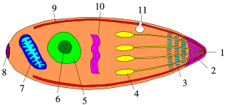
Apicomplexans, a group of intracellular parasites, have life cycle stages that allow them to survive the wide variety of environments they are exposed to during their complex life cycle. Each stage in the life cycle of an apicomplexan organism is typified by a cellular variety with a distinct morphology and biochemistry.

Dactylosoma is a genus of parasitic alveolates of the phylum Apicomplexa.

Leucocytozoon caulleryi is a species of the genus Leucocytozoon, a genus of parasitic alveolates.

The genus Schellackia comprises obligate unicellular eukaryotic parasites within the phylum Apicomplexa, and infects numerous species of lizards and amphibians worldwide. Schellackia is transmitted via insect vectors, primarily mites and mosquitoes, which take up the parasite in blood meals. These vectors then subsequently infect reptilian and amphibian which consume the infected insects. The parasites deform erythrocytes of the host into crescents, and can be visualized using a blood smear.

Atoxoplasma is a genus of parasitic alveolates in the phylum Apicomplexa. The species in this genus infect birds. They are spread by the orofaecal route.

Nephroisospora is a genus of parasites that infects bats

The Ophryocystidae are a family of parasites in the phylum Apicomplexa. Species in this family infect insects.

References

  1. 1 2 3 4 Valkiunas G (2004). "Life Cycle and Morphology of Leucocytozoidae Species". Avian Malaria Parasites and Other Haemosporidia (1 ed.). CRC Press. pp. 36–45. doi:10.1201/9780203643792. ISBN   978-0-415-30097-1.
  2. Bennett, GF (1993). "Phylogenetic distribution and possible evolution of the avian species of the Haemoproteidae". Syst. Parasitol. 26 (1): 39–44. doi:10.1007/bf00009646. S2CID   35811974.
  3. Greiner EC, Kocan AA (1977). "Leucocytozoon (Haemosporida; Leucocytozoidae) of the falconiformes". Can. J. Zool. 55 (5): 761–70. doi:10.1139/z77-099. PMID   406030.
  4. Evans, M; Otter, A (18 July 1998). "Fatal combined infection with Haemoproteus noctuae and Leucocytozoon ziemanni in juvenile snowy owls (Nyctea scandiaca)". The Veterinary Record. 143 (3): 72–6. doi:10.1136/vr.143.3.72. PMID   9717222. S2CID   23546240.
  5. Atchley, F.O. (1951). "Leucocytozoon andrewsi n. sp., from chickens observed in a survey of blood parasites in domestic animals in South Carolina". J. Parasitol. 37 (5): 483–8. doi:10.2307/3273257. JSTOR   3273257.
  6. Peirce, M.A.; Adlard, R.D.; Lederer, R. (2005). "A new species of Leucocytozoon Berestneff, 1904 (Apicomplexa: Leucocytozoidae) from the avian family Artamidae". Syst. Parasitol. 60 (2): 151–4. doi:10.1007/s11230-004-1387-4. PMID   15841351. S2CID   1135988.
  7. Savage, A.F.; Ariey, F.; Greiner, E.C. (2006). "Leucocytozoon atkinsoni n. sp. (Apicomplexa: Leucocytozoidae) from the avian family Timaliidae". Syst. Parasitol. 64 (2): 105–9. doi:10.1007/s11230-005-9023-5. PMID   16612655. S2CID   11870669.
  8. Barraclough, R.K.; Robert, V.; Peirce, M.A. (2008). "New species of haematozoa from the avian families Campephagidae and Apodidae". Parasite. 15 (2): 105–110. doi: 10.1051/parasite/2008152105 . ISSN   1252-607X. PMID   18642502. Open Access logo PLoS transparent.svg
  9. Savage, A.F.; Greiner, E.C. (2004). "Hematozoa of the avian family Brachypteraciidae (the ground-rollers)". J. Parasitol. 90 (6): 1468–72. doi:10.1645/ge-227r. PMID   15715245. S2CID   3260365.
  10. Gill, H.; Paperna, I. (2005). "Leucocytozoonosis in the Israeli sparrow, Passer domesticus biblicus Hartert 1904". Parasitol. Res. 96 (6): 373–7. doi:10.1007/s00436-005-1352-4. PMID   15940526. S2CID   23198260.
  11. 1 2 Savage, A.F. (2004). "Hematozoa of the avian family Philepittidae (the asities and sunbird asities)". J. Parasitol. 90 (6): 1473–4. doi:10.1645/ge-228r. PMID   15715246. S2CID   24081599.
  12. Bennett, G.F.; Khan, R.A.; Campbell, A.G. (1974). "Leucocytozoon grusi sp. n. (Sporozoa: Leucocytozoidae) from a sandhill crane, Grus canadensis (L.)". J. Parasitol. 60 (2): 359–363. doi:10.2307/3278486. JSTOR   3278486. PMID   4206882.
  13. Valkiünas, G.; Iezhova, T.A.; Mironov, S.V. (2002). "Leucocytozoon hamiltoni n. sp. (Haemosporida, Leucocytozoidae) from the Bukharan great tit Parus bokharensis". J. Parasitol. 88 (3): 577–581. doi:10.2307/3285453. JSTOR   3285453. PMID   12099430.
  14. Greiner, E.C. (1976). "Leucocytozoon maccluri sp. n. (Haemosphorida: Leucocytozoidae) from a Thailand thrush, Zoothera marginata Blyth". J. Parasitol. 62 (4): 545–7. doi:10.2307/3279409. JSTOR   3279409. PMID   822146.
  15. Bunbury, N.; Barton, E.; Jones, C.G.; Greenwood, A.G.; Tyler, K.M.; Bell, D.J. (2007). "Avian blood parasites in an endangered columbid: Leucocytozoon marchouxi in the Mauritian Pink Pigeon Columba mayeri". Parasitology. 134 (6): 797–804. CiteSeerX   10.1.1.391.9645 . doi:10.1017/s0031182006002149. PMID   17201998. S2CID   8703315.
  16. 1 2 Jones, H.I.; Sehgal, R.N.; Smith, T.B. (2005). "Leucocytozoon (Apicomplexa: Leucocytozoidae) from West African birds, with descriptions of two species". J. Parasitol. 91 (2): 397–401. doi:10.1645/ge-3409. PMID   15986615. S2CID   7661872.
  17. Nooroong, Pornpiroon; Watthanadirek, Amaya; Minsakorn, Sutthida; Poolsawat, Napassorn; Junsiri, Witchuta; Srionrod, Nitipon; Sangchuai, Siriphan; Chawengkirttikul, Runglawan; Anuracpreeda, Panat (2022). "Molecular genetic diversity and bioinformatic analysis of Leucocytozoon sabrazesi based on the mitochondrial genes cytb, coxI and coxIII and co-infection of Plasmodium spp". Parasite. 29: 22. doi:10.1051/parasite/2022022. ISSN   1776-1042. PMC   9044968 . PMID   35475784. Open Access logo PLoS transparent.svg
  18. Ortego J, Cordero PJ (2009). "PCR-based detection and genotyping of haematozoa (Protozoa) parasitizing eagle owls, Bubo bubo". Parasitol. Res. 104 (2): 467–70. doi:10.1007/s00436-008-1207-x. PMID   18818949. S2CID   29793321.
  19. Sato Y, Tamada A, Mochizuki Y, Nakamura S, Okano E, Yoshida C, Ejiri H, Omori S, Yukawa M, Murata K (2009). "Molecular detection of Leucocytozoon lovati from probable vectors, black flies (Simuliudae) collected in the alpine regions of Japan". Parasitol. Res. 104 (2): 251–5. doi:10.1007/s00436-008-1183-1. PMID   18791737. S2CID   10885934.
  20. Wenyon CM (April 1910). "Some remarks on the genus Leucocytozoon". Parasitology. 3 (1): 63–72. doi:10.1017/S0031182000001931. S2CID   84506447.
  21. 1 2 3 4 5 Valkiunas G (2004). "Brief historical summary". Avian Malaria Parasites and Other Haemosporidia (1 ed.). CRC Press. pp. 9–16. doi:10.1201/9780203643792. ISBN   978-0-415-30097-1.