Lipoprotein(a) was discovered in 1963 by Kåre Berg.[7] The human gene encoding apolipoprotein(a) was successfully cloned in 1987.[8]
Structure
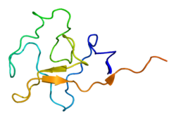
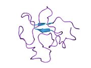
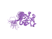
Lipoprotein(a) [Lp(a)] consists of an LDL-like particle and the specific apolipoprotein(a), which is bound covalently to the apoB contained in the outer shell of the particle. Lp(a) plasma concentrations are highly heritable and mainly controlled by the LPA gene located on chromosome 6q26-27. Apo(a) proteins vary in size due to a size polymorphism [KIV-2 VNTR], which is caused by a variable number of kringle IV repeats in the LPA gene. This size variation at the gene level is expressed on the protein level as well, resulting in apo(a) proteins with 10 to more than 50 kringle IV repeats (each of the variable kringle IV consists of 114 amino acids).[8][9] These variable apo(a) sizes are known as "apo(a) isoforms".
There is a general inverse correlation between the size of the apo(a) isoform and the Lp(a) plasma concentration.[10] One theory explaining this correlation involves different rates of protein synthesis. Specifically, the larger the isoform, the more apo(a) precursor protein accumulates intracellularly in the endoplasmic reticulum. Lp(a) is not fully synthesised until the precursor protein is released from the cell, so the slower rate of production for the larger isoforms limits the plasma concentration.[11][12]
Lp(a) concentrations can vary by more than one thousand between individuals, from <0.2 to >200mg/dL. This range of concentrations is observed in all populations studied by scientists so far. The mean and median concentrations differ among world populations. Most prominently, there is a two- to threefold higher mean Lp(a) plasma concentration in populations of African descent compared to Asian, Oceanic, or European populations. The general inverse correlation between apo(a) isoform size and Lp(a) plasma concentration is observed in all populations.[10] However, it was also discovered that mean Lp(a) associated with certain apo(a) isoforms varies between populations.
In addition to size effects, mutations in the LPA promoter may lead to a decreased apo(a) production.[13]
Function and pathology
Lp(a) is assembled at the hepatocyte cell membrane surface, which is similar to typical LDL particles. However, there are other possible locations of assembly. The particles mainly exist in plasma.[14][15][16][17]
Lp(a) contributes to the process of atherogenesis. The structure of apolipoprotein(a) is similar to plasminogen and tPA (tissue plasminogen activator) and it competes with plasminogen for its binding site, leading to reduced fibrinolysis. Also, because Lp(a) stimulates secretion of PAI-1, it leads to thrombogenesis.[18][19][20] It also may enhance coagulation by inhibiting the function of tissue factor pathway inhibitor.[21]
Moreover, Lp(a) carries atherosclerosis-causing cholesterol and binds atherogenic pro-inflammatory oxidised phospholipids as a preferential carrier of oxidised phospholipids in human plasma,[22] which attracts inflammatory cells to vessel walls and leads to smooth muscle cell proliferation.[23][24][25] Moreover, Lp(a) also is hypothesised to be involved in wound healing and tissue repair by interacting with components of the vascular wall and extracellular matrix.[26][27] Apo(a), a distinct feature of the Lp(a) particle, binds to immobilized fibronectin and endows Lp(a) with the serine-proteinase-type proteolytic activity.[28]
Nonetheless, individuals without Lp(a) or with very low Lp(a) levels seem to be healthy.[citation needed] Thus, plasma Lp(a) is not vital, at least under normal environmental conditions.[citation needed] Since apo(a)/Lp(a) appeared rather recently in mammalian evolution – only old world monkeys and humans have been shown to harbour Lp(a) – its function might not be vital, but just evolutionarily advantageous under certain environmental conditions, e.g. in case of exposure to certain infectious diseases.[13]
Another possibility, suggested by Linus Pauling, is that Lp(a) is a primate adaptation to L-gulonolactone oxidase (GULO) deficiency, found only in certain lines of mammals. GULO is required for converting glucose to ascorbic acid (vitamin C), which is needed to repair arteries; following the loss of GULO, those primates who adopted diets less abundant in vitamin C may have used Lp(a) as an ascorbic-acid surrogate to repair arterial walls.[29]
Catabolism and clearance
The half-life of Lp(a) in circulation is approximately three to four days.[15] The mechanism and sites of Lp(a) catabolism are largely unknown. Uptake via the LDL receptor is not a major pathway of Lp(a) metabolism.[30][31] The kidney has been identified as playing a role in Lp(a) clearance from plasma.[32]
Disease
High Lp(a) in blood correlates with coronary heart disease (CHD), cardiovascular disease (CVD), atherosclerosis, thrombosis, and stroke.[33] However, the association between Lp(a) levels and stroke is not as strong as that between Lp(a) and cardiovascular disease.[3] Lp(a) concentrations may be affected by disease states (for example kidney failure), but are only slightly affected by diet, exercise, and other environmental factors.
Most commonly prescribed lipid-reducing drugs have little or no effect on Lp(a) concentration. Results using statin medications have been mixed in most trials, although a meta-analysis published in 2012 suggests that atorvastatin may be of benefit.[34]
Niacin (Vitamin B3) has been shown to reduce the levels of Lp(a) significantly in individuals with high levels of low-molecular weight Lp(a).[35][36]
High Lp(a) correlates with early atherosclerosis independently of other cardiac risk factors, including LDL. In patients with advanced cardiovascular disease, Lp(a) indicates a coagulant risk of plaque thrombosis. Apo(a) contains domains that are very similar to plasminogen (PLG). Lp(a) accumulates in the vessel wall and inhibits the binding of PLG to the cell surface, reducing plasmin generation, which increases clotting. This inhibition of PLG by Lp(a) also promotes the proliferation of smooth muscle cells. These unique features of Lp(a) suggest that Lp(a) causes generation of clots and atherosclerosis.[37]
In one homogeneous tribal population of Tanzania, vegetarians have higher levels of Lp(a) than fish eaters, raising the possibility that pharmacologic amounts of fish oil supplements may help lower the levels of Lp(a).[38] Researchers in studies in 1995 and 1998 concluded that regular consumption of moderate amounts of alcohol led to a significant decline in plasma levels of Lp(a).[39] Other studies did not report this.
Diagnostic testing
Numerous studies confirming a strong correlation between elevated Lp(a) and heart disease have led to the consensus that Lp(a) is an important independent predictor of cardiovascular disease.[3] Animal studies have shown that Lp(a) may directly contribute to atherosclerotic damage by increasing plaque size, inflammation, instability, and smooth muscle cell growth.[40] Genetic data also support the theory that Lp(a) causes cardiovascular disease.[4]
The European Atherosclerosis Society currently recommends that patients with a moderate or high risk of cardiovascular disease should have their Lp(a) levels checked. Any patient with one of the following risk factors should be screened:
premature cardiovascular disease
familial hypercholesterolaemia
family history of premature cardiovascular disease
≥3% ten-year risk of fatal cardiovascular disease according to the European guidelines
≥10% ten-year risk of fatal and/or non-fatal cardiovascular disease according to the U.S. guidelines[3]
If the level is elevated, treatment should be initiated to bring the level below 50mg/dL. In addition, the patient's other cardiovascular risk factors (including LDL levels) should be managed optimally.[3] Apart from the total Lp(a) plasma concentration, the apo(a) isoform might be an important risk parameter as well.[41][42]
Prior studies of the relationship between Lp(a) and ethnicity have shown inconsistent results. Lp(a) levels seem to differ in different populations. For example, in some African populations, Lp(a) levels are higher on average than in other groups, so that using a risk threshold of 30mg/dl could classify over 50% of the individuals as higher risk.[43][44][45][46] Some part of this complexity may be related to the different genetic factors involved in determining Lp(a) levels. One recent study showed that in different ethnic groups, different genetic alterations were associated with increased Lp(a) levels.[47]
More recent data suggest that prior studies were underpowered. The Atherosclerosis Risk in Communities (ARIC) Study followed 3467 African Americans and 9851 whites for 20 years. The researchers found that an elevated Lp(a) conferred the same risk in each group. African Americans had roughly three times the level of Lp(a), however, and Lp(a) also predicted an increased risk of stroke.[48]
Approximate levels of risk are indicated by the results below, although at present there are a variety of different methods by which to measure Lp(a). A standardized international reference material has been developed and is accepted by the WHO Expert Committee on Biological Standardization and the International Federation of Clinical Chemistry and Laboratory Medicine. Although further standardization is still needed, development of a reference material is an importance step toward standardizing results.[49][50]
Lp(a) appears with different isoforms (per kringle repeats) of apolipoprotein; 40% of the variation in Lp(a) levels when measured in mg/dl can be attributed to different isoforms. Lighter Lp(a) are also associated with disease. Thus, a test with simple quantitative results may not provide a complete assessment of risk.[52]
Treatment
The current simplest treatment for elevated Lp(a) is to take 1–3 grams of niacin daily, typically in an extended-release form. Niacin therapy may reduce Lp(a) levels by 20–30%.[53]
A meta-analysis suggested that atorvastatin may lower Lp(a) levels.[34] In severe cases, such as familial hypercholesterolemia or treatment-resistant hypercholesterolemia, LDL apheresis may dramatically reduce Lp(a). The goal of the treatment is to reduce levels to below 50mg/dL. Cost is prohibitively high.[3]
A meta-analysis of six clinical trials confirmed that flaxseed supplementation modestly lowers Lp(a) levels.[54]
Testosterone is known to reduce Lp(a) levels.[55] Testosterone replacement therapy also appears to be associated with lower Lp(a) levels.[56][57] Estrogen replacement therapy in post-menopausal women will reduce Lp(a).[58]Raloxifene has not been shown to reduce Lp(a), while tamoxifen has.[59]
L-carnitine may also reduce Lp(a) levels. A systematic review and meta-analysis found a significant reduction with oral but not intravenous carnitine.[60] Other medications that are in various stages of development include thyromimetics, cholesterol-ester-transfer protein (CETP inhibitors), anti-sense oligonucleopeptides (such as Pelacarsen and Olpasiran), and proprotein convertase subtilisin/kexin type 9 (PCSK9) inhibitors.[61][62]
The American Academy of Pediatrics now recommends that all children between the ages of nine and eleven years old be screened for hyperlipidemia. Lp(a) levels should be considered in particular in children with a family history of early heart disease or high blood cholesterol levels. However, there have not been enough studies to determine which therapies might be beneficial.[63]
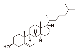
Cholesterol is the principal sterol of all higher animals, distributed in body tissues, especially the brain and spinal cord, and in animal fats and oils.
High-density lipoprotein (HDL) is one of the five major groups of lipoproteins. Lipoproteins are complex particles composed of multiple proteins which transport all fat molecules (lipids) around the body within the water outside cells. They are typically composed of 80–100 proteins per particle. HDL particles enlarge while circulating in the blood, aggregating more fat molecules and transporting up to hundreds of fat molecules per particle.
Low-density lipoprotein (LDL) is one of the five major groups of lipoprotein that transport all fat molecules around the body in extracellular water. These groups, from least dense to most dense, are chylomicrons, very low-density lipoprotein (VLDL), intermediate-density lipoprotein (IDL), low-density lipoprotein (LDL) and high-density lipoprotein (HDL). LDL delivers fat molecules to cells. LDL is involved in atherosclerosis, a process in which it is oxidized within the walls of arteries.
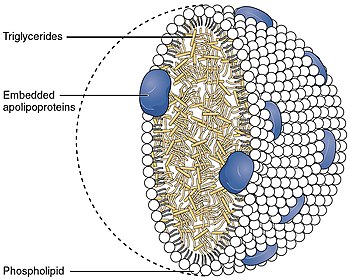
A lipoprotein is a biochemical assembly whose primary function is to transport hydrophobic lipid molecules in water, as in blood plasma or other extracellular fluids. They consist of a triglyceride and cholesterol center, surrounded by a phospholipid outer shell, with the hydrophilic portions oriented outward toward the surrounding water and lipophilic portions oriented inward toward the lipid center. A special kind of protein, called apolipoprotein, is embedded in the outer shell, both stabilising the complex and giving it a functional identity that determines its role.
Hypercholesterolemia, also called high cholesterol, is the presence of high levels of cholesterol in the blood. It is a form of hyperlipidemia, hyperlipoproteinemia, and dyslipidemia.
Dyslipidemia is a metabolic disorder characterized by abnormally high or low amounts of any or all lipids or lipoproteins in the blood. Dyslipidemia is a risk factor for the development of atherosclerotic cardiovascular diseases (ASCVD), which include coronary artery disease, cerebrovascular disease, and peripheral artery disease. Although dyslipidemia is a risk factor for ASCVD, abnormal levels don't mean that lipid lowering agents need to be started. Other factors, such as comorbid conditions and lifestyle in addition to dyslipidemia, is considered in a cardiovascular risk assessment. In developed countries, most dyslipidemias are hyperlipidemias; that is, an elevation of lipids in the blood. This is often due to diet and lifestyle. Prolonged elevation of insulin resistance can also lead to dyslipidemia. Likewise, increased levels of O-GlcNAc transferase (OGT) may cause dyslipidemia.
Apolipoproteins are proteins that bind lipids to form lipoproteins. They transport lipids in blood, cerebrospinal fluid and lymph.
Hyperlipidemia is abnormally high levels of any or all lipids or lipoproteins in the blood. The term hyperlipidemia refers to the laboratory finding itself and is also used as an umbrella term covering any of various acquired or genetic disorders that result in that finding. Hyperlipidemia represents a subset of dyslipidemia and a superset of hypercholesterolemia. Hyperlipidemia is usually chronic and requires ongoing medication to control blood lipid levels.
The low-density lipoprotein receptor (LDL-R) is a mosaic protein of 839 amino acids that mediates the endocytosis of cholesterol-rich low-density lipoprotein (LDL). It is a cell-surface receptor that recognizes apolipoprotein B100 (ApoB100), which is embedded in the outer phospholipid layer of very low-density lipoprotein (VLDL), their remnants—i.e. intermediate-density lipoprotein (IDL), and LDL particles. The receptor also recognizes apolipoprotein E (ApoE) which is found in chylomicron remnants and IDL. In humans, the LDL receptor protein is encoded by the LDLR gene on chromosome 19. It belongs to the low density lipoprotein receptor gene family. It is most significantly expressed in bronchial epithelial cells and adrenal gland and cortex tissue.
Campesterol is a phytosterol whose chemical structure is similar to that of cholesterol, and is one of the ingredients for E number E499.
Apolipoprotein E (Apo-E) is a protein involved in the metabolism of fats in the body of mammals. A subtype is implicated in Alzheimer's disease and cardiovascular diseases. It is encoded in humans by the gene APOE.
Apolipoprotein B (ApoB) is a protein that in humans is encoded by the APOB gene. It is commonly used to detect risk of atherosclerotic cardiovascular disease.
The lipid hypothesis is a medical theory postulating a link between blood cholesterol levels and the occurrence of cardiovascular disease. A summary from 1976 described it as: "measures used to lower the plasma lipids in patients with hyperlipidemia will lead to reductions in new events of coronary heart disease". It states, more concisely, that "decreasing blood cholesterol [...] significantly reduces coronary heart disease".
Familial hypercholesterolemia (FH) is a genetic disorder characterized by high cholesterol levels, specifically very high levels of low-density lipoprotein cholesterol, in the blood and early cardiovascular diseases. The most common mutations diminish the number of functional LDL receptors in the liver or produce abnormal LDL receptors that never go to the cell surface to function properly. Since the underlying body biochemistry is slightly different in individuals with FH, their high cholesterol levels are less responsive to the kinds of cholesterol control methods which are usually more effective in people without FH. Nevertheless, treatment is usually effective.
Apolipoprotein AI(Apo-AI) is a protein that in humans is encoded by the APOA1 gene. As the major component of HDL particles, it has a specific role in lipid metabolism.
Apolipoprotein C-IV, also known as apolipoprotein C4, is a protein that in humans is encoded by the APOC4 gene.
Apolipoprotein D (ApoD) is a protein that in humans is encoded by the APOD gene. Unlike other lipoproteins, which are mainly produced in the liver, apolipoprotein D is mainly produced in the brain and testes. It is a 29 kDa glycoprotein discovered in 1963 as a component of the high-density lipoprotein (HDL) fraction of human plasma. It is the major component of human mammary cyst fluid. The human gene encoding it was cloned in 1986 and the deduced protein sequence revealed that ApoD is a member of the lipocalin family, small hydrophobic molecule transporters. ApoD is 169 amino acids long, including a secretion peptide signal of 20 amino acids. It contains two glycosylation sites and the molecular weight of the mature protein varies from 20 to 32 kDa.
Apolipoprotein A-V is a protein that in humans is encoded by the APOA5 gene on chromosome 11. It is significantly expressed in liver. The protein encoded by this gene is an apolipoprotein and an important determinant of plasma triglyceride levels, a major risk factor for coronary artery disease. It is a component of several lipoprotein fractions including VLDL, HDL, chylomicrons. It is believed that apoA-V affects lipoprotein metabolism by interacting with LDL-R gene family receptors. Considering its association with lipoprotein levels, APOA5 is implicated in metabolic syndrome. The APOA5 gene also contains one of 27 SNPs associated with increased risk of coronary artery disease.
Proprotein convertase subtilisin/kexin type 9 (PCSK9) is an enzyme encoded by the PCSK9 gene in humans on chromosome 1. It is the 9th member of the proprotein convertase family of proteins that activate other proteins. Similar genes (orthologs) are found across many species. As with many proteins, PCSK9 is inactive when first synthesized, because a section of peptide chains blocks their activity; proprotein convertases remove that section to activate the enzyme. The PCSK9 gene also contains one of 27 loci associated with increased risk of coronary artery disease.
Lipoprotein-associated phospholipase A2 (Lp-PLA2) also known as platelet-activating factor acetylhydrolase (PAF-AH) is a phospholipase A2 enzyme that in humans is encoded by the PLA2G7 gene. Lp-PLA2 is a 45-kDa protein of 441 amino acids. It is one of several PAF acetylhydrolases.
1 2 Sandholzer C, Hallman DM, Saha N, Sigurdsson G, Lackner C, Császár A, etal. (1991). "Effects of the apolipoprotein(a) size polymorphism on the lipoprotein(a) concentration in 7 ethnic groups". Hum. Genet. 86 (6): 607–14. doi:10.1007/BF00201550. PMID2026424. S2CID19657929.
↑ Lobentanz EM, Krasznai K, Gruber A, Brunner C, Müller HJ, Sattler J, etal. (April 1998). "Intracellular metabolism of human apolipoprotein(a) in stably transfected Hep G2 cells". Biochemistry. 37 (16): 5417–25. doi:10.1021/bi972761t. PMID9548923.
1 2 Pati N, Rouf A, Pati U (February 2000). "Simultaneous mutations (A/G(-418) and C/T(-384)) in the apo(a) promoter of individuals with low Lp(a) levels". Molecular Genetics and Metabolism. 69 (2): 165–7. doi:10.1006/mgme.1999.2956. PMID10720444.
↑ Dieplinger H, Utermann G (June 1999). "The seventh myth of lipoprotein(a): where and how is it assembled?". Current Opinion in Lipidology. 10 (3): 275–83. doi:10.1097/00041433-199906000-00010. PMID10431664.
↑ Koschinsky ML, Marcovina SM (April 2004). "Structure-function relationships in apolipoprotein(a): insights into lipoprotein(a) assembly and pathogenicity". Current Opinion in Lipidology. 15 (2): 167–74. doi:10.1097/00041433-200404000-00009. PMID15017359. S2CID45103589.
↑ Pan S, Kleppe LS, Witt TA, Mueske CS, Simari RD (September 2004). "The effect of vascular smooth muscle cell-targeted expression of tissue factor pathway inhibitor in a murine model of arterial thrombosis". Thrombosis and Haemostasis. 92 (3): 495–502. doi:10.1160/TH04-01-0006. PMID15351845. S2CID1212821.
↑ Kostner GM, Bihari-Varga M (August 1990). "Is the atherogenicity of Lp(a) caused by its reactivity with proteoglycans?". European Heart Journal. 11 Suppl E: 184–9. doi:10.1093/eurheartj/11.suppl_e.184. PMID2146124.
↑ Christian Wilde (2003). Hidden Causes of Heart Attack and Stroke: Inflammation, Cardiology's New Frontier. Abigon Press. pp.182–183. ISBN978-0-9724959-0-5.
1 2 Takagi H, Umemoto T (January 2012). "Atorvastatin decreases lipoprotein(a): a meta-analysis of randomized trials". Int. J. Cardiol. 154 (2): 183–6. doi:10.1016/j.ijcard.2011.09.060. PMID21996415.
↑ Sahebkar A, Reiner Ž, Simental-Mendía LE, Ferretti G, Cicero AF (2016). "Effect of extended-release niacin on plasma lipoprotein(a) levels: A systematic review and meta-analysis of randomized placebo-controlled trials". Metabolism: Clinical and Experimental. 65 (11): 1664–1678. doi:10.1016/j.metabol.2016.08.007. PMID27733255.
↑ Artemeva NV, Safarova MS, Ezhov MV, Afanasieva OI, Dmitrieva OA, Pokrovsky SN (May 2015). "Lowering of lipoprotein(a) level under niacin treatment is dependent on apolipoprotein(a) phenotype". Atherosclerosis. Supplements. 18: 53–8. doi:10.1016/j.atherosclerosissup.2015.02.008. PMID25936305.
↑ Boerwinkle E, Menzel HJ, Kraft HG, Utermann G (April 1989). "Genetics of the quantitative Lp(a) lipoprotein trait. III. Contribution of Lp(a) glycoprotein phenotypes to normal lipid variation". Hum. Genet. 82 (1): 73–8. doi:10.1007/BF00288277. PMID2523852. S2CID912295.
↑ Parhofer KG (2011). "Lipoprotein(a): medical treatment options for an elusive molecule". Curr. Pharm. Des. 17 (9): 871–6. doi:10.2174/138161211795428777 (inactive 2024-04-11). PMID21476974.{{cite journal}}: CS1 maint: DOI inactive as of April 2024 (link)
↑ Anagnostis P, Galanis P, Chatzistergiou V, Stevenson JC, Godsland IF, Lambrinoudaki I, etal. (May 2017). "The effect of hormone replacement therapy and tibolone on lipoprotein (a) concentrations in postmenopausal women: A systematic review and meta-analysis". Maturitas. 99: 27–36. doi:10.1016/j.maturitas.2017.02.009. hdl:10044/1/48763. PMID28364865.
↑ Parhofer KG (2011). "Lipoprotein(a): medical treatment options for an elusive molecule". Current Pharmaceutical Design. 17 (9): 871–6. doi:10.2174/138161211795428777 (inactive 2024-04-11). PMID21476974.{{cite journal}}: CS1 maint: DOI inactive as of April 2024 (link)
This page is based on this Wikipedia article Text is available under the CC BY-SA 4.0 license; additional terms may apply. Images, videos and audio are available under their respective licenses.