Long thoracic nerve

Last updated

Long thoracic nerve
Nerves of the left upper extremity.gif
Nerves of the left upper extremity. (Long thoracic labeled vertically at shoulder, to left of artery.)
Gray808.png
The right brachial plexus with its short branches, viewed from in front. (Long thoracic labeled at center, third from top.)
Details
From Brachial plexus (C5-C7)
Innervates Serratus anterior muscle
Identifiers
Latin nervus thoracicus longus
TA98 A14.2.03.012
TA2 6410
FMA 65275
Anatomical terms of neuroanatomy

The long thoracic nerve (also: external respiratory nerve of Bell or posterior thoracic nerve) is a branch of the brachial plexus derived from cervical nerves C5-C7 that innervates the serratus anterior muscle.

Contents

Structure

Origin

The long thoracic nerve arises from the anterior rami of cervical spinal nerves C5, C6, and C7. [1] [2] [3] The root from C7 may occasionally be absent. [4] The roots from C5 and C6 pierce through the scalenus medius, while the C7 root passes in front of the muscle.[ citation needed ]

Course and relations

The long thoracic nerve descends through the cervicoaxillary canal. It is posterior to the brachial plexus, [3] and the axillary artery and vein. [4] This takes it deep to the clavicle. [2] It rests on the outer surface of the serratus anterior muscle. It extends along the side of the thorax to the lower border of the serratus anterior muscle, supplying fibres to each of the muscle's digitations. [5] [6]

Function

The long thoracic nerve innervates the serratus anterior muscle. [1] It supplies filaments to each of its digitations (finger-like projections). [5] [6]

Clinical significance

Due to its long, relatively superficial course, the long thoracic nerve is susceptible to injury, either through direct trauma or stretch. [7] Mechanisms of injury include:

Symptoms are often minimal – if symptomatic, a posterior shoulder or scapular burning type of pain may be reported. Some injuries, particularly lesions, can paralyse the serratus anterior muscle to produce a winged scapula. [3] [12] This is most prominent when the arm is lifted forward or when the patient pushes the outstretched arm against a wall. However, even winging may not be evident until the trapezius stretches enough to reveal an injury several weeks later. [13]

See also

Related Research Articles

<span class="mw-page-title-main">Radial nerve</span> Nerve in the human body that supplies the posterior portion of the upper limb

The radial nerve is a nerve in the human body that supplies the posterior portion of the upper limb. It innervates the medial and lateral heads of the triceps brachii muscle of the arm, as well as all 12 muscles in the posterior osteofascial compartment of the forearm and the associated joints and overlying skin.

<span class="mw-page-title-main">Median nerve</span> Nerve of the upper limb

The median nerve is a nerve in humans and other animals in the upper limb. It is one of the five main nerves originating from the brachial plexus.

<span class="mw-page-title-main">Brachial plexus</span> Network of nerves

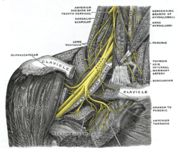
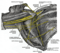
The brachial plexus is a network of nerves formed by the anterior rami of the lower four cervical nerves and first thoracic nerve. This plexus extends from the spinal cord, through the cervicoaxillary canal in the neck, over the first rib, and into the armpit, it supplies afferent and efferent nerve fibers to the chest, shoulder, arm, forearm, and hand.

<span class="mw-page-title-main">Axillary nerve</span> Nerve of the human body near the armpit

The axillary nerve or the circumflex nerve is a nerve of the human body, that originates from the brachial plexus at the level of the axilla (armpit) and carries nerve fibers from C5 and C6. The axillary nerve travels through the quadrangular space with the posterior circumflex humeral artery and vein to innervate the deltoid and teres minor.

<span class="mw-page-title-main">Dorsal scapular nerve</span> Branch of the brachial plexus that supplies rhomboid muscles and levator scapulae

The dorsal scapular nerve is a branch of the brachial plexus, usually derived from the ventral ramus of cervical nerve C5. It provides motor innervation to the rhomboid major muscle, rhomboid minor muscle, and levator scapulae muscle.

<span class="mw-page-title-main">Subclavian artery</span> Major arteries of the upper thorax, below the clavicle

In human anatomy, the subclavian arteries are paired major arteries of the upper thorax, below the clavicle. They receive blood from the aortic arch. The left subclavian artery supplies blood to the left arm and the right subclavian artery supplies blood to the right arm, with some branches supplying the head and thorax. On the left side of the body, the subclavian comes directly off the aortic arch, while on the right side it arises from the relatively short brachiocephalic artery when it bifurcates into the subclavian and the right common carotid artery.

<span class="mw-page-title-main">Infrahyoid muscles</span> Group of muscles

The infrahyoid muscles, or strap muscles, are a group of four pairs of muscles in the anterior (frontal) part of the neck. The four infrahyoid muscles are the sternohyoid, sternothyroid, thyrohyoid and omohyoid muscles.

<span class="mw-page-title-main">Cervical plexus</span> Network of nerves in the neck

The cervical plexus is a nerve plexus of the anterior rami of the first four cervical spinal nerves C1-C4. The cervical plexus provides motor innervation to some muscles of the neck, and the diaphragm; it provides sensory innervation to parts of the head, neck, and chest.

<span class="mw-page-title-main">Epineurium</span> Outermost layer of a nerve

The epineurium is the outermost layer of dense irregular connective tissue surrounding a peripheral nerve. It usually surrounds multiple nerve fascicles as well as blood vessels which supply the nerve. Smaller branches of these blood vessels penetrate into the perineurium. In addition to blood vessels which supply the nerve, lymphocytes and fibroblasts are also present and contribute to the production of collagen fibers that form the backbone of the epineurium. In addition to providing structural support, lymphocytes and fibroblasts also play a vital role in maintenance and repair of the surrounding tissues.

<span class="mw-page-title-main">Teres major muscle</span> Muscle of the upper limb

The teres major muscle is a muscle of the upper limb. It attaches to the scapula and the humerus and is one of the seven scapulohumeral muscles. It is a thick but somewhat flattened muscle.

<span class="mw-page-title-main">Intercostal nerves</span> Nerves in the thorax and abdomen

The intercostal nerves are part of the somatic nervous system, and arise from the anterior rami of the thoracic spinal nerves from T1 to T11. The intercostal nerves are distributed chiefly to the thoracic pleura and abdominal peritoneum, and differ from the anterior rami of the other spinal nerves in that each pursues an independent course without plexus formation.

<span class="mw-page-title-main">Medial cord</span>

The medial cord is the part of the brachial plexus formed by of the anterior division of the lower trunk (C8-T1). Its name comes from it being medial to the axillary artery as it passes through the axilla. The other cords of the brachial plexus are the posterior cord and lateral cord.

<span class="mw-page-title-main">Medial pectoral nerve</span>

The medial pectoral nerve is (typically) a branch of the medial cord of the brachial plexus and is derived from spinal nerve roots C8-T1. It provides motor innervation to the pectoralis minor muscle, and the lower half of the pectoralis major muscle. It runs along the inferior border of the pectoralis minor muscle.

<span class="mw-page-title-main">Thoracodorsal nerve</span> Nerve that supplies the latissimus dorsi

The thoracodorsal nerve is a nerve present in humans and other animals, also known as the middle subscapular nerve or the long subscapular nerve. It supplies the latissimus dorsi muscle.

<span class="mw-page-title-main">Anterior superior iliac spine</span> Bony projection of the iliac bone

The anterior superior iliac spine (ASIS) is a bony projection of the iliac bone, and an important landmark of surface anatomy. It refers to the anterior extremity of the iliac crest of the pelvis. It provides attachment for the inguinal ligament, and the sartorius muscle. The tensor fasciae latae muscle attaches to the lateral aspect of the superior anterior iliac spine, and also about 5 cm away at the iliac tubercle.

<span class="mw-page-title-main">Lower subscapular nerve</span>

The lower subscapular nerve, also known as the inferior subscapular nerve, is the third branch of the posterior cord of the brachial plexus. It innervates the inferior portion of the subscapularis muscle and the teres major muscle.

<span class="mw-page-title-main">Lateral pectoral nerve</span>

The lateral pectoral nerve arises from the lateral cord of the brachial plexus, and through it from the C5-7.

<span class="mw-page-title-main">Bicipital aponeurosis</span> Distal end of the biceps muscle

The bicipital aponeurosis is a broad aponeurosis of the biceps brachii, which is located in the cubital fossa of the elbow. It separates superficial from deep structures in much of the fossa.

<span class="mw-page-title-main">Piriformis nerve</span>

The piriformis nerve, also known as the nerve to piriformis, is the peripheral nerve that provides motor innervation to the piriformis muscle.

<span class="mw-page-title-main">Nerve to obturator internus</span> Human nerve

The nerve to obturator internus is a mixed nerve providing motor innervation to the obturator internus muscle and gemellus superior muscle, and sensory innervation to the hip joint. It is a branch of the sacral plexus. It is one of the group of deep gluteal nerves.

References

  1. 1 2 Rakocevic, G. (January 1, 2014), "Thoracic Nerve, Long", in Aminoff, Michael J.; Daroff, Robert B. (eds.), Encyclopedia of the Neurological Sciences (Second Edition), Oxford: Academic Press, pp. 448–450, doi:10.1016/b978-0-12-385157-4.00699-0, ISBN   978-0-12-385158-1 , retrieved October 25, 2020
  2. 1 2 Tubbs, R. Shane; Goodrich, Dylan; Watanabe, Koichi; Loukas, Marios (January 1, 2015), Tubbs, R. Shane; Rizk, Elias; Shoja, Mohammadali M.; Loukas, Marios (eds.), "Chapter 43 - Anatomic Landmarks for Selected Nerves of the Head, Neck, and Upper and Lower Limbs", Nerves and Nerve Injuries, San Diego: Academic Press, pp. 575–588, doi:10.1016/b978-0-12-410390-0.00045-7, ISBN   978-0-12-410390-0 , retrieved October 25, 2020
  3. 1 2 3 4 Ryan, Monique M.; Jones, H. Royden (January 1, 2015), Darras, Basil T.; Jones, H. Royden; Ryan, Monique M.; De Vivo, Darryl C. (eds.), "Chapter 14 – Mononeuropathies", Neuromuscular Disorders of Infancy, Childhood, and Adolescence (Second Edition), San Diego: Academic Press, pp. 243–273, doi:10.1016/b978-0-12-417044-5.00014-7, ISBN   978-0-12-417044-5 , retrieved October 25, 2020
  4. 1 2 Tomberlin, John (January 1, 2009), Wilk, Kevin E.; Reinold, Michael M.; Andrews, James R. (eds.), "CHAPTER 53 - Neurodynamic Techniques for the Athlete's Shoulder", The Athlete's Shoulder (Second Edition), Philadelphia: Churchill Livingstone, pp. 707–717, doi:10.1016/b978-044306701-3.50056-6, ISBN   978-0-443-06701-3 , retrieved October 25, 2020
  5. 1 2 Fischer, J. (2012). Anatomy of the Axilla. Fischer's Mastery of Surgery, 2 Volume Set. Retrieved September 20, 2015 from http://www.r2library.com/Resource/Title/1608317404/ch0046s1193
  6. 1 2 Bertelli, Jayme Augusto (2005). "Long Thoracic Nerve: Anatomy and Functional Assessment". The Journal of Bone and Joint Surgery. American Volume. 87 (5): 993–8. doi:10.2106/JBJS.D.02383. ISSN   0021-9355. PMID   15866961.
  7. Ostrowska, Monika; de Carvalho, Mamede (January 1, 2015), Tubbs, R. Shane; Rizk, Elias; Shoja, Mohammadali M.; Loukas, Marios (eds.), "Chapter 34 - Injuries of the Nerves of the Thorax", Nerves and Nerve Injuries, San Diego: Academic Press, pp. 525–543, doi:10.1016/b978-0-12-802653-3.00083-x, ISBN   978-0-12-802653-3 , retrieved October 25, 2020
  8. 1 2 "An Isolated Long Thoracic Nerve Injury in a Navy Airman". Military Medicine. 2004.
  9. Belmonte, Roser; Monleon, Sandra; Bofill, Neus; Alvarado, Martha Ligia; Espadaler, Josep; Royo, Inmaculada (2015). "Long thoracic nerve injury in breast cancer patients treated with axillary lymph node dissection". Supportive Care in Cancer. 23 (1): 169–175. doi:10.1007/s00520-014-2338-5. ISSN   1433-7339. PMID   25035064. S2CID   25547808.
  10. Berthold, Justin B.; Burg, Timothy M.; Nussbaum, Ryan Paul (February 1, 2017). "Long Thoracic Nerve Injury Caused by Overhead Weight Lifting Leading to Scapular Dyskinesis and Medial Scapular Winging". Journal of Osteopathic Medicine. 117 (2): 133–137. doi: 10.7556/jaoa.2017.025 . ISSN   2702-3648. PMID   28134956. S2CID   25777654.
  11. Monteiro dos Santos, Ricardo Barreto; dos Santos, Saulo Monteiro; Carneiro Leal, Flávio José Câmara; Lins, Otávio Gomes; Magalhães, Carmem; Mertens Fittipaldi, Ricardo Bruno (April 17, 2015). "Parsonage–Turner syndrome". Revista Brasileira de Ortopedia. 50 (3): 336–341. doi:10.1016/j.rboe.2015.04.002. ISSN   2255-4971. PMC   4519651 . PMID   26229940.
  12. Le Nail, L. R.; Bacle, G.; Marteau, E.; Corcia, P.; Favard, L.; Laulan, J. (June 2014). "Isolated paralysis of the serratus anterior muscle: surgical release of the distal segment of the long thoracic nerve in 52 patients". Orthopaedics & Traumatology, Surgery & Research. 100 (4 Suppl): S243–248. doi: 10.1016/j.otsr.2014.03.004 . ISSN   1877-0568. PMID   24703793.
  13. Preston, David C.; Shapiro, Barbara E. (January 1, 2013), Preston, David C.; Shapiro, Barbara E. (eds.), "31 - Proximal Neuropathies of the Shoulder and Arm", Electromyography and Neuromuscular Disorders (Third Edition), London: W.B. Saunders, pp. 487–500, doi:10.1016/b978-1-4557-2672-1.00031-3, ISBN   978-1-4557-2672-1 , retrieved October 25, 2020

Additional images