Lunate sulcus

Last updated
Lunate sulcus
Gray726.svg
Lateral surface of left cerebral hemisphere, viewed from the side.
Details
Location Occipital lobe
Function Sulcus
Identifiers
Latin sulcus lunatus
NeuroLex ID birnlex_4017
TA98 A14.1.09.134
TA2 5483
FMA 83788
Anatomical terms of neuroanatomy

In brain anatomy, the lunate sulcus or simian sulcus also known as the sulcus lunatus is a fissure in the occipital lobe [1] variably found in humans and more often larger when present in apes and monkeys. [2] The lunate sulcus marks the transition between V1 and V2. [3]

Contents

The lunate sulcus lies further back in human brains but has a more forward location in chimpanzees. [4] The evolutionary expansion of the frontal areas of the lunate sulcus would have caused a shift in the particular location of the fissure. [4] [5] It has been hypothesized that evolutionary pressures resulted in the human brain undergoing internal reorganization to develop the capability of human language. [6] Furthermore, this reorganization must have been implemented during early maturity and is likely responsible for eidetic imagery in some adolescents. [6]

During early development, the neural connections in prefrontal cortex and posterior parietal lobe rapidly expand to allow capability for human language, while visual memory capacity of human brain would become limited. [7] Biological studies have demonstrated that the lunate sulcus is subject to white matter growth, and dental fossil and tomography studies have shown that the brain organization of Australopithecus africanus is pongid-like. [8]

History

Comparison of the Affenspalte homologue, renamed the lunate sulcus by Sir Grafton Elliot Smith, in the primary visual cortex of chimpanzee versus human brains. The Affenspalte, or lunate sulcus, is marked by the red line. Lunate sulcus.png
Comparison of the Affenspalte homologue, renamed the lunate sulcus by Sir Grafton Elliot Smith, in the primary visual cortex of chimpanzee versus human brains. The Affenspalte, or lunate sulcus, is marked by the red line.

The lunate sulcus was first identified during the early 1900s in the human brain as a homologue of the Affenspalte, a major sulcus defining the primary visual cortex (V1) in apes and other monkey species, by anatomist and Egyptologist Sir Grafton Elliot Smith. [9] Based on Smith’s observations from studying over 400 Egyptian and ape brains, he noticed that the sulcal patterns between humans and apes were very similar. [9] His methodology involved mapping cortical areas via simple visual inspection of endocasts from mummies, as well as of fresh whole and sectioned brains. [9] Paleoneurologists and scientists study endocasts in order to gather information about brain size and shape, as well as sulcal patterns resulting from pressure-induced impressions by the brain’s surface. Comparison of data gathered from endocasts and the brains of living hominoids allows scientists to study the evolution of the human brain, both anatomically and cognitively. Ultimately, Smith argued that the lunate sulcus was responsible for delineating the rostrolateral boundary of the V1 in both humans and non-human primates, and even pointed out the specific location of the lunate sulcus in chimpanzee versus human brains. [9] Notably, Smith noticed that the position of the lunate sulcus was more posterior in human, especially those of European descent, as compared to monkey brains. [9] Based on this observation, he was the first to hypothesize that the caudal shift of the lunate sulcus in Homo sapiens was due to the evolutionary rapid overgrowth of the cerebral cortex that is unique to human neurodevelopment. [9]

Smith’s observation that the caudal shift of the lunate sulcus could also be used as a predictor for determining both the evolutionary posterolateral shift of the occipital lobes/V1 and the corresponding expansion of the neighboring parietotemporo-occipital visual association cortices was supported by recent research. [9] [10] However, some scientists today disagree with Smith’s assertion that a lunate sulcus exists in humans, arguing that there is only an Affenspalte which is solely unique to apes. Specifically, in a high-resolution MRI study conducted by Allen et al. (2006), the researchers scanned and analyzed 220 human brains and found no sign of the lunate sulcus homologue. Based on this finding, they suggested that the claim asserting humans have a lunate sulcus homologue fails to account for and show appreciation of the extensive evolutionary reorganization of the visual cortex in humans. [1]

Evolution

Analyzing variability in the location of gross anatomical landmarks, like sulci, is an accepted method for studying evolutionary hominin brain reorganization. Notably, the position of the lunate sulcus in the occipital lobe has been studied in humans, early hominin endocasts, apes, and other monkey species by researchers seeking to make inferences about the morphological evolution of brain regions associated with human visual versus cognitive behaviors. [10] [11] However, some scientists remain skeptical about whether the lunate sulcus is a valid and reliable indicator for studying volumetric changes in the V1 due to the inconsistencies of the sulcus’ presence and lack of histological correspondence with cytoarchitectonic boundaries in hominoids. [12] Despite this, previous allometry studies have suggested that the lunate sulcus shifts from a lateral-anterior to a medial-posterior position as brain size increases. [13] [14] Such shifts have been accredited with predicting whether the lunate sulcus will occur or not based on an increase or reduction in V1 volume, thus providing an explanation for inconsistencies in its presence and position in the occipital lobes. [13] [15] Moreover, a study conducted by de Sousa et al. (2010) compared the volumes of the V1 relative to the position of the lunate sulcus in three-dimensional reconstructed non-human hominoid brains in order to determine if an allometric relationship existed between V1 volume and lunate sulcus position. The researchers found that the position of the lunate sulcus does accurately predict V1 volume in apes, and that V1 volume in humans is smaller than would be expected based on our large brain size. [10] Furthermore, other research suggests a more posteriorly positioned lunate sulcus from the early hominin fossil record. [4] [5] Based on all these findings, de Sousa et al. (2010) concluded V1 reduction began during early hominin evolution given the more lateral-anterior position of the lunate sulcus in human and other primate brains today.

Related Research Articles

Visual cortex Region of the brain that processes visual information

The visual cortex of the brain is the area of the cerebral cortex that processes visual information. It is located in the occipital lobe. Sensory input originating from the eyes travels through the lateral geniculate nucleus in the thalamus and then reaches the visual cortex. The area of the visual cortex that receives the sensory input from the lateral geniculate nucleus is the primary visual cortex, also known as visual area 1 (V1), Brodmann area 17, or the striate cortex. The extrastriate areas consist of visual areas 2, 3, 4, and 5.

Cerebral cortex Outer layer of the cerebrum of the mammalian brain

The cerebral cortex, also known as the cerebral mantle, is the outer layer of neural tissue of the cerebrum of the brain in humans and other mammals. The cerebral cortex mostly consists of the six-layered neocortex, with just 10% consisting of allocortex. It is separated into two cortices, by the longitudinal fissure that divides the cerebrum into the left and right cerebral hemispheres. The two hemispheres are joined beneath the cortex by the corpus callosum. The cerebral cortex is the largest site of neural integration in the central nervous system. It plays a key role in attention, perception, awareness, thought, memory, language, and consciousness.

Parietal lobe Part of the brain responsible for sensory input and some language processing

The parietal lobe is one of the four major lobes of the cerebral cortex in the brain of mammals. The parietal lobe is positioned above the temporal lobe and behind the frontal lobe and central sulcus.

Precuneus Part of the brain

The precuneus is the portion of the superior parietal lobule on the medial surface of each brain hemisphere. It is located in front of the cuneus. The precuneus is bounded in front by the marginal branch of the cingulate sulcus, at the rear by the parietooccipital sulcus, and underneath by the subparietal sulcus. It is involved with episodic memory, visuospatial processing, reflections upon self, and aspects of consciousness.

Central sulcus

The central sulcus is a sulcus, or groove, in the cerebral cortex in the brains of vertebrates. Also called the central fissure, or the fissure of Rolando or the Rolandic fissure, after Luigi Rolando. It is sometimes confused with the longitudinal fissure.

Occipital lobe Part of the brain at the back of the head

The occipital lobe is one of the four major lobes of the cerebral cortex in the brain of mammals. The name derives from its position at the back of the head, from the Latin ob, "behind," and caput, "the head."

Brodmann area 10

Brodmann area 10 is the anterior-most portion of the prefrontal cortex in the human brain. BA10 was originally defined broadly in terms of its cytoarchitectonic traits as they were observed in the brains of cadavers, but because modern functional imaging cannot precisely identify these boundaries, the terms anterior prefrontal cortex, rostral prefrontal cortex and frontopolar prefrontal cortex are used to refer to the area in the most anterior part of the frontal cortex that approximately covers BA10—simply to emphasize the fact that BA10 does not include all parts of the prefrontal cortex.

Brodmann area 40

Brodmann area 40 (BA40) is part of the parietal cortex in the human brain. The inferior part of BA40 is in the area of the supramarginal gyrus, which lies at the posterior end of the lateral fissure, in the inferior lateral part of the parietal lobe.

Lobes of the brain Parts of the cerebrum

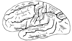
The lobes of the brain are the major identifiable zones of the cerebral cortex, and they comprise the surface of each hemisphere of the cerebrum. The two hemispheres, which are only roughly symmetrical in structure, are today considered as having six lobes each. The lobes are large areas that are anatomically distinguishable, and are also functionally distinct to some degree. Each lobe of the brain has numerous ridges, or gyri, and furrows, the sulci that constitute further subzones of the cortex. The expression "lobes of the brain" usually refers only to those of the cerebrum, not to the distinct areas of the cerebellum.

Inferior temporal gyrus One of three gyri of the temporal lobe of the brain

The inferior temporal gyrus is one of three gyri of the temporal lobe and is located below the middle temporal gyrus, connected behind with the inferior occipital gyrus; it also extends around the infero-lateral border on to the inferior surface of the temporal lobe, where it is limited by the inferior sulcus. This region is one of the higher levels of the ventral stream of visual processing, associated with the representation of objects, places, faces, and colors. It may also be involved in face perception, and in the recognition of numbers.

Parieto-occipital sulcus

The parieto-occipital sulcus is a deep sulcus in the cerebral cortex that marks the boundary between the cuneus and precuneus, and also between the parietal and occipital lobes. Only a small part can be seen on the lateral surface of the hemisphere, its chief part being on the medial surface.

Sulcus (neuroanatomy) Fold in the surface of the brain

In neuroanatomy, a sulcus is a depression or groove in the cerebral cortex. It surrounds a gyrus, creating the characteristic folded appearance of the brain in humans and other mammals. The larger sulci are usually called fissures.

Superior longitudinal fasciculus Association fiber tract of the brain

The superior longitudinal fasciculus (SLF) is an association tract in the brain that is composed of three separate components. It is present in both hemispheres and can be found lateral to the centrum semiovale and connects the frontal, occipital, parietal, and temporal lobes. This bundle of tracts (fasciculus) passes from the frontal lobe through the operculum to the posterior end of the lateral sulcus where they either radiate to and synapse on neurons in the occipital lobe, or turn downward and forward around the putamen and then radiate to and synapse on neurons in anterior portions of the temporal lobe.

Ralph Leslie Holloway, Jr. is a physical anthropologist at Columbia University and research associate with the American Museum of Natural History. Since obtaining his Ph.D from the University of California, Berkeley in 1964, Holloway has served as a professor of anthropology at Columbia. Holloway's interests lie in craniology, producing endocasts, primate behavior, biology of gender, sexual dimorphism in the corpus callosum, and other topics.

Dean Falk American academic Neuroanthropologist

Dean Falk is an American academic neuroanthropologist who specializes in the evolution of the brain and cognition in higher primates. She is the Hale G. Smith Professor of Anthropology and a Distinguished Research Professor at Florida State University.

The Riddoch syndrome is a term coined by Zeki and Ffytche (1998) in a paper published in Brain. The term acknowledges the work of George Riddoch who was the first to describe a condition in which a form of visual impairment, caused by lesions in the occipital lobe, leaves the sufferer blind but able to distinguish visual stimuli with specific characteristics when these appear in the patient's blind field. The most common stimuli that can be perceived consciously are the presence and direction of fast moving objects ; in his work these moving objects were described as "vague and shadowy". Riddoch concluded from his observations that "movement may be recognized as a special visual perception".

The principles that govern the evolution of brain structure are not well understood. Brain to body size scales allometrically. Small bodied mammals have relatively large brains compared to their bodies whereas large mammals have smaller brain to body ratios. If brain weight is plotted against body weight for primates, the regression line of the sample points can indicate the brain power of a primate species. Lemurs for example fall below this line which means that for a primate of equivalent size, we would expect a larger brain size. Humans lie well above the line indicating that humans are more encephalized than lemurs. In fact, humans are more encephalized than all other primates.

The neuroanatomy of memory encompasses a wide variety of anatomical structures in the brain.

Paleoneurobiology

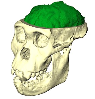
Paleoneurobiology is the study of brain evolution by analysis of brain endocasts to determine endocranial traits and volumes. Considered a subdivision of neuroscience, paleoneurobiology combines techniques from other fields of study including paleontology and archaeology. It reveals specific insight concerning human evolution. The cranium is unique in that it grows in response to the growth of brain tissue rather than genetic guidance, as is the case with bones that support movement. Fossil skulls and their endocasts can be compared to each other, to the skulls and fossils of recently deceased individuals, and even compared to those of other species to make inferences about functional anatomy, physiology and phylogeny. Paleoneurobiology is in large part influenced by developments in neuroscience as a whole; without substantial knowledge about current functionality, it would be impossible to make inferences about the functionality of ancient brains.

Occipital gyri Three parallel gyri of the occipital lobe of the brain

The occipital gyri (OcG) are three gyri in parallel, along the lateral portion of the occipital lobe, also referred to as a composite structure in the brain. The gyri are the superior occipital gyrus, the middle occipital gyrus, and the inferior occipital gyrus, and these are also known as the occipital face area. The superior and inferior occipital sulci separates the three occipital gyri.

References

  1. 1 2 Allen JS, Bruss J, Damasio H (August 2006). "Looking for the lunate sulcus: a magnetic resonance imaging study in modern humans". Anat Rec A. 288 (8): 867–76. doi: 10.1002/ar.a.20362 . PMID   16835937.
  2. Srijit D, Shipra P (2008). "Unilateral absence of the lunate sulcus: an anatomical perspective". Rom J Morphol Embryol. 49 (2): 257–8. PMID   18516336.
  3. Schmolesky, M. "The organization of the retina and visual system".{{cite journal}}: Cite journal requires |journal= (help)
  4. 1 2 3 Rincon, Paul (2004). "Human brain began evolving early". BBC.
  5. 1 2 Bruner, E (2014). Human paleoneurology. Springer.
  6. 1 2 Ko, Kwang Hyun (2015). "Brain Reorganization Allowed for the Development of Human Language: Lunate Sulcus". International Journal of Biology. 7 (3): 59. doi: 10.5539/ijb.v7n3p59 .
  7. Gogtay, N (2004). "Dynamic mapping of human cortical development during childhood through early adulthood". Proceedings of the National Academy of Sciences. 101 (21): 8174–8179. doi: 10.1073/pnas.0402680101 . PMC   419576 . PMID   15148381.
  8. Dean, C (2001). "Growth processes in teeth distinguish modern humans from Homo erectus and earlier hominins". Nature. 414 (6864): 628–631. Bibcode:2001Natur.414..628D. doi:10.1038/414628a. PMID   11740557. S2CID   4428635.
  9. 1 2 3 4 5 6 7 8 Falk, Dean (May 2014). "Interpreting sulci on hominin endocasts: old hypothesis and new findings". Frontiers in Human Neuroscience. 8: 134. doi: 10.3389/fnhum.2014.00134 . PMC   4013485 . PMID   24822043.
  10. 1 2 3 De Sousa, Alexandra A.; Sherwood, Chet C.; Mohlberg, Hartmut; Amunts, Katrin; Schleicher, Axel; MacLeod, Carol E.; Hof, Patrick R.; Frahm, Heiko; Zilles, Karl (April 2010). "Hominoid visual brain structure volumes and the position of the lunate sulcus". Journal of Human Evolution. 58 (4): 281–292. doi:10.1016/j.jhevol.2009.11.011. PMID   20172590.
  11. Holloway L, Broadfield C, Yuan S, Tobias V (2003). "The lunate sulcus and early hominid brain evolution: Toward the end of a controversy". American Journal of Physical Anthropology. 120: 117. doi: 10.1002/ajpa.10249 .
  12. Amunts K, Schleicher A, Zilles K (2007b). "Cytoarchitecture of the cerebral cortex: More than localization". NeuroImage. 37 (4): 1061–5, discussion 1066–8. doi:10.1016/j.neuroimage.2007.02.037. PMID   17870622. S2CID   3359887.
  13. 1 2 Jerison J (1975). "Fossil evidence of evolution of the human brain". Annual Review of Anthropology. 4: 27–58. doi:10.1146/annurev.an.04.100175.000331.
  14. Kaas, JH (2000). "Why is brain size so important: design problems and solutions as neocortex gets bigger or smaller". Brain and Mind. 1: 7–23. doi:10.1023/A:1010028405318. S2CID   142597701.
  15. Preuss M and Coleman Q (2002). "Human-specific organization of primary visual cortex: Alternating compartments of dense Cat-301 and calbindin immunore- activity in layer 4A". Cerebral Cortex. 12 (7): 671–691. doi: 10.1093/cercor/12.7.671 . PMID   12050080.