M13 bacteriophage

Last updated
Escherichia virus M13
M13B.svg
Blue: Coat Protein pIII; Brown: Coat Protein pVI; Red: Coat Protein pVII; Limegreen: Coat Protein pVIII; Fuchsia: Coat Protein pIX; Purple: Single Stranded DNA
Virus classification OOjs UI icon edit-ltr.svg
(unranked): Virus
Realm: Monodnaviria
Kingdom: Loebvirae
Phylum: Hofneiviricota
Class: Faserviricetes
Order: Tubulavirales
Family: Inoviridae
Genus: Inovirus
Species:
Escherichia virus M13

M13 is one of the Ff phages (fd and f1 are others), a member of the family filamentous bacteriophage (inovirus). Ff phages are composed of circular single-stranded DNA (ssDNA), which in the case of the m13 phage is 6407 nucleotides long and is encapsulated in approximately 2700 copies of the major coat protein p8, and capped with about 5 copies each of four different minor coat proteins (p3 and p6 at one end and p7 and p9 at the other end). [1] [2] [3] The minor coat protein p3 attaches to the receptor at the tip of the F pilus of the host Escherichia coli . The life cycle is relatively short, with the early phage progeny exiting the cell ten minutes after infection. Ff phages are chronic phage, releasing their progeny without killing the host cells. The infection causes turbid plaques in E. coli lawns, of intermediate opacity in comparison to regular lysis plaques. However, a decrease in the rate of cell growth is seen in the infected cells. The replicative form of M13 is circular double-stranded DNA similar to plasmids that are used for many recombinant DNA processes, and the virus has also been used for phage display, directed evolution, nanostructures and nanotechnology applications. [4] [5] [6]

Contents

Phage particles

The phage coat is primarily assembled from a 50 amino acid protein called p8, which is encoded by gene 8 in the phage genome. For a wild type M13 particle, it takes approximately 2700 copies of p8 to make the coat about 900 nm long. The coat's dimensions are flexible because the number of p8 copies adjusts to accommodate the size of the single stranded genome it packages. [7] The phage appear to be limited to approximately twice the natural DNA content. However, deletion of a phage protein (p3) prevents full escape from the host E. coli, and phage that are 10-20X the normal length with several copies of the phage genome can be seen shedding from the E. coli host.

At one end of the filament are up to five copies of the surface exposed protein (p9) and a more buried companion protein (p7). If p8 forms the shaft of the phage, p9 and p7 form the "blunt" end that is seen in micrographs. These proteins are very small, containing only 33 and 32 amino acids respectively, though some additional residues can be added to the N-terminal portion of each which are then presented on the outside of the coat. At the other end of the phage particle are five copies of the surface exposed (p3) and its less exposed accessory protein (p6). These form the rounded tip of the phage and are the first proteins to interact with the E. coli host during infection. Protein p3 is also the last point of contact with the host as a new phage buds from the bacterial surface. [8] [9] [10] The production of phage particles causes a host cell to grow and divide, but it does not lead to lysis of the cell. [8]

Replication in E. coli

Entry of the virus into a host cell is mediated by the p3 protein, specifically the N domains, binding to the primary and secondary receptors of the host cell. [11] After the positive single strand DNA has entered the cell, it is duplicated to form the double stranded DNA that is then used to transcribe the mRNA that will build the proteins. [8]

Below are steps involved with replication of M13 in E. coli.

Phage proteins in the cytoplasm are p2, p10 and p5, and they are part of the replication process of DNA. The other phage proteins are synthesized and inserted into the cytoplasmic or outer membranes.

Unusually, the major coat protein can insert post-translation into membranes, even those lacking translocation structures, and even into liposomes with no protein content. [12]

Research

George Smith, among others, showed that fragments of EcoRI endonuclease could be fused in the unique Bam site of f1 filamentous phage and thereby expressed in gene 3 whose protein p3 was externally accessible. M13 does not have this unique Bam site in gene 3. M13 had to be engineered to have accessible insertion sites, making it limited in its flexibility in handling different sized inserts. Because the M13 phage display system allows great flexibility in the location and number of recombinant proteins on the phage, it is a popular tool to construct or serve as a scaffold for nanostructures. [13] For example, the phage can be engineered to have a different protein on each end and along its length. This can be used to assemble structures like gold or cobalt oxide nano-wires for batteries [14] or to pack carbon nanotubes into straight bundles for use in photovoltaics. [15] The M13 capsid is also the first intact virus structure to ever be solved entirely by solid state NMR. [16]

See also

Related Research Articles

<span class="mw-page-title-main">Bacteriophage</span> Virus that infects and replicates within bacteria

A bacteriophage, also known informally as a phage, is a virus that infects and replicates within bacteria and archaea. The term was derived from "bacteria" and the Greek φαγεῖν, meaning "to devour". Bacteriophages are composed of proteins that encapsulate a DNA or RNA genome, and may have structures that are either simple or elaborate. Their genomes may encode as few as four genes and as many as hundreds of genes. Phages replicate within the bacterium following the injection of their genome into its cytoplasm.

<span class="mw-page-title-main">Lambda phage</span> Bacteriophage that infects Escherichia coli

Enterobacteria phage λ is a bacterial virus, or bacteriophage, that infects the bacterial species Escherichia coli. It was discovered by Esther Lederberg in 1950. The wild type of this virus has a temperate life cycle that allows it to either reside within the genome of its host through lysogeny or enter into a lytic phase, during which it kills and lyses the cell to produce offspring. Lambda strains, mutated at specific sites, are unable to lysogenize cells; instead, they grow and enter the lytic cycle after superinfecting an already lysogenized cell.

A phagemid or phasmid is a DNA-based cloning vector, which has both bacteriophage and plasmid properties. These vectors carry, in addition to the origin of plasmid replication, an origin of replication derived from bacteriophage. Unlike commonly used plasmids, phagemid vectors differ by having the ability to be packaged into the capsid of a bacteriophage, due to their having a genetic sequence that signals for packaging. Phagemids are used in a variety of biotechnology applications; for example, they can be used in a molecular biology technique called "Phage Display".

<i>Escherichia virus T4</i> Species of bacteriophage

Escherichia virus T4 is a species of bacteriophages that infect Escherichia coli bacteria. It is a double-stranded DNA virus in the subfamily Tevenvirinae from the family Myoviridae. T4 is capable of undergoing only a lytic life cycle and not the lysogenic life cycle. The species was formerly named T-even bacteriophage, a name which also encompasses, among other strains, Enterobacteria phage T2, Enterobacteria phage T4 and Enterobacteria phage T6.

<span class="mw-page-title-main">Phage display</span> Biological technique to evolve proteins using bacteriophages

Phage display is a laboratory technique for the study of protein–protein, protein–peptide, and protein–DNA interactions that uses bacteriophages to connect proteins with the genetic information that encodes them. In this technique, a gene encoding a protein of interest is inserted into a phage coat protein gene, causing the phage to "display" the protein on its outside while containing the gene for the protein on its inside, resulting in a connection between genotype and phenotype. The proteins that the phages are displaying can then be screened against other proteins, peptides or DNA sequences, in order to detect interaction between the displayed protein and those of other molecules. In this way, large libraries of proteins can be screened and amplified in a process called in vitro selection, which is analogous to natural selection.

<span class="mw-page-title-main">Filamentous bacteriophage</span> Family of viruses

Filamentous bacteriophages are a family of viruses (Inoviridae) that infect bacteria, or bacteriophages. They are named for their filamentous shape, a worm-like chain, about 6 nm in diameter and about 1000-2000 nm long. This distinctive shape reflects their method of replication: the coat of the virion comprises five types of viral protein, which are located in the inner membrane of the host bacterium during phage assembly, and these proteins are added to the nascent virion's DNA as it is extruded through the membrane. The simplicity of filamentous phages makes them an appealing model organism for research in molecular biology, and they have also shown promise as tools in nanotechnology and immunology.

Microviridae is a family of bacteriophages with a single-stranded DNA genome. The name of this family is derived from the ancient Greek word μικρός (mikrós), meaning "small". This refers to the size of their genomes, which are among the smallest of the DNA viruses. Enterobacteria, intracellular parasitic bacteria, and spiroplasma serve as natural hosts. There are 22 species in this family, divided among seven genera and two subfamilies.

<span class="mw-page-title-main">Phi X 174</span> A single-stranded DNA virus that infects bacteria

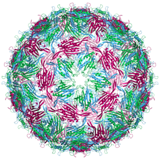
The phi X 174 bacteriophage is a single-stranded DNA (ssDNA) virus that infects Escherichia coli. This virus was isolated in 1935 by Nicolas Bulgakov in Félix d'Hérelle's laboratory at the Pasteur Institute, from samples collected in Paris sewers. Its characterization and the study of its replication mechanism were carried out from the 1950s onwards. It was the first DNA-based genome to be sequenced. This work was completed by Fred Sanger and his team in 1977. In 1962, Walter Fiers and Robert Sinsheimer had already demonstrated the physical, covalently closed circularity of ΦX174 DNA. Nobel prize winner Arthur Kornberg used ΦX174 as a model to first prove that DNA synthesized in a test tube by purified enzymes could produce all the features of a natural virus, ushering in the age of synthetic biology. In 1972–1974, Jerard Hurwitz, Sue Wickner, and Reed Wickner with collaborators identified the genes required to produce the enzymes to catalyze conversion of the single stranded form of the virus to the double stranded replicative form. In 2003, it was reported by Craig Venter's group that the genome of ΦX174 was the first to be completely assembled in vitro from synthesized oligonucleotides. The ΦX174 virus particle has also been successfully assembled in vitro. In 2012, it was shown how its highly overlapping genome can be fully decompressed and still remain functional.

<i>Pseudomonas virus phi6</i> Species of virus

Φ6 is the best-studied bacteriophage of the virus family Cystoviridae. It infects Pseudomonas bacteria. It has a three-part, segmented, double-stranded RNA genome, totalling ~13.5 kb in length. Φ6 and its relatives have a lipid membrane around their nucleocapsid, a rare trait among bacteriophages. It is a lytic phage, though under certain circumstances has been observed to display a delay in lysis which may be described as a "carrier state".

<span class="mw-page-title-main">T7 phage</span> Species of virus

Bacteriophage T7 is a bacteriophage, a virus that infects bacteria. It infects most strains of Escherichia coli and relies on these hosts to propagate. Bacteriophage T7 has a lytic life cycle, meaning that it destroys the cell it infects. It also possesses several properties that make it an ideal phage for experimentation: its purification and concentration have produced consistent values in chemical analyses; it can be rendered noninfectious by exposure to UV light; and it can be used in phage display to clone RNA binding proteins.

<span class="mw-page-title-main">Bacteriophage MS2</span> Species of virus

Bacteriophage MS2, commonly called MS2, is an icosahedral, positive-sense single-stranded RNA virus that infects the bacterium Escherichia coli and other members of the Enterobacteriaceae. MS2 is a member of a family of closely related bacterial viruses that includes bacteriophage f2, bacteriophage Qβ, R17, and GA.

<span class="mw-page-title-main">Tectivirus</span> Family of viruses

Tectiviridae is a family of viruses with 10 species in five genera. Bacteria serve as natural hosts. Tectiviruses have no head-tail structure, but are capable of producing tail-like tubes of ~ 60×10 nm upon adsorption or after chloroform treatment. The name is derived from Latin tectus.

<i>Corticovirus</i> Genus of viruses

Corticovirus is a genus of viruses in the family Corticoviridae. Corticoviruses are bacteriophages; that is, their natural hosts are bacteria. The genus contains two species. The name is derived from Latin cortex, corticis. However, prophages closely related to PM2 are abundant in the genomes of aquatic bacteria, suggesting that the ecological importance of corticoviruses might be underestimated. Bacteriophage PM2 was first described in 1968 after isolation from seawater sampled from the coast of Chile.

<span class="mw-page-title-main">Bacteriophage Qbeta</span> Species of virus

Bacteriophage Qbeta, commonly referred to as Qbeta or Qβ, is a species consisting of several strains of positive-strand RNA virus which infects bacteria that have F-pili, most commonly Escherichia coli. Its linear genome is packaged into an icosahedral capsid with a diameter of 28 nm. Bacteriophage Qβ enters its host cell after binding to the side of the F-pilus.

<span class="mw-page-title-main">Bacteriophage P2</span> Species of virus

Bacteriophage P2, scientific name Escherichia virus P2, is a temperate phage that infects E. coli. It is a tailed virus with a contractile sheath and is thus classified in the genus Peduovirus, subfamily Peduovirinae, family Myoviridae within order Caudovirales. This genus of viruses includes many P2-like phages as well as the satellite phage P4.

The CTXφ bacteriophage is a filamentous bacteriophage. It is a positive-strand DNA virus with single-stranded DNA (ssDNA).

<span class="mw-page-title-main">Ff phages</span> Group of viruses

Ff phages is a group of almost identical filamentous phage including phages f1, fd, M13 and ZJ/2, which infect bacteria bearing the F fertility factor. The virion is a flexible filament measuring about 6 by 900 nm, comprising a cylindrical protein tube protecting a single-stranded circular DNA molecule at its core. The phage codes for only 11 gene products, and is one of the simplest viruses known. It has been widely used to study fundamental aspects of molecular biology. George Smith and Greg Winter used f1 and fd for their work on phage display for which they were awarded a share of the 2018 Nobel Prize in Chemistry. Early experiments on Ff phages used M13 to identify gene functions, and M13 was also developed as a cloning vehicle, so the name M13 is sometimes used as an informal synonym for the whole group of Ff phages.

<i>Spiroplasma phage 1-R8A2B</i> Species of virus

Spiroplasma phage 1-R8A2B is a filamentous bacteriophage in the genus Vespertiliovirus of the family Plectroviridae, part of the group of single-stranded DNA viruses. The virus has many synonyms, such as SpV1-R8A2 B, Spiroplasma phage 1, and Spiroplasma virus 1, SpV1. SpV1-R8A2 B infects Spiroplasma citri. Its host itself is a prokaryotic pathogen for citrus plants, causing Citrus stubborn disease.

Phage-assisted continuous evolution (PACE) is a phage-based technique for the automated directed evolution of proteins. It relies on relating the desired activity of a target protein with the fitness of an infectious bacteriophage which carries the protein's corresponding gene. Proteins with greater desired activity hence confer greater infectivity to their carrier phage. More infectious phage propagate more effectively, selecting for advantageous mutations. Genetic variation is generated using error-prone polymerases on the phage vectors, and over time the protein accumulates beneficial mutations. This technique is notable for performing hundreds of rounds of selection with minimal human intervention.

Bacteriophage AP205 is a plaque-forming bacteriophage that infects Acinetobacter bacteria. Bacteriophage AP205 is a protein-coated virus with a positive single-stranded RNA genome. It is a member of the family Fiersviridae, consisting of particles that infect Gram-negative bacteria such as E. coli.

References

  1. Smeal SW, Schmitt MA, Pereira RR, Prasad A, Fisk JD (January 2017). "Simulation of the M13 life cycle I: Assembly of a genetically-structured deterministic chemical kinetic simulation". Virology. 500: 259–274. doi: 10.1016/j.virol.2016.08.017 . PMID   27644585.
  2. Rakonjac J, Das B, Derda R (2016). "Editorial: Filamentous Bacteriophage in Bio/Nano/Technology, Bacterial Pathogenesis and Ecology". Frontiers in Microbiology. 7: 2109. doi: 10.3389/fmicb.2016.02109 . PMC   5179506 . PMID   28066406.
  3. Roux S, Krupovic M, Daly RA, Borges AL, Nayfach S, Schulz F, et al. (November 2019). "Cryptic inoviruses revealed as pervasive in bacteria and archaea across Earth's biomes". Nature Microbiology. 4 (11): 1895–1906. doi:10.1038/s41564-019-0510-x. PMC   6813254 . PMID   31332386.
  4. Khalil AS, Ferrer JM, Brau RR, Kottmann ST, Noren CJ, Lang MJ, Belcher AM (March 2007). "Single M13 bacteriophage tethering and stretching". Proceedings of the National Academy of Sciences of the United States of America. 104 (12): 4892–7. doi: 10.1073/pnas.0605727104 . PMC   1829235 . PMID   17360403.
  5. Suthiwangcharoen N, Li T, Li K, Thompson P, You S, Wang Q (May 2011). "M13 bacteriophage-polymer nanoassemblies as drug delivery vehicles". Nano Research. 4 (5): 483–93. doi:10.1007/s12274-011-0104-2. S2CID   97544776.
  6. Esvelt KM, Carlson JC, Liu DR (April 2011). "A system for the continuous directed evolution of biomolecules". Nature. 472 (7344): 499–503. Bibcode:2011Natur.472..499E. doi:10.1038/nature09929. PMC   3084352 . PMID   21478873.
  7. Sattar S, Bennett NJ, Wen WX, Guthrie JM, Blackwell LF, Conway JF, Rakonjac J (2015). "Ff-nano, short functionalized nanorods derived from Ff (f1, fd, or M13) filamentous bacteriophage". Frontiers in Microbiology. 6: 316. doi: 10.3389/fmicb.2015.00316 . PMC   4403547 . PMID   25941520.
  8. 1 2 3 Smeal SW, Schmitt MA, Pereira RR, Prasad A, Fisk JD (January 2017). "Simulation of the M13 life cycle I: Assembly of a genetically-structured deterministic chemical kinetic simulation". Virology. 500: 259–274. doi: 10.1016/j.virol.2016.08.017 . PMID   27644585.
  9. Moon; Choi; Jeong; Sohn; Han; Oh (2019-10-11). "Research Progress of M13 Bacteriophage-Based Biosensors". Nanomaterials. 9 (10): 1448. doi: 10.3390/nano9101448 . ISSN   2079-4991. PMC   6835900 . PMID   31614669.
  10. Haase, Maximilian; Tessmer, Lutz; Köhnlechner, Lilian; Kuhn, Andreas (2022-05-27). "The M13 Phage Assembly Machine Has a Membrane-Spanning Oligomeric Ring Structure". Viruses. 14 (6): 1163. doi: 10.3390/v14061163 . ISSN   1999-4915. PMC   9228878 . PMID   35746635.
  11. Bennett, Nicholas J.; Gagic, Dragana; Sutherland-Smith, Andrew J.; Rakonjac, Jasna (2011). "Characterization of a Dual-Function Domain That Mediates Membrane Insertion and Excision of Ff Filamentous Bacteriophage". Journal of Molecular Biology. 411 (5): 972–985. doi:10.1016/j.jmb.2011.07.002. ISSN   0022-2836. PMID   21763316.
  12. Rapoport TA, Jungnickel B, Kutay U (1996). "Protein transport across the eukaryotic endoplasmic reticulum and bacterial inner membranes". Annual Review of Biochemistry. 65 (1). Annual Reviews: 271–303. doi:10.1146/annurev.bi.65.070196.001415. PMID   8811181.
  13. Huang Y, Chiang CY, Lee SK, Gao Y, Hu EL, De Yoreo J, Belcher AM (July 2005). "Programmable assembly of nanoarchitectures using genetically engineered viruses". Nano Letters. 5 (7): 1429–34. Bibcode:2005NanoL...5.1429H. doi:10.1021/nl050795d. PMID   16178252.
  14. Nam KT, Kim DW, Yoo PJ, Chiang CY, Meethong N, Hammond PT, et al. (May 2006). "Virus-enabled synthesis and assembly of nanowires for lithium ion battery electrodes". Science. 312 (5775): 885–8. Bibcode:2006Sci...312..885N. doi:10.1126/science.1122716. PMID   16601154. S2CID   5105315.
  15. Dang X, Yi H, Ham MH, Qi J, Yun DS, Ladewski R, et al. (April 2011). "Virus-templated self-assembled single-walled carbon nanotubes for highly efficient electron collection in photovoltaic devices". Nature Nanotechnology. 6 (6): 377–84. Bibcode:2011NatNa...6..377D. doi:10.1038/nnano.2011.50. PMID   21516089.
  16. Morag O, Sgourakis NG, Baker D, Goldbourt A (January 2015). "The NMR–Rosetta capsid model of M13 bacteriophage reveals a quadrupled hydrophobic packing epitope". Proceedings of the National Academy of Sciences. 112 (4): 971–976. doi:10.1073/pnas.1415393112. PMC   4313819 . PMID   25587134.

Further reading