Mad2

Last updated
Mitotic spindle checkpoint component Mad2
Identifiers
Organism S. cerevisiae S288c
SymbolMad2
Alt. symbolsYJL030W
Entrez 853422
RefSeq (mRNA) NM_001181464
RefSeq (Prot) NP_012504
UniProt P40958
Other data
Chromosome X: 0.39 - 0.39 Mb
Search for
Structures Swiss-model
Domains InterPro

Mad2 (mitotic arrest deficient 2) is an essential spindle checkpoint protein. The spindle checkpoint system is a regulatory system that restrains progression through the metaphase-to-anaphase transition. The Mad2 gene was first identified in the yeast S. cerevisiae in a screen for genes which when mutated would confer sensitivity to microtubule poisons. [1] The human orthologues of Mad2 (MAD2L1 and MAD2L2) were first cloned in a search for human cDNAs that would rescue the microtubule poison-sensitivity of a yeast strain in which a kinetochore binding protein was missing. [2] The protein was shown to be present at unattached kinetochores and antibody inhibition studies demonstrated it was essential to execute a block in the metaphase-to-anaphase transition in response to the microtubule poison nocodazole. [2] Subsequent cloning of the Xenopus laevis orthologue, facilitated by the sharing of the human sequence, allowed for the characterization of the mitotic checkpoint in egg extracts. [3]

Contents

Metaphase-to-anaphase transition

In response to kinetochores that are not bi-oriented, the checkpoint is turned on. APC/Cdc20 remains inactive preventing securin destruction and separase activation. MCC on.png
In response to kinetochores that are not bi-oriented, the checkpoint is turned on. APC/Cdc20 remains inactive preventing securin destruction and separase activation.

Progression from metaphase to anaphase is marked by sister chromatid separation. The cell cycle surveillance mechanism that prevents sister-chromatid separation and transition into anaphase is called the spindle checkpoint. As a safeguard against chromosome segregation errors, the spindle assembly checkpoint (SAC) delays anaphase until all sister chromatid pairs have become bipolarly attached.

Once microtubules attach to kinetochores, chromosomes are aligned on the metaphase plate, and proper bi-orientation has been achieved, the SAC stopping mechanisms are removed. Entrance into anaphase is mediated by APCCdc20 activation. APCCdc20 is a ubiquitin-protein ligase that tags the protein, securin, for destruction. Securin destruction liberates and activates its bound protease partner, separase. Separase bound to securin remains inhibited; however, when inhibition is relieved, activated separase cleaves the cohesin complex which links the sister chromatids together. [5]

Without Cdc20, the anaphase-promoting complex (APC) cannot become activated and anaphase is not triggered. Mad2 was shown to inhibit the activity of the APC by direct physical interaction [6] in a ternary complex with Cdc20. [7] Kinetochores that remain unattached to microtubules catalyze the sequestration of Cdc20 by Mad2. In fact, when metaphase mammalian cells are treated with the spindle-depolymerizing agent nocodazole, Mad2 proteins become localized at the kinetochores of all sister-chromatid pairs. [5]

Mad2 conformers

Mad2conformers.png

Mad2 is capable of forming multimers and adopts at least two structural conformations. Open Mad2 differs from closed Mad2 in the positioning of the 50 residue C-terminal segment. This “safety belt” is held tightly against the right side of the protein in the open conformation. Upon loosening, the safety belt can be re-positioned around a binding partner. In the closed conformation, the safety belt wraps around the bound ligand and interacts with a different region of Mad2. Binding partners of Mad2 include either Cdc20 or Mad1. Mad1 and Cdc20 bind Mad2 in an identical fashion. Mad2 uses the same site to bind either Mad1 or Cdc20 and, thus, can only bind one of the two proteins at a time. [5]

Mad2 activation in the spindle assembly checkpoint

Template Model: Mad2 already bound to Mad1 is the receptor for free Open Mad2. Open Mad2 binds Cdc20 and then dissociates and can "breed" further Closed Mad2:Cdc20 halt signals. Mad2exchange.png
Template Model: Mad2 already bound to Mad1 is the receptor for free Open Mad2. Open Mad2 binds Cdc20 and then dissociates and can “breed” further Closed Mad2:Cdc20 halt signals.

Since unattached kinetochores establish and maintain the SAC, Mad2 is recruited to prevent these misaligned sister chromatids from separating. When the checkpoint/braking process is activated, Mad2 binds Mad1 to form Closed-Mad2-Mad1 complexes. Given that Mad1:Mad2 is a stable complex and Cdc20 and Mad1 bind Mad 2 in the very same binding site, it is highly unlikely that Closed Mad2 releases Mad1 to bind Cdc20.

A model, which accounts for Mad2 adopting a conformation capable of binding Cdc20, relies upon the formation of Mad1-Mad2 core complex first. In this model, external Open Mad2 is recruited to the Mad1:Mad2 template. This Mad1:Mad2 interaction is thought to enable a conformational change which allows the peripherally bound Open Mad2 to interact with Cdc20. Cdc20:Mad2 then dissociates and Mad1:Mad2 is enabled to bind a free cytosolic Mad2 again. [8]

It is speculated that once formed, Cdc20:Mad2 complexes can amplify the anaphase wait signal by stimulating further conversion of cytosolic Open Mad2 and free Cdc20 into more Cdc20:Closed Mad2 complexes. This diffusible signal propagation away from the kinetochore complexes could account for how vacancy of just one tiny kinetochore site can completely shut down the metaphase-to-anaphase transition. [9]

Future work

Much remains to be explained about spindle checkpoint signaling and the contribution of other spindle checkpoint assembly proteins such as Bub1, BubR1, and Bub3. BubR1 and Bub3 can also form complexes with Cdc20, but it remains to be seen if these proteins facilitate Cdc20 binding to Open Mad2. [9]

It is also unclear how p31comet antagonizes the checkpoint and promotes the dissociation of Mad2-Cdc20. De Antoni et al. in conjunction with the “Mad2 Template” suggest that p31comet competes with Open Mad2 for binding to Closed Mad2:Mad1. Testing is underway in order to illuminate how p31comet may silence the spindle checkpoint. [10]

Related Research Articles

<span class="mw-page-title-main">Anaphase</span> Stage of a cell division

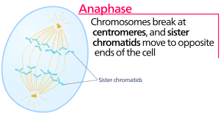
Anaphase is the stage of mitosis after the process of metaphase, when replicated chromosomes are split and the newly-copied chromosomes are moved to opposite poles of the cell. Chromosomes also reach their overall maximum condensation in late anaphase, to help chromosome segregation and the re-formation of the nucleus.

<span class="mw-page-title-main">Spindle apparatus</span> Feature of biological cell structure

In cell biology, the spindle apparatus is the cytoskeletal structure of eukaryotic cells that forms during cell division to separate sister chromatids between daughter cells. It is referred to as the mitotic spindle during mitosis, a process that produces genetically identical daughter cells, or the meiotic spindle during meiosis, a process that produces gametes with half the number of chromosomes of the parent cell.

<span class="mw-page-title-main">Anaphase-promoting complex</span> Cell-cycle regulatory complex

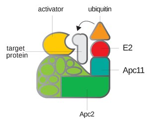
Anaphase-promoting complex is an E3 ubiquitin ligase that marks target cell cycle proteins for degradation by the 26S proteasome. The APC/C is a large complex of 11–13 subunit proteins, including a cullin (Apc2) and RING (Apc11) subunit much like SCF. Other parts of the APC/C have unknown functions but are highly conserved.

<span class="mw-page-title-main">Metaphase</span> Stage of cell division

Metaphase is a stage of mitosis in the eukaryotic cell cycle in which chromosomes are at their second-most condensed and coiled stage. These chromosomes, carrying genetic information, align in the equator of the cell between the spindle poles at the metaphase plate, before being separated into each of the two daughter nuclei. This alignment marks the beginning of metaphase. Metaphase accounts for approximately 4% of the cell cycle's duration.

<span class="mw-page-title-main">Spindle checkpoint</span> Cell cycle checkpoint

The spindle checkpoint, also known as the metaphase-to-anaphase transition, the spindle assembly checkpoint (SAC), the metaphase checkpoint, or the mitotic checkpoint, is a cell cycle checkpoint during metaphase of mitosis or meiosis that prevents the separation of the duplicated chromosomes (anaphase) until each chromosome is properly attached to the spindle. To achieve proper segregation, the two kinetochores on the sister chromatids must be attached to opposite spindle poles. Only this pattern of attachment will ensure that each daughter cell receives one copy of the chromosome. The defining biochemical feature of this checkpoint is the stimulation of the anaphase-promoting complex by M-phase cyclin-CDK complexes, which in turn causes the proteolytic destruction of cyclins and proteins that hold the sister chromatids together.

<span class="mw-page-title-main">Kinetochore</span> Protein complex that allows microtubules to attach to chromosomes during cell division

A kinetochore is a disc-shaped protein structure associated with duplicated chromatids in eukaryotic cells where the spindle fibers attach during cell division to pull sister chromatids apart. The kinetochore assembles on the centromere and links the chromosome to microtubule polymers from the mitotic spindle during mitosis and meiosis. The term kinetochore was first used in a footnote in a 1934 Cytology book by Lester W. Sharp and commonly accepted in 1936. Sharp's footnote reads: "The convenient term kinetochore has been suggested to the author by J. A. Moore", likely referring to John Alexander Moore who had joined Columbia University as a freshman in 1932.

<span class="mw-page-title-main">Separase</span> Mammalian protein found in Homo sapiens

Separase, also known as separin, is a cysteine protease responsible for triggering anaphase by hydrolysing cohesin, which is the protein responsible for binding sister chromatids during the early stage of anaphase. In humans, separin is encoded by the ESPL1 gene.

Securin is a protein involved in control of the metaphase-anaphase transition and anaphase onset. Following bi-orientation of chromosome pairs and inactivation of the spindle checkpoint system, the underlying regulatory system, which includes securin, produces an abrupt stimulus that induces highly synchronous chromosome separation in anaphase.

<span class="mw-page-title-main">Cohesin</span> Protein complex that regulates the separation of sister chromatids during cell division

Cohesin is a protein complex that mediates sister chromatid cohesion, homologous recombination, and DNA looping. Cohesin is formed of SMC3, SMC1, SCC1 and SCC3. Cohesin holds sister chromatids together after DNA replication until anaphase when removal of cohesin leads to separation of sister chromatids. The complex forms a ring-like structure and it is believed that sister chromatids are held together by entrapment inside the cohesin ring. Cohesin is a member of the SMC family of protein complexes which includes Condensin, MukBEF and SMC-ScpAB.

<span class="mw-page-title-main">CDC20</span> Protein-coding gene in the species Homo sapiens

The cell division cycle protein 20 homolog is an essential regulator of cell division that is encoded by the CDC20 gene in humans. To the best of current knowledge its most important function is to activate the anaphase promoting complex (APC/C), a large 11-13 subunit complex that initiates chromatid separation and entrance into anaphase. The APC/CCdc20 protein complex has two main downstream targets. Firstly, it targets securin for destruction, enabling the eventual destruction of cohesin and thus sister chromatid separation. It also targets S and M-phase (S/M) cyclins for destruction, which inactivates S/M cyclin-dependent kinases (Cdks) and allows the cell to exit from mitosis. A closely related protein, Cdc20homologue-1 (Cdh1) plays a complementary role in the cell cycle.

<span class="mw-page-title-main">BUB1</span> Protein-coding gene in the species Homo sapiens

Mitotic checkpoint serine/threonine-protein kinase BUB1 also known as BUB1 is an enzyme that in humans is encoded by the BUB1 gene.

<span class="mw-page-title-main">BUB1B</span> Protein-coding gene in the species Homo sapiens

Mitotic checkpoint serine/threonine-protein kinase BUB1 beta is an enzyme that in humans is encoded by the BUB1B gene. Also known as BubR1, this protein is recognized for its mitotic roles in the spindle assembly checkpoint (SAC) and kinetochore-microtubule interactions that facilitate chromosome migration and alignment. BubR1 promotes mitotic fidelity and protects against aneuploidy by ensuring proper chromosome segregation between daughter cells. BubR1 is proposed to prevent tumorigenesis.

<span class="mw-page-title-main">MAD2L1</span> Protein-coding gene in the species Homo sapiens

Mitotic spindle assembly checkpoint protein MAD2A is a protein that in humans is encoded by the MAD2L1 gene.

<span class="mw-page-title-main">CDC27</span> Protein-coding gene in the species Homo sapiens

Cell division cycle protein 27 homolog is a protein that in humans is encoded by the CDC27 gene.

<span class="mw-page-title-main">CDC16</span> Protein-coding gene in the species Homo sapiens

Cell division cycle protein 16 homolog is a protein that in humans is encoded by the CDC16 gene.

<span class="mw-page-title-main">BUB3</span> Protein-coding gene in the species Homo sapiens

Mitotic checkpoint protein BUB3 is a protein that in humans is encoded by the BUB3 gene.

<span class="mw-page-title-main">TRIP13</span> Protein-coding gene in the species Homo sapiens

TRIP13 is a mammalian gene that encodes the thyroid receptor-interacting protein 13. In budding yeast, the analog for TRIP13 is PCH2. TRIP13 is a member of the AAA+ ATPase family, a family known for mechanical forces derived from ATP hydrolase reactions. The TRIP13 gene has been shown to interact with a variety of proteins and implicated in a few diseases, notably interacting with the ligand binding domain of thyroid hormone receptors, and may play a role in early-stage non-small cell lung cancer. However, recent evidence implicates TRIP13 in various cell cycle phases, including meiosis G2/Prophase and during the Spindle Assembly checkpoint (SAC). Evidence shows regulation to occur through the HORMA domains, including Hop1, Rev7, and Mad2. Of note, Mad2's involvement in the SAC is shown to be affected by TRIP13 Due to TRIP13's role in cell cycle arrest and progression, it may present opportunity as a therapeutic candidate for cancers.

A series of biochemical switches control transitions between and within the various phases of the cell cycle. The cell cycle is a series of complex, ordered, sequential events that control how a single cell divides into two cells, and involves several different phases. The phases include the G1 and G2 phases, DNA replication or S phase, and the actual process of cell division, mitosis or M phase. During the M phase, the chromosomes separate and cytokinesis occurs.

<span class="mw-page-title-main">Mad1</span>

Mad1 is a non-essential protein which in yeast has a function in the spindle assembly checkpoint (SAC). This checkpoint monitors chromosome attachment to spindle microtubules and prevents cells from starting anaphase until the spindle is built up. The name Mad refers to the observation that mutant cells are mitotic arrest deficient (MAD) during microtubule depolymerization. Mad1 recruits the anaphase inhibitor Mad2 to unattached kinetochores and is essential for Mad2-Cdc20 complex formation in vivo but not in vitro. In vivo, Mad1 acts as a competitive inhibitor of the Mad2-Cdc20 complex. Mad1 is phosphorylated by Mps1 which then leads together with other activities to the formation of the mitotic checkpoint complex (MCC). Thereby it inhibits the activity of the anaphase-promoting complex/cyclosome (APC/C). Homologues of Mad1 are conserved in eukaryotes from yeast to mammals.

Mitotic exit is an important transition point that signifies the end of mitosis and the onset of new G1 phase for a cell, and the cell needs to rely on specific control mechanisms to ensure that once it exits mitosis, it never returns to mitosis until it has gone through G1, S, and G2 phases and passed all the necessary checkpoints. Many factors including cyclins, cyclin-dependent kinases (CDKs), ubiquitin ligases, inhibitors of cyclin-dependent kinases, and reversible phosphorylations regulate mitotic exit to ensure that cell cycle events occur in correct order with fewest errors. The end of mitosis is characterized by spindle breakdown, shortened kinetochore microtubules, and pronounced outgrowth of astral (non-kinetochore) microtubules. For a normal eukaryotic cell, mitotic exit is irreversible.

References

  1. Li R, Murray AW (1991). "Feedback control of mitosis in budding yeast". Cell. 66 (3): 519–31. doi:10.1016/0092-8674(81)90015-5. PMID   1651172. S2CID   11306198.
  2. 1 2 Li Y, Benezra R (1996). "Identification of a human mitotic checkpoint gene: hsMAD2". Science. 274 (5285): 246–8. Bibcode:1996Sci...274..246L. doi:10.1126/science.274.5285.246. PMID   8824189. S2CID   41334564.
  3. Chen RH, Waters JC, Salmon ED, Murray AW (October 1996). "Association of spindle assembly checkpoint component XMAD2 with unattached kinetochores". Science. 274 (5285): 242–6. Bibcode:1996Sci...274..242C. doi:10.1126/science.274.5285.242. PMID   8824188. S2CID   20142555.
  4. 1 2 Yu H (April 2006). "Structural activation of Mad2 in the mitotic spindle checkpoint: the two-state Mad2 model versus the Mad2 template model". J. Cell Biol. 173 (2): 153–7. doi:10.1083/jcb.200601172. PMC   2063805 . PMID   16636141.
  5. 1 2 3 Morgan DL (2007). The cell cycle: principles of control. London: Published by New Science Press in association with Oxford University Press. ISBN   978-0-87893-508-6.
  6. Li Y, Gorbea C, Mahaffey D, Rechsteiner M, Benezra R (November 1997). "MAD2 associates with the cyclosome/anaphase-promoting complex and inhibits its activity". Proc. Natl. Acad. Sci. U.S.A. 94 (23): 12431–6. Bibcode:1997PNAS...9412431L. doi: 10.1073/pnas.94.23.12431 . PMC   24983 . PMID   9356466.
  7. Wassmann K, Benezra R (September 1998). "Mad2 transiently associates with an APC/p55Cdc complex during mitosis". Proc. Natl. Acad. Sci. U.S.A. 95 (19): 11193–8. Bibcode:1998PNAS...9511193W. doi: 10.1073/pnas.95.19.11193 . PMC   21618 . PMID   9736712.
  8. Hardwick KG (February 2005). "Checkpoint signalling: Mad2 conformers and signal propagation". Curr. Biol. 15 (4): R122–4. doi: 10.1016/j.cub.2005.02.008 . PMID   15723780.
  9. 1 2 Nasmyth K (March 2005). "How do so few control so many?". Cell. 120 (6): 739–46. doi: 10.1016/j.cell.2005.03.006 . PMID   15797376.
  10. De Antoni A, Pearson CG, Cimini D, Canman JC, Sala V, Nezi L, Mapelli M, Sironi L, Faretta M, Salmon ED, Musacchio A (February 2005). "The Mad1/Mad2 complex as a template for Mad2 activation in the spindle assembly checkpoint". Curr. Biol. 15 (3): 214–25. doi: 10.1016/j.cub.2005.01.038 . PMID   15694304. S2CID   3224122.