Magnetic nanoparticles

Last updated

Magnetic nanoparticles (MNPs) are a class of nanoparticle that can be manipulated using magnetic fields. Such particles commonly consist of two components, a magnetic material, often iron, nickel and cobalt, and a chemical component that has functionality. While nanoparticles are smaller than 1 micrometer in diameter (typically 1–100 nanometers), the larger microbeads are 0.5–500 micrometer in diameter. Magnetic nanoparticle clusters that are composed of a number of individual magnetic nanoparticles are known as magnetic nanobeads with a diameter of 50–200 nanometers. [1] [2] Magnetic nanoparticle clusters are a basis for their further magnetic assembly into magnetic nanochains. [3] The magnetic nanoparticles have been the focus of much research recently because they possess attractive properties which could see potential use in catalysis including nanomaterial-based catalysts, [4] biomedicine [5] and tissue specific targeting, [6] magnetically tunable colloidal photonic crystals, [7] microfluidics, [8] magnetic resonance imaging, [9] magnetic particle imaging, [10] data storage, [11] [12] environmental remediation, [13] nanofluids, [14] [15] optical filters, [16] defect sensor, [17] magnetic cooling [18] [19] and cation sensors. [20]

Contents

Properties

The physical and chemical properties of magnetic nanoparticles largely depend on the synthesis method and chemical structure. In most cases, the particles range from 1 to 100 nm in size and may display superparamagnetism. [21]

Types of magnetic nanoparticles

Oxides: ferrites

Ferrite nanoparticles or iron oxide nanoparticles (iron oxides in crystal structure of maghemite or magnetite) are the most explored magnetic nanoparticles up to date. Once the ferrite particles become smaller than 128 nm [22] they become superparamagnetic which prevents self agglomeration since they exhibit their magnetic behavior only when an external magnetic field is applied. The magnetic moment of ferrite nanoparticles can be greatly increased by controlled clustering of a number of individual superparamagnetic nanoparticles into superparamagnetic nanoparticle clusters, namely magnetic nanobeads. [1] With the external magnetic field switched off, the remanence falls back to zero. Just like non-magnetic oxide nanoparticles, the surface of ferrite nanoparticles is often modified by surfactants, silica, [1] silicones or phosphoric acid derivatives to increase their stability in solution. [23]

Ferrites with a shell

TEM image of a maghemite magnetic nanoparticle cluster with silica shell. Maghemite silica nanoparticle cluster.jpg
TEM image of a maghemite magnetic nanoparticle cluster with silica shell.

The surface of a maghemite or magnetite magnetic nanoparticle is relatively inert and does not usually allow strong covalent bonds with functionalization molecules. However, the reactivity of the magnetic nanoparticles can be improved by coating a layer of silica onto their surface. [25] The silica shell can be easily modified with various surface functional groups via covalent bonds between organo-silane molecules and silica shell. [26] In addition, some fluorescent dye molecules can be covalently bonded to the functionalized silica shell. [27]

Ferrite nanoparticle clusters with narrow size distribution consisting of superparamagnetic oxide nanoparticles (~ 80 maghemite superparamagnetic nanoparticles per bead) coated with a silica shell have several advantages over metallic nanoparticles: [1]

Metallic

Metallic nanoparticles may be beneficial for some technical applications due to their higher magnetic moment whereas oxides (maghemite, magnetite) would be beneficial for biomedical applications. This also implies that for the same moment, metallic nanoparticles can be made smaller than their oxide counterparts. On the other hand, metallic nanoparticles have the great disadvantage of being pyrophoric and reactive to oxidizing agents to various degrees. This makes their handling difficult and enables unwanted side reactions which makes them less appropriate for biomedical applications. Colloid formation for metallic particles is also much more challenging.

Metallic with a shell

Cobalt nanoparticle with graphene shell (note: The individual graphene layers are visible) Cobalt-graphene-nanoparticle.tif
Cobalt nanoparticle with graphene shell (note: The individual graphene layers are visible)

The metallic core of magnetic nanoparticles may be passivated by gentle oxidation, surfactants, polymers and precious metals. [21] In an oxygen environment, Co nanoparticles form an anti-ferromagnetic CoO layer on the surface of the Co nanoparticle. Recently, work has explored the synthesis and exchange bias effect in these Co core CoO shell nanoparticles with a gold outer shell. [29] Nanoparticles with a magnetic core consisting either of elementary Iron or Cobalt with a nonreactive shell made of graphene have been synthesized recently. [30] The advantages compared to ferrite or elemental nanoparticles are:

Synthesis

Several methods exist for preparing magnetic nanoparticle.

Co-precipitation

Co-precipitation is a facile and convenient way to synthesize iron oxides (either Fe3O4 or γ-Fe2O3) from aqueous Fe2+/Fe3+ salt solutions by the addition of a base under inert atmosphere at room temperature or at elevated temperature. The size, shape, and composition of the magnetic nanoparticles very much depends on the type of salts used (e.g.chlorides, sulfates, nitrates), the Fe2+/Fe3+ ratio, the reaction temperature, the pH value and ionic strength of the media, [21] and the mixing rate with the base solution used to provoke the precipitation. [31] The co-precipitation approach has been used extensively to produce ferrite nanoparticles of controlled sizes and magnetic properties. [32] [33] [34] [35] A variety of experimental arrangements have been reported to facilitate continuous and large–scale co–precipitation of magnetic particles by rapid mixing. [36] [37] Recently, the growth rate of the magnetic nanoparticles was measured in real-time during the precipitation of magnetite nanoparticles by an integrated AC magnetic susceptometer within the mixing zone of the reactants. [38]

Thermal decomposition

Magnetic nanocrystals with smaller size can essentially be synthesized through the thermal decomposition of alkaline organometallic compounds in high-boiling organic solvents containing stabilizing surfactants. [21] [39] [40]

Microemulsion

Using the microemulsion technique, metallic cobalt, cobalt/platinum alloys, and gold-coated cobalt/platinum nanoparticles have been synthesized in reverse micelles of cetyltrimethlyammonium bromide, using 1-butanol as the cosurfactant and octane as the oil phase., [21] [41]

Flame spray synthesis

Using flame spray pyrolysis [30] [42] and varying the reaction conditions, oxides, metal or carbon coated nanoparticles are produced at a rate of > 30 g/h .

Flame-spray-synthesis.JPG

Various flame spray conditions and their impact on the resulting nanoparticles

Flame-spray-synthesis-2.JPG

Operational layout differences between conventional and reducing flame spray synthesis

Potential applications

A wide variety of potential applications have been envisaged. Since magnetic nanoparticles are expensive to produce, there is interest in their recycling or for highly specialized applications.

The potential and versatility of magnetic chemistry arises from the fast and easy separation of the magnetic nanoparticles, eliminating tedious and costly separation processes usually applied in chemistry. Furthermore, the magnetic nanoparticles can be guided via a magnetic field to the desired location which could, for example, enable pinpoint precision in fighting cancer.

Medical diagnostics and treatments

Magnetic nanoparticles have been examined for use in an experimental cancer treatment called magnetic hyperthermia [43] in which an alternating magnetic field (AMF) is used to heat the nanoparticles. To achieve sufficient magnetic nanoparticle heating, the AMF typically has a frequency between 100–500 kHz, although significant research has been done at lower frequencies as well as frequencies as high as 10 MHz, with the amplitude of the field usually between 8-16kAm−1. [44]

Affinity ligands such as epidermal growth factor (EGF), folic acid, aptamers, lectins etc. can be attached to the magnetic nanoparticle surface with the use of various chemistries. This enables targeting of magnetic nanoparticles to specific tissues or cells. [45] This strategy is used in cancer research to target and treat tumors in combination with magnetic hyperthermia or nanoparticle-delivered cancer drugs. Despite research efforts, however, the accumulation of nanoparticles inside of cancer tumors of all types is sub-optimal, even with affinity ligands. Willhelm et al. conducted a broad analysis of nanoparticle delivery to tumors and concluded that the median amount of injected dose reaching a solid tumor is only 0.7%. [46] The challenge of accumulating large amounts of nanoparticles inside of tumors is arguably the biggest obstacle facing nanomedicine in general. While direct injection is used in some cases, intravenous injection is most often preferred to obtain a good distribution of particles throughout the tumor. Magnetic nanoparticles have a distinct advantage in that they can accumulate in desired regions via magnetically guided delivery, although this technique still needs further development to achieve optimal delivery to solid tumors.

Another potential treatment of cancer includes attaching magnetic nanoparticles to free-floating cancer cells, allowing them to be captured and carried out of the body. The treatment has been tested in the laboratory on mice and will be looked at in survival studies. [47] [48]

Magnetic nanoparticles can be used for the detection of cancer. Blood can be inserted onto a microfluidic chip with magnetic nanoparticles in it. These magnetic nanoparticles are trapped inside due to an externally applied magnetic field as the blood is free to flow through. The magnetic nanoparticles are coated with antibodies targeting cancer cells or proteins. The magnetic nanoparticles can be recovered and the attached cancer-associated molecules can be assayed to test for their existence.

Magnetic nanoparticles can be conjugated with carbohydrates and used for detection of bacteria. Iron oxide particles have been used for the detection of Gram negative bacteria like Escherichia coli and for detection of Gram positive bacteria like Streptococcus suis [49] [50]

Core-shell magnetic nanoparticles, particularly cobalt ferrite, possess antimicrobial properties against hazardous prokaryotic (E. coli, Staphylococcus aureus) and eukaryotic (Candida parapsilosis, Candida albicans) microorganisms. It is known that the size of the magnetic nanoparticles performs a critical role, as the smaller the particles, the more significant the antimicrobial effect. [51]

Other diagnostic uses can be achieved by conjugation of the nanoparticles with oligonucleotides that can either be complementary to a DNA or RNA sequence of interest to detect them, such as pathogenic DNA or products of DNA amplification reactions in the presence of pathogenic DNA, [52] or an aptamer recognizing a molecule of interest. This can lead to detection of pathogens such as virus or bacteria in humans or dangerous chemicals or other substances in the body. [53]

Magnetic immunoassay

Magnetic immunoassay [54] (MIA) is a novel type of diagnostic immunoassay utilizing magnetic nanobeads as labels in lieu of conventional, enzymes, radioisotopes or fluorescent moieties. This assay involves the specific binding of an antibody to its antigen, where a magnetic label is conjugated to one element of the pair. The presence of magnetic nanobeads is then detected by a magnetic reader (magnetometer) which measures the magnetic field change induced by the beads. The signal measured by the magnetometer is proportional to the analyte (virus, toxin, bacteria, cardiac marker, etc.) quantity in the initial sample.

Waste water treatment

Thanks to the easy separation by applying a magnetic field and the very large surface to volume ratio, magnetic nanoparticles have a potential for treatment of contaminated water. [55] In this method, attachment of EDTA-like chelators to carbon coated metal nanomagnets results in a magnetic reagent for the rapid removal of heavy metals from solutions or contaminated water by three orders of magnitude to concentrations as low as micrograms per Litre. Magnetic nanobeads or nanoparticle clusters composed of FDA-approved oxide superparamagnetic nanoparticles (e.g. maghemite, magnetite) hold much potential for waste water treatment since they express excellent biocompatibility which concerning the environmental impacts of the material is an advantage compared to metallic nanoparticles.

Electrochemical sensing

Magneto-electrochemical assays are based on the use of magnetic nanoparticles in electrochemical sensing either by being distributed through a sample where they can collect and preconcentrate the analyte and handled by a magnetic field or by modifying an electrode surface enhancing its conductivity and the affinity with the analyte. Coated-magnetic nanoparticles have a key aspect in electrochemical sensing not only because it facilitates the collecting of analyte but also it allows MNPs to be part of the sensor transduction mechanism. [56] For the manipulation of MNPs in electrochemical sensing has been used magnetic electrode shafts [57] or disposable screen-printed electrodes integrating permanent bonded magnets, [58] aiming to replace magnetic supports or any external magnetic field.

Supported enzymes and peptides

Enzymes, proteins, and other biologically and chemically active substances have been immobilized on magnetic nanoparticles. [59] The immobilization of enzymes on inexpensive, non-toxic and easily synthesized iron magnetic nanoparticles (MNP) has shown great promise due to more stable proteins, better product yield, ease of protein purification and multiple usage as a result of their magnetic susceptibility. [60] They are of interest as possible supports for solid phase synthesis. [61]

This technology is potentially relevant to cellular labelling/cell separation, detoxification of biological fluids, tissue repair, drug delivery, magnetic resonance imaging, hyperthermia and magnetofection. [62]

Random versus site-directed enzyme immobilization

Enzymes immobilized on magnetic nanoparticles (MNP) via random multipoint attachment, result in a heterogeneous protein population with reduced activity due to restriction of substrate access to the active site. Methods based on chemical modifications are now available where MNP can be linked to a protein molecule via a single specific amino acid (such as N- or C- termini), thus avoiding reduction in activity due to the free access of the substrate to the active site. Moreover, site-directed immobilization also avoids modifying catalytic residues. One such common method involves using Alkyne-Azide Click chemistry as both groups are absent in proteins. [63]

Catalyst support

Magnetic nanoparticles are of potential use as a catalyst or catalyst supports. [64] [65] In chemistry, a catalyst support is the material, usually a solid with a high surface area, to which a catalyst is affixed. The reactivity of heterogeneous catalysts occurs at the surface atoms. Consequently, great effort is made to maximize the surface area of a catalyst by distributing it over the support. The support may be inert or participate in the catalytic reactions. Typical supports include various kinds of carbon, alumina, and silica. Immobilizing the catalytic center on top of nanoparticles with a large surface to volume ratio addresses this problem. In the case of magnetic nanoparticles it adds the property of facile a separation. An early example involved a rhodium catalysis attached to magnetic nanoparticles . [66]

Magnetic-chemistry-1.tif

In another example, the stable radical TEMPO was attached to the graphene-coated cobalt nanoparticles via a diazonium reaction. The resulting catalyst was then used for the chemoselective oxidation of primary and secondary alcohols. [67]

Magnetic-chemistry-2.tif

The catalytic reaction can be conducted in a continuous flow reactor instead of a batch reactor with no remains of the catalyst in the end product. Graphene coated cobalt nanoparticles have been used for that experiment since they exhibit a higher magnetization than Ferrite nanoparticles, which is essential for a fast and clean separation via external magnetic field. [68]

Magnetic-chemistry-3.tif

Biomedical imaging

There are many applications for iron-oxide based nanoparticles in concert with magnetic resonance imaging. [69] Magnetic CoPt nanoparticles are being used as an MRI contrast agent for transplanted neural stem cell detection. [70]

Cancer therapy

In magnetic fluid hyperthermia, [71] nanoparticles of different types like Iron oxide, magnetite, maghemite or even gold are injected in tumor and then subjected under a high frequency magnetic field. These nanoparticles produce heat that typically increases tumor temperature to 40-46 °C, which can kill cancer cells. [72] [73] [74] Another major potential of magnetic nanoparticles is the ability to combine heat (hyperthermia) and drug release for a cancer treatment. Numerous studies have shown particle constructs that can be loaded with a drug cargo and magnetic nanoparticles. [75] The most prevalent construct is the "Magnetoliposome", which is a liposome with magnetic nanoparticles typically embedded in the lipid bilayer. Under an alternating magnetic field, the magnetic nanoparticles are heated, and this heat permeabilizes the membrane. This causes release of the loaded drug. This treatment option has a lot of potential as the combination of hyperthermia and drug release is likely to treat tumors better than either option alone, but it is still under development.

Information storage

A promising candidate for high-density storage is the face-centered tetragonal phase FePt alloy. Grain sizes can be as small as 3 nanometers. If it's possible to modify the MNPs at this small scale, the information density that can be achieved with this media could easily surpass 1 Terabyte per square inch. [12]

Genetic engineering

Magnetic nanoparticles can be used for a variety of genetics applications. One application is the rapid isolation of DNA [76] and mRNA. In one application, the magnetic bead is attached to a poly T tail. When mixed with mRNA, the poly A tail of the mRNA will attach to the bead's poly T tail and the isolation takes place simply by placing a magnet on the side of the tube and pouring out the liquid. Magnetic beads have also been used in plasmid assembly. Rapid genetic circuit construction has been achieved by the sequential addition of genes onto a growing genetic chain, using nanobeads as an anchor. This method has been shown to be much faster than previous methods, taking less than an hour to create functional multi-gene constructs in vitro. [77]

Physical modeling

There are a variety of mathematical models to describe the dynamics of the rotations of magnetic nanoparticles. [78] [79] Simple models include the Langevin function and the Stoner-Wohlfarth model which describe the magnetization of a nanoparticle at equilibrium. The Debye/Rosenszweig model can be used for low amplitude or high frequency oscillations of particles, which assumes linear response of the magnetization to an oscillating magnetic field. [80] Non-equilibrium approaches include the Langevin equation formalism and the Fokker-Planck equation formalism, and these have been developed extensively to model applications such as magnetic nanoparticle hyperthermia, magnetic nanoparticle imaging (MPI), [81] magnetic spectroscopy [82] and biosensing [83] etc.

See also

Related Research Articles

<span class="mw-page-title-main">Iron(III) oxide</span> Chemical compound

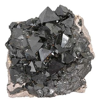
Iron(III) oxide or ferric oxide is the inorganic compound with the formula Fe2O3. It is one of the three main oxides of iron, the other two being iron(II) oxide (FeO), which is rare; and iron(II,III) oxide (Fe3O4), which also occurs naturally as the mineral magnetite. As the mineral known as hematite, Fe2O3 is the main source of iron for the steel industry. Fe2O3 is readily attacked by acids. Iron(III) oxide is often called rust, since rust shares several properties and has a similar composition; however, in chemistry, rust is considered an ill-defined material, described as hydrous ferric oxide.

<span class="mw-page-title-main">Iron oxide</span> Class of chemical compounds composed of iron and oxygen

Iron oxides are chemical compounds composed of iron and oxygen. Several iron oxides are recognized. Often they are non-stoichiometric. Oxyhydroxides are a related class of compounds, perhaps the best known of which is rust.

<span class="mw-page-title-main">Magnetite</span> Iron ore mineral

Magnetite is a mineral and one of the main iron ores, with the chemical formula Fe2+Fe3+2O4. It is one of the oxides of iron, and is ferrimagnetic; it is attracted to a magnet and can be magnetized to become a permanent magnet itself. With the exception of extremely rare native iron deposits, it is the most magnetic of all the naturally occurring minerals on Earth. Naturally magnetized pieces of magnetite, called lodestone, will attract small pieces of iron, which is how ancient peoples first discovered the property of magnetism.

<span class="mw-page-title-main">Ferrofluid</span> Special type of liquid which is attracted by poles of a magnet

Ferrofluid is a liquid that is attracted to the poles of a magnet. It is a colloidal liquid made of nanoscale ferromagnetic or ferrimagnetic particles suspended in a carrier fluid. Each magnetic particle is thoroughly coated with a surfactant to inhibit clumping. Large ferromagnetic particles can be ripped out of the homogeneous colloidal mixture, forming a separate clump of magnetic dust when exposed to strong magnetic fields. The magnetic attraction of tiny nanoparticles is weak enough that the surfactant's Van der Waals force is sufficient to prevent magnetic clumping or agglomeration. Ferrofluids usually do not retain magnetization in the absence of an externally applied field and thus are often classified as "superparamagnets" rather than ferromagnets.

<span class="mw-page-title-main">Maghemite</span> Iron oxide with a spinel ferrite structure

Maghemite (Fe2O3, γ-Fe2O3) is a member of the family of iron oxides. It has the same formula as hematite, but the same spinel ferrite structure as magnetite (Fe3O4) and is also ferrimagnetic. It is sometimes spelled as "maghaemite".

<span class="mw-page-title-main">Nanoparticle</span> Particle with size less than 100 nm

A nanoparticle or ultrafine particle is a particle of matter 1 to 100 nanometres (nm) in diameter. The term is sometimes used for larger particles, up to 500 nm, or fibers and tubes that are less than 100 nm in only two directions. At the lowest range, metal particles smaller than 1 nm are usually called atom clusters instead.

<span class="mw-page-title-main">Iron(II,III) oxide</span> Chemical compound

Iron(II,III) oxide, or black iron oxide, is the chemical compound with formula Fe3O4. It occurs in nature as the mineral magnetite. It is one of a number of iron oxides, the others being iron(II) oxide (FeO), which is rare, and iron(III) oxide (Fe2O3) which also occurs naturally as the mineral hematite. It contains both Fe2+ and Fe3+ ions and is sometimes formulated as FeO ∙ Fe2O3. This iron oxide is encountered in the laboratory as a black powder. It exhibits permanent magnetism and is ferrimagnetic, but is sometimes incorrectly described as ferromagnetic. Its most extensive use is as a black pigment (see: Mars Black). For this purpose, it is synthesized rather than being extracted from the naturally occurring mineral as the particle size and shape can be varied by the method of production.

Magnetic particle imaging (MPI) is an emerging non-invasive tomographic technique that directly detects superparamagnetic nanoparticle tracers. The technology has potential applications in diagnostic imaging and material science. Currently, it is used in medical research to measure the 3-D location and concentration of nanoparticles. Imaging does not use ionizing radiation and can produce a signal at any depth within the body. MPI was first conceived in 2001 by scientists working at the Royal Philips Research lab in Hamburg. The first system was established and reported in 2005. Since then, the technology has been advanced by academic researchers at several universities around the world. The first commercial MPI scanners have recently become available from Magnetic Insight and Bruker Biospin.

<span class="mw-page-title-main">Ferrite (magnet)</span> Ferrimagnetic ceramic material composed of rust and a metallic element

A ferrite is a ceramic material made by mixing and firing iron(III) oxide with one or more additional metallic elements, such as strontium, barium, manganese, nickel, and zinc. They are ferrimagnetic, meaning they are attracted by magnetic fields and can be magnetized to become permanent magnets. Unlike other ferromagnetic materials, most ferrites are not electrically conductive, making them useful in applications like magnetic cores for transformers to suppress eddy currents. Ferrites can be divided into two families based on their resistance to being demagnetized.

<span class="mw-page-title-main">Nanochemistry</span> Combination of chemistry and nanoscience

Nanochemistry is an emerging sub-discipline of the chemical and material sciences that deals with the development of new methods for creating nanoscale materials. The term "nanochemistry" was first used by Ozin in 1992 as 'the uses of chemical synthesis to reproducibly afford nanomaterials from the atom "up", contrary to the nanoengineering and nanophysics approach that operates from the bulk "down"'. Nanochemistry focuses on solid-state chemistry that emphasizes synthesis of building blocks that are dependent on size, surface, shape, and defect properties, rather than the actual production of matter. Atomic and molecular properties mainly deal with the degrees of freedom of atoms in the periodic table. However, nanochemistry introduced other degrees of freedom that controls material's behaviors by transformation into solutions. Nanoscale objects exhibit novel material properties, largely as a consequence of their finite small size. Several chemical modifications on nanometer-scaled structures approve size dependent effects.

Magnetofection is a transfection method that uses magnetic fields to concentrate particles containing vectors to target cells in the body. Magnetofection has been adapted to a variety of vectors, including nucleic acids, non-viral transfection systems, and viruses. This method offers advantages such as high transfection efficiency and biocompatibility which are balanced with limitations.

Magnetic-targeted carriers, also known as MTCs or magnetic vehicles, are micro- or nanoparticles that carry an anticancer drug to the target site by using an external magnetic field and field gradient to direct the desired drug. Usually, the complex involves microscopic beads of activated carbon, which bind the anticancer drug. A magnet applied from outside the body then can direct the drug to the tumor site. This can keep a larger dose of the drug at the tumor site for a longer period of time, and help protect healthy tissue from the side effects of chemotherapy.

<span class="mw-page-title-main">Janus particles</span> Type of nanoparticle or microparticle

Janus particles are special types of nanoparticles or microparticles whose surfaces have two or more distinct physical properties. This unique surface of Janus particles allows two different types of chemistry to occur on the same particle. The simplest case of a Janus particle is achieved by dividing the particle into two distinct parts, each of them either made of a different material, or bearing different functional groups. For example, a Janus particle may have one half of its surface composed of hydrophilic groups and the other half hydrophobic groups, the particles might have two surfaces of different color, fluorescence, or magnetic properties. This gives these particles unique properties related to their asymmetric structure and/or functionalization.

Magnetic Nanorings are a form of magnetic nanoparticles, typically made of iron oxide in the shape of a ring. They have multiple applications in the medical field and computer engineering. In experimental trials, they provide a more localized form of cancer treatment by attacking individual cells instead of a general cancerous region of the body, as well as a clearer image of tumors by improving accuracy of cancer cell identification. They also allow for a more efficient and smaller, MRAM, which helps reduce the size of the technology houses it. Magnetic nanorings can be produced in various compositions, shapes, and sizes by using hematite nanorings as the base structure.

<span class="mw-page-title-main">Iron oxide nanoparticle</span>

Iron oxide nanoparticles are iron oxide particles with diameters between about 1 and 100 nanometers. The two main forms are composed of magnetite and its oxidized form maghemite. They have attracted extensive interest due to their superparamagnetic properties and their potential applications in many fields including molecular imaging.

Cuprospinel is a mineral. Cuprospinel is an inverse spinel with the chemical formula CuFe2O4, where copper substitutes some of the iron cations in the structure. Its structure is similar to that of magnetite, Fe3O4, yet with slightly different chemical and physical properties due to the presence of copper.

<span class="mw-page-title-main">Cobalt oxide nanoparticle</span>

In materials and electric battery research, cobalt oxide nanoparticles usually refers to particles of cobalt(II,III) oxide Co
3
O
4
of nanometer size, with various shapes and crystal structures.

<span class="mw-page-title-main">Iron–platinum nanoparticle</span>

Iron–platinum nanoparticles are 3D superlattices composed of an approximately equal atomic ratio of Fe and Pt. Under standard conditions, FePt NPs exist in the face-centered cubic phase but can change to a chemically ordered face-centered tetragonal phase as a result of thermal annealing. Currently there are many synthetic methods such as water-in-oil microemulsion, one-step thermal synthesis with metal precursors, and exchanged-coupled assembly for making FePt NPs. An important property of FePt NPs is their superparamagnetic character below 10 nanometers. The superparamagnetism of FePt NPs has made them attractive candidates to be used as MRI/CT scanning agents and a high-density recording material.

Magnetoelastic filaments are one-dimensional composite structures that exhibit both magnetic and elastic properties. Interest in these materials tends to focus on the ability to precisely control mechanical events using an external magnetic field. Like piezoelectricity materials, they can be used as actuators, but do not need to be physically connected to a power source. The conformations adopted by magnetoelastic filaments are dictated by the competition between its elastic and magnetic properties.

Praseodymium(III,IV) oxide is the inorganic compound with the formula Pr6O11 that is insoluble in water. It has a cubic fluorite structure. It is the most stable form of praseodymium oxide at ambient temperature and pressure.

References

  1. 1 2 3 4 Tadic, Marin; Kralj, Slavko; Jagodic, Marko; Hanzel, Darko; Makovec, Darko (December 2014). "Magnetic properties of novel superparamagnetic iron oxide nanoclusters and their peculiarity under annealing treatment". Applied Surface Science. 322: 255–264. Bibcode:2014ApSS..322..255T. doi:10.1016/j.apsusc.2014.09.181.
  2. Magnetic Nanomaterials, Editors: S H Bossmann, H Wang, Royal Society of Chemistry, Cambridge 2017, https://pubs.rsc.org/en/content/ebook/978-1-78801-037-5
  3. 1 2 Kralj, Slavko; Makovec, Darko (27 October 2015). "Magnetic Assembly of Superparamagnetic Iron Oxide Nanoparticle Clusters into Nanochains and Nanobundles". ACS Nano. 9 (10): 9700–9707. doi:10.1021/acsnano.5b02328. PMID   26394039.
  4. A.-H. Lu; W. Schmidt; N. Matoussevitch; H. Bönnemann; B. Spliethoff; B. Tesche; E. Bill; W. Kiefer; F. Schüth (August 2004). "Nanoengineering of a Magnetically Separable Hydrogenation Catalyst". Angewandte Chemie International Edition. 43 (33): 4303–4306. doi:10.1002/anie.200454222. PMID   15368378.
  5. A. K. Gupta; M. Gupta (June 2005). "Synthesis and surface engineering of iron oxide nanoparticles for biomedical applications". Biomaterials. 26 (18): 3995–4021. doi:10.1016/j.biomaterials.2004.10.012. PMID   15626447.
  6. Ramaswamy, B; Kulkarni, SD; Villar, PS; Smith, RS; Eberly, C; Araneda, RC; Depireux, DA; Shapiro, B (24 June 2015). "Movement of magnetic nanoparticles in brain tissue: mechanisms and safety". Nanomedicine: Nanotechnology, Biology and Medicine. 11 (7): 1821–9. doi:10.1016/j.nano.2015.06.003. PMC   4586396 . PMID   26115639.
  7. He, Le; Wang, Mingsheng; Ge, Jianping; Yin, Yadong (18 September 2012). "Magnetic Assembly Route to Colloidal Responsive Photonic Nanostructures". Accounts of Chemical Research. 45 (9): 1431–1440. doi:10.1021/ar200276t. PMID   22578015.
  8. Kavre, Ivna; Kostevc, Gregor; Kralj, Slavko; Vilfan, Andrej; Babič, Dušan (13 August 2014). "Fabrication of magneto-responsive microgears based on magnetic nanoparticle embedded PDMS". RSC Advances. 4 (72): 38316–38322. Bibcode:2014RSCAd...438316K. doi:10.1039/C4RA05602G.
  9. Mornet, S.; Vasseur, S.; Grasset, F.; Veverka, P.; Goglio, G.; Demourgues, A.; Portier, J.; Pollert, E.; Duguet, E. (July 2006). "Magnetic nanoparticle design for medical applications". Progress in Solid State Chemistry. 34 (2–4): 237–247. doi:10.1016/j.progsolidstchem.2005.11.010.
  10. B. Gleich; J. Weizenecker (2005). "Tomographic imaging using the nonlinear response of magnetic particles". Nature. 435 (7046): 1214–1217. Bibcode:2005Natur.435.1214G. doi:10.1038/nature03808. PMID   15988521. S2CID   4393678.
  11. Hyeon, Taeghwan (3 April 2003). "Chemical synthesis of magnetic nanoparticles". Chemical Communications (8): 927–934. doi:10.1039/B207789B. PMID   12744306. S2CID   27657072.
  12. 1 2 Natalie A. Frey and Shouheng Sun Magnetic Nanoparticle for Information Storage Applications
  13. Elliott, Daniel W.; Zhang, Wei-xian (December 2001). "Field Assessment of Nanoscale Bimetallic Particles for Groundwater Treatment". Environmental Science & Technology. 35 (24): 4922–4926. Bibcode:2001EnST...35.4922E. doi:10.1021/es0108584. PMID   11775172.
  14. J. Philip; Shima.P.D. B. Raj (2006). "Nanofluid with tunable thermal properties". Applied Physics Letters. 92 (4): 043108. Bibcode:2008ApPhL..92d3108P. doi:10.1063/1.2838304.
  15. Chaudhary, V.; Wang, Z.; Ray, A.; Sridhar, I.; Ramanujan, R. V. (2017). "Self pumping magnetic cooling". Journal of Physics D: Applied Physics. 50 (3): 03LT03. Bibcode:2017JPhD...50cLT03C. doi: 10.1088/1361-6463/aa4f92 .
  16. J.Philip; T.J.Kumar; P.Kalyanasundaram; B.Raj (2003). "Tunable Optical Filter". Measurement Science and Technology. 14 (8): 1289–1294. Bibcode:2003MeScT..14.1289P. doi:10.1088/0957-0233/14/8/314. S2CID   250923543.
  17. Mahendran, V. (2012). "Nanofluid based opticalsensor for rapid visual inspection of defects in ferromagnetic materials". Appl. Phys. Lett. 100 (7): 073104. Bibcode:2012ApPhL.100g3104M. doi:10.1063/1.3684969.
  18. Chaudhary, V.; Ramanujan, R. V. (11 October 2016). "Magnetocaloric Properties of Fe-Ni-Cr Nanoparticles for Active Cooling". Scientific Reports. 6 (1): 35156. Bibcode:2016NatSR...635156C. doi:10.1038/srep35156. PMC   5057077 . PMID   27725754.
  19. Chaudhary, V.; Chen, X.; Ramanujan, R.V. (February 2019). "Iron and manganese based magnetocaloric materials for near room temperature thermal management". Progress in Materials Science. 100: 64–98. doi:10.1016/j.pmatsci.2018.09.005. hdl: 10356/142917 . S2CID   139870597.
  20. Philip, V. Mahendran; Felicia, Leona J. (2013). "A Simple, In-Expensive and Ultrasensitive Magnetic Nanofluid Based Sensor for Detection of Cations, Ethanol and Ammonia". Journal of Nanofluids. 2 (2): 112–119. doi:10.1166/jon.2013.1050.
  21. 1 2 3 4 5 A.-H. Lu; E. L. Salabas; F. Schüth (2007). "Magnetic Nanoparticles: Synthesis, Protection, Functionalization, and Application". Angew. Chem. Int. Ed. 46 (8): 1222–1244. doi:10.1002/anie.200602866. PMID   17278160.
  22. An-Hui Lu, An-Hui; E. L. Salabas; Ferdi Schüth (2007). "Magnetic Nanoparticles: Synthesis, Protection, Functionalization, and Application". Angew. Chem. Int. Ed. 46 (8): 1222–1244. doi:10.1002/anie.200602866. PMID   17278160.
  23. Kim, DK, G.; Mikhaylova, M; et al. (2003). "Anchoring of Phosphonate and Phosphinate Coupling Molecules on Titania Particles". Chemistry of Materials. 15 (8): 1617–1627. doi:10.1021/cm001253u.
  24. http://nanos-sci.com/technology.html Properties and use of magnetic nanoparticle clusters (magnetic nanobeads)
  25. Kralj, Slavko; Makovec, Darko; Čampelj, Stanislav; Drofenik, Miha (July 2010). "Producing ultra-thin silica coatings on iron-oxide nanoparticles to improve their surface reactivity". Journal of Magnetism and Magnetic Materials. 322 (13): 1847–1853. Bibcode:2010JMMM..322.1847K. doi:10.1016/j.jmmm.2009.12.038.
  26. Kralj, Slavko; Drofenik, Miha; Makovec, Darko (16 December 2010). "Controlled surface functionalization of silica-coated magnetic nanoparticles with terminal amino and carboxyl groups". Journal of Nanoparticle Research. 13 (7): 2829–2841. Bibcode:2011JNR....13.2829K. doi:10.1007/s11051-010-0171-4. S2CID   97708934.
  27. Kralj, Slavko; Rojnik, Matija; Romih, Rok; Jagodič, Marko; Kos, Janko; Makovec, Darko (7 September 2012). "Effect of surface charge on the cellular uptake of fluorescent magnetic nanoparticles". Journal of Nanoparticle Research. 14 (10): 1151. Bibcode:2012JNR....14.1151K. doi:10.1007/s11051-012-1151-7. S2CID   94550418.
  28. 1 2 R.N. Grass, Robert N.; E.K. Athanassiou; W.J. Stark (2007). "Covalently Functionalized Cobalt Nanoparticles as a Platform for Magnetic Separations in Organic Synthesis". Angew. Chem. Int. Ed. 46 (26): 4909–12. doi:10.1002/anie.200700613. PMID   17516598.
  29. Johnson, Stephanie H.; C.L. Johnson; S.J. May; S. Hirsch; M.W. Cole; J.E. Spanier (2010). "Co@CoO@Au core-multi-shell nanocrystals". Journal of Materials Chemistry. 20 (3): 439–443. doi:10.1039/b919610b.
  30. 1 2 R. N. Grass, Robert N.; W. J. Stark (2006). "Gas phase synthesis of fcc-cobalt nanoparticles". J. Mater. Chem. 16 (19): 1825. doi:10.1039/B601013J. S2CID   97850340.
  31. Fang, Mei; Ström, Valter; Olsson, Richard T.; Belova, Lyubov; Rao, K. V. (2011). "Rapid mixing: A route to synthesize magnetite nanoparticles with high moment". Appl. Phys. Lett. 99 (22): 222501. Bibcode:2011ApPhL..99v2501F. doi:10.1063/1.3662965.
  32. G.Gnanaprakash; S.Ayyappan; T.Jayakumar; John Philip; Baldev Raj (2006). "A simple method to produce magnetic nanoparticles with enhanced alpha to gamma-Fe2O3 phase transition temperature". Nanotechnology. 17 (23): 5851–5857. Bibcode:2006Nanot..17.5851G. doi:10.1088/0957-4484/17/23/023. S2CID   94579266.
  33. G. Gnanaprakash; John Philip; T. Jayakumar; Baldev Raj (2007). "Effect of Digestion Time and Alkali Addition Rate on the Physical Properties of Magnetite Nanoparticles". J. Phys. Chem. B. 111 (28): 7978–7986. doi:10.1021/jp071299b. PMID   17580856.
  34. S.Ayyappan, John Philip & Baldev Raj (2009). "Solvent polarity effect on physical properties of CoFe2O3 nanoparticles". J. Phys. Chem. C. 113 (2): 590–596. doi:10.1021/jp8083875.
  35. S. Ayyappan; S. Mahadevan; P. Chandramohan; M. P.Srinivasan; John Philip; Baldev Raj (2010). "Influence of Co2 Ion Concentration on the Size, Magnetic Properties, and Purity of CoFe2O4 Spinel Ferrite Nanoparticles". J. Phys. Chem. C. 114 (14): 6334–6341. doi:10.1021/jp911966p.
  36. Fun Chin, Suk; Iyer, K. Swaminathan; Raston, Colin L.; Saunders, Martin (2008). "Size Selective Synthesis of Superparamagnetic Nanoparticles in Thin Fluids under Continuous Flow Conditions" (PDF). Adv. Funct. Mater. 18 (6): 922–927. doi:10.1002/adfm.200701101. S2CID   138114978.
  37. Raston, CL; Saunders, M; Smith, N; Woodward, R (7 May 2006). "Synthesis of Magnetic Nanoparticles Using Spinning Disc Processing". TechConnect Briefs. 1 (2006): 343–346.
  38. Ström, Valter; Olsson, Richard T.; Rao, K. V. (2010). "Real-time monitoring of the evolution of magnetism during precipitation of superparamagnetic nanoparticles for bioscience applications". Journal of Materials Chemistry. 20 (20): 4168. doi:10.1039/c0jm00043d.
  39. Sharifi, Ibrahim; Zamanian, Ali; Behnamghader, Aliasghar (2016-08-15). "Synthesis and characterization of Fe0.6Zn0.4Fe2O4 ferrite magnetic nanoclusters using simple thermal decomposition method". Journal of Magnetism and Magnetic Materials. 412: 107–113. Bibcode:2016JMMM..412..107S. doi:10.1016/j.jmmm.2016.03.091. ISSN   0304-8853.
  40. Monfared, A. H.; Zamanian, A.; Beygzadeh, M.; Sharifi, I.; Mozafari, M. (2017-02-05). "A rapid and efficient thermal decomposition approach for the synthesis of manganese-zinc/oleylamine core/shell ferrite nanoparticles". Journal of Alloys and Compounds. 693: 1090–1095. doi: 10.1016/j.jallcom.2016.09.253 . ISSN   0925-8388.
  41. S S.Rana; J. Philip; B.Raj (2010). "Micelle based synthesis of Cobalt Ferrite nanoparticles and its characterization using Fourier Transform Infrared Transmission Spectrometry and Thermogravimetry". Materials Chemistry and Physics. 124: 264–269. doi:10.1016/j.matchemphys.2010.06.029.
  42. E. K. Athanassiou, Evagelos K.; R. N. Grass; W. J. Stark (2010). "Chemical Aerosol Engineering as a Novel Tool for Material Science: From Oxides to Salt and Metal Nanoparticles". Aerosol Science and Technology. 44 (2): 161–72. Bibcode:2010AerST..44..161A. doi:10.1080/02786820903449665. S2CID   97163337.
  43. Rabias, I.; et al. (2010). "Rapid magnetic heating treatment by highly charged maghemite nanoparticles on Wistar rats exocranial glioma tumors at microliter volume". Biomicrofluidics. 4 (2): 024111. doi:10.1063/1.3449089. PMC   2917883 . PMID   20697578.
  44. Kumar, CS; Mohammad, F (2011). "Magnetic nanomaterials for hyperthermia-based therapy and controlled drug delivery". Adv. Drug Deliv. Rev. 63 (9): 789–808. doi:10.1016/j.addr.2011.03.008. PMC   3138885 . PMID   21447363.
  45. Kralj, Slavko; Rojnik, Matija; Kos, Janko; Makovec, Darko (26 April 2013). "Targeting EGFR-overexpressed A431 cells with EGF-labeled silica-coated magnetic nanoparticles". Journal of Nanoparticle Research. 15 (5): 1666. Bibcode:2013JNR....15.1666K. doi:10.1007/s11051-013-1666-6. S2CID   135831754.
  46. Wilhelm, Stefan; Tavares, Anthony J.; Dai, Qin; Ohta, Seiichi; Audet, Julie; Dvorak, Harold F.; Chan, Warren C. W. (2016). "Analysis of nanoparticle delivery to tumours". Nature Reviews Materials. 1 (5): 16014. Bibcode:2016NatRM...116014W. doi:10.1038/natrevmats.2016.14.
  47. Scarberry KE, Dickerson EB, McDonald JF, Zhang ZJ (2008). "Magnetic Nanoparticle-Peptide Conjugates for in Vitro and in Vivo Targeting and Extraction of Cancer Cells". Journal of the American Chemical Society. 130 (31): 10258–62. doi:10.1021/ja801969b. PMID   18611005.
  48. Using Magnetic Nanoparticles to Combat Cancer Newswise, Retrieved on July 17, 2008.
  49. Parera Pera N; Kouki A.; Finne J.; Pieters R. J. (2010). "Detection of pathogenic Streptococcus suis bacteria using magnetic glycoparticles". Organic & Biomolecular Chemistry. 8 (10): 2425–2429. doi:10.1039/C000819B. PMID   20448902. S2CID   44593515.
  50. Barden, David (30 March 2010). "An attractive method for bacteria detection". Highlights in Chemical Biology. Archived from the original on 21 October 2012.
  51. Ramanavičius, Simonas; Žalnėravičius, Rokas; Niaura, Gediminas; Drabavičius, Audrius; Jagminas, Arūnas (2018-07-01). "Shell-dependent antimicrobial efficiency of cobalt ferrite nanoparticles". Nano-Structures & Nano-Objects. 15: 40–47. doi:10.1016/j.nanoso.2018.03.007. ISSN   2352-507X. S2CID   139265717.
  52. Göransson, Jenny; Zardán Gómez De La Torre, Teresa; Strömberg, Mattias; Russell, Camilla; Svedlindh, Peter; Strømme, Maria; Nilsson, Mats (2010-11-15). "Sensitive Detection of Bacterial DNA by Magnetic Nanoparticles". Analytical Chemistry. 82 (22): 9138–9140. doi: 10.1021/ac102133e . ISSN   0003-2700. PMID   20977277.
  53. Jo, Hunho; Ban, Changill (May 2016). "Aptamer–nanoparticle complexes as powerful diagnostic and therapeutic tools". Experimental & Molecular Medicine. 48 (5): e230. doi:10.1038/emm.2016.44. ISSN   2092-6413. PMC   4910152 . PMID   27151454.
  54. Luc Lenglet; Petr Nikitin; Clayton Péquignot (July–August 2008). "Magnetic immunoassays: A new paradigm in POCT". IVD Technology. Archived from the original on 2008-08-30.
  55. F.M. Koehler, Fabian M.; M. Rossier; M. Waelle; E.K. Athanassiou; L.K. Limbach; R.N. Grass; D. Günther; W.J. Stark (2009). "Magnetic EDTA: Coupling heavy metal chelators to metal nanomagnets for rapid removal of cadmium, lead and copper from contaminated water". Chem. Commun. 32 (32): 4862–4. doi:10.1039/B909447D. PMID   19652806. S2CID   33582926.
  56. Gloag, Lucy; Mehdipour, Milad; Chen, Dongfei; Tilley, Richard D.; Gooding, J. Justin (2019). "Advances in the Application of Magnetic Nanoparticles for Sensing". Advanced Materials. 31 (48): 1904385. Bibcode:2019AdM....3104385G. doi: 10.1002/adma.201904385 . ISSN   1521-4095. PMID   31538371.
  57. Yang, Guangming; Zhao, Faqiong; Zeng, Baizhao (2014-07-20). "Magnetic entrapment for fast and sensitive determination of metronidazole with a novel magnet-controlled glassy carbon electrode". Electrochimica Acta. 135: 154–160. doi:10.1016/j.electacta.2014.04.162. ISSN   0013-4686.
  58. Papavasileiou, Anastasios V.; Panagiotopoulos, Ioannis; Prodromidis, Mamas I. (2020-11-10). "All-screen-printed graphite sensors integrating permanent bonded magnets. Fabrication, characterization and analytical utility". Electrochimica Acta. 360: 136981. doi:10.1016/j.electacta.2020.136981. ISSN   0013-4686. S2CID   225022388.
  59. Huang-Hao Yang, Huang-Hao; Shu-Qiong Zhang; Xiao-Lan Chen; Zhi-Xia Zhuang; Jin-Gou Xu; Xiao-Ru Wang (2004). "Magnetite-Containing Spherical Silica Nanoparticles for Biocatalysis and Bioseparations". Analytical Chemistry. 76 (5): 1316–1321. doi:10.1021/ac034920m. PMID   14987087.
  60. Siddiqui KS, Shemsi AM, Guerriero G, Najnin T, Taha, Ertan H, 2017. Biotechnological improvements of cold-adapted enzymes: commercialization via an integrated approach. In: Margesin, Rosa (Ed.), Psychrophiles: From Biodiversity to Biotechnology, Springer-Verlag, pp. 477–512.
  61. K.Norén, Katarina; M. Kempe (2009). "Multilayered Magnetic Nanoparticles as a Support in Solid-Phase Peptide Synthesis". International Journal of Peptide Research and Therapeutics. 15 (4): 287–292. doi:10.1007/s10989-009-9190-3. S2CID   40277196.
  62. Gupta AK, Ajay Kumar; Gupta M (2005). "Synthesis and surface engineering of iron oxide nanoparticles for biomedical applications". Biomaterials. 26 (18): 3995–4021. doi:10.1016/j.biomaterials.2004.10.012. PMID   15626447.
  63. Shemsi, AM, Khanday F, Qureshi AH, Khalil A, Guerriero G, *Siddiqui KS (2019). Site-directed chemically-modified magnetic enzymes: fabrication, improvements, biotechnological applications and future prospects. Biotechnol. Adv. 37:357-381
  64. A. Schätz, Alexander; O. Reiser; W.J. Stark (2010). "Nanoparticles as Semi-Heterogeneous Catalyst Supports". Chem. Eur. J. 16 (30): 8950–67. doi:10.1002/chem.200903462. PMID   20645330.
  65. F. Panahi; F. Bahrami; A. Khalafi-nezhad (2017). "Magnetic nanoparticles grafted l-carnosine dipeptide: remarkable catalytic activity in water at room temperature". Journal of Iranian Chemical Society. 14 (10): 2211–20. doi:10.1007/s13738-017-1157-2. S2CID   103858148.
  66. Tae-Jong Yoon, Tae-Jong; Woo Lee; Yoon-Seuk Oh; Jin-Kyu Lee (2003). "Magnetic nanoparticles as a catalyst vehicle for simple and easy recycling". New Journal of Chemistry. 27 (2): 227.229. doi:10.1039/B209391J.
  67. A. Schätz, Alexander; R. N. Grass; W. J. Stark; O. Reiser (2008). "TEMPO Supported on Magnetic C/Co-Nanoparticles: A Highly Active and Recyclable Organocatalyst". Chemistry: A European Journal. 14 (27): 8262–8266. doi:10.1002/chem.200801001. PMID   18666291.
  68. A. Schätz, Alexander; R. N. Grass; Q. Kainz; W. J. Stark; O. Reiser (2010). "Cu(II)−Azabis(oxazoline) Complexes Immobilized on Magnetic Co/C Nanoparticles: Kinetic Resolution of 1,2-Diphenylethane-1,2-diol under Batch and Continuous-Flow Conditions". Chemistry of Materials. 22 (2): 305–310. doi:10.1021/cm9019099.
  69. Colombo, M; et al. (2012). "Biological Applications of Magnetic Nanoparticles". Chem Soc Rev. 41 (11): 4306–34. doi:10.1039/c2cs15337h. PMID   22481569.
  70. Xiaoting Meng, Xiaoting; Hugh C. Seton; Le T. Lu; Ian A. Prior; Nguyen T. K. Thanh; Bing Song (2011). "Magnetic CoPt nanoparticles as MRI contrast agent for transplanted neural stem cells detection". Nanoscale. 3 (3): 977–984. Bibcode:2011Nanos...3..977M. doi:10.1039/C0NR00846J. PMID   21293831.
  71. Sharifi, Ibrahim; Shokrollahi, H.; Amiri, S. (2012-03-01). "Ferrite-based magnetic nanofluids used in hyperthermia applications". Journal of Magnetism and Magnetic Materials. 324 (6): 903–915. Bibcode:2012JMMM..324..903S. doi:10.1016/j.jmmm.2011.10.017. ISSN   0304-8853.
  72. Javidi, Mehrdad; Heydari, Morteza; Attar, Mohammad Mahdi; Haghpanahi, Mohammad; Karimi, Alireza; Navidbakhsh, Mahdi; Amanpour, Saeid (2014). "Cylindrical agar gel with fluid flow subjected to an alternating magnetic field during hyperthermia". International Journal of Hyperthermia. 31 (1): 33–39. doi: 10.3109/02656736.2014.988661 . PMID   25523967. S2CID   881157.
  73. Javidi, M; Heydari, M; Karimi, A; Haghpanahi, M; Navidbakhsh, M; Razmkon, A (2014). "Evaluation of the effects of injection velocity and different gel concentrations on nanoparticles in hyperthermia therapy". J Biomed Phys Eng. 4 (4): 151–62. PMC   4289522 . PMID   25599061.
  74. Heydari, Morteza; Javidi, Mehrdad; Attar, Mohammad Mahdi; Karimi, Alireza; Navidbakhsh, Mahdi; Haghpanahi, Mohammad; Amanpour, Saeid (2015). "Magnetic Fluid Hyperthermia in a Cylindrical Gel Contains Water Flow". Journal of Mechanics in Medicine and Biology . 15 (5): 1550088. doi:10.1142/S0219519415500888.
  75. Estelrich, Joan; et al. (2015). "Iron Oxide Nanoparticles for Magnetically-Guided and Magnetically-Responsive Drug Delivery". Int. J. Mol. Sci. 16 (12): 8070–8101. doi: 10.3390/ijms16048070 . PMC   4425068 . PMID   25867479.
  76. Ernst, Constanze; Bartel, Alexander; Elferink, Johannes Wilhelmus; Huhn, Jennifer; Eschbach, Erik; Schönfeld, Kirsten; Feßler, Andrea T.; Oberheitmann, Boris; Schwarz, Stefan (2019). "Improved DNA extraction and purification with magnetic nanoparticles for the detection of methicillin-resistant Staphylococcus aureus". Veterinary Microbiology. 230: 45–48. doi:10.1016/j.vetmic.2019.01.009. PMID   30827403. S2CID   73465453.
  77. A Elaissari; J Chatterjee; M Hamoudeh; H Fessi (2010). "Chapter 14. Advances in the Preparation and Biomedical Applications of Magnetic Colloids". In Roque Hidalgo-Ålvarez (ed.). Structure and Functional Properties of Colloidal Systems. CRC Press. pp. 315–337. doi:10.1201/9781420084474-c14. ISBN   978-1-4200-8447-4.
  78. Reeves, Daniel B. (2017). "Nonlinear Nonequilibrium Simulations of Magnetic Nanoparticles". Magnetic Characterization Techniques for Nanomaterials. Springer, Berlin, Heidelberg. pp. 121–156. doi:10.1007/978-3-662-52780-1_4. ISBN   978-3-662-52779-5.
  79. Reeves, Daniel B.; Weaver, John B. (2014). "Approaches for Modeling Magnetic Nanoparticle Dynamics". Critical Reviews in Biomedical Engineering. 42 (1): 85–93. arXiv: 1505.02450 . doi:10.1615/CritRevBiomedEng.2014010845. ISSN   0278-940X. PMC   4183932 . PMID   25271360.
  80. Carrey, J.; Mehdaoui, B.; Respaud, M. (15 April 2011). "Simple models for dynamic hysteresis loop calculations of magnetic single-domain nanoparticles: Application to magnetic hyperthermia optimization" (PDF). Journal of Applied Physics. 109 (8): 083921–083921–17. arXiv: 1007.2009 . Bibcode:2011JAP...109h3921C. doi:10.1063/1.3551582. ISSN   0021-8979. S2CID   119228529.
  81. Weizenecker, J.; Gleich, B.; Rahmer, J.; Dahnke, H.; Borgert, J. (2009). "Three-dimensional real-time in vivo magnetic particle imaging". Physics in Medicine and Biology. 54 (5): L1–L10. Bibcode:2009PMB....54L...1W. doi:10.1088/0031-9155/54/5/L01. ISSN   0031-9155. PMID   19204385. S2CID   2635545.
  82. Reeves, Daniel B.; Weaver, John B. (15 December 2012). "Simulations of magnetic nanoparticle Brownian motion". Journal of Applied Physics. 112 (12): 124311. Bibcode:1998JChPh.109.4281T. doi:10.1063/1.4770322. ISSN   0021-8979. PMC   3537703 . PMID   23319830.
  83. Zhang, Xiaojuan; Reeves, Daniel B.; Perreard, Irina M.; Kett, Warren C.; Griswold, Karl E.; Gimi, Barjor; Weaver, John B. (15 December 2013). "Molecular sensing with magnetic nanoparticles using magnetic spectroscopy of nanoparticle Brownian motion". Biosensors and Bioelectronics. 50: 441–446. doi:10.1016/j.bios.2013.06.049. PMC   3844855 . PMID   23896525.

Bibliography