Marshall syndrome

Last updated
Marshall syndrome
Other namesDeafness, myopia, cataract, saddle nose-Marshall type [1]
Autosomal dominant - en.svg
Marshall syndrome and Stickler syndrome is inherited in an autosomal dominant pattern.

Marshall syndrome is a genetic disorder of the connective tissue [2] that can cause hearing loss. The three most common areas to be affected are the eyes, which are uncommonly large, joints and the mouth and facial structures. Marshall syndrome and Stickler syndrome closely resemble each other; in fact they are so similar, some say they are the same. [3] [4] The condition is named for D. Weber. [5]

Contents

Presentation

Eyes

Myopia is the most common eye problem in Marshall syndrome. Cataracts also occur more frequently and detached retina less frequently than in Stickler syndrome. Myopia also is the most common problem with the eyes in Stickler syndrome. In the latter syndrome, extreme myopia may lead to severe eye problems such as detached retina more frequently than in Marshall syndrome.[ citation needed ]

Joints

The joint changes include hyperextensibility (double-jointedness) and arthritis. Babies and young children with Stickler syndrome usually have very hyperextensible joints. As an affected child gets older, they may experience pain and stiffness from overuse of a joint. Osteoarthritis of the large joints often develops during the third or fourth decade. The joint changes in Marshall syndrome are of the same type but to a lesser degree. There also may be changes in the bones that show up on X-ray but they generally are not a problem.[ citation needed ]

Orofacial structure

The most severe problem associated with Stickler syndrome is Pierre Robin syndrome. This refers to a cleft palate resulting from a very small lower jaw. During early fetal life, the roof of the mouth is normally open and the sides of the palate have to come together to close. If the jaw is too small, there is not enough room for the tongue, which is then pushed up and gets in the way of the closing palate. Sometimes the chin is so small the baby has problems with eating and breathing if the tongue blocks the back of the throat. Cleft palate is found less frequently in Marshall Syndrome than in Stickler syndrome but still more frequently than in the general population.[ citation needed ]

The facial features of Marshall Syndrome include a flat midface, the appearance of large eyes, short upturned nose, and a round face. The facial features of Stickler syndrome are less prominent but include a rather long flat face, and depressed nasal bridge.[ citation needed ]

Hearing loss

The hearing loss associated with Stickler syndrome can be progressive and usually involves the high frequencies. Sensorineural hearing loss has been reported in as many as 100% and as low as 20% of affected individuals. A conductive loss due to otitis can magnify an existing sensorineural loss and is a frequent problem for children with Stickler or Marshall Syndrome.[ citation needed ]

Genetics

Stickler syndrome and Marshall syndrome have an autosomal dominant pattern of inheritance. However, there is a great deal of variation within and among families with regard to gene expression. Some may be more severely affected and others may be very mildly affected. Often these syndromes are not recognized in a family until a baby is born with Pierre Robin syndrome or some members have detached retinas or cataracts at a young age. [6]

Both syndromes where correlated with mutations in the COL11A1 gene. [7]

Diagnosis

Diagnosis is made based on features as well as by the very early onset of serious eye and ear disease. Because Marshall syndrome is an autosomal dominant hereditary disease, physicians can also note the characteristic appearance of the biological parent of the child. A genetic test can be performed in Stickler syndrome diagnosis is made by doing a genetic test for the several collagen genes as well as other genes that are associated with Stickler as well as considering the presence and severity of Spondyloepiphyseal Dysplasia with association of reduced height and growth rate as well as other connective tissue features like skin hyperextensibility and poor wound healing. It is an area of active research, also the genetic testing being expensive supports that the diagnosis is made depending on the features.[ citation needed ]

Treatment

There is no medical treatment for either syndrome but there are some recommendations that can help with prevention or early identification of some of the problems. Children with either syndrome should have their hearing tested, and adults should be aware that the hearing loss may not develop until the adult years. Yearly visits to an ophthalmologist or other eye care professional who has been informed of the diagnosis of Stickler or Marshall syndrome is important for all affected individuals. Children should have the opportunity to have myopia corrected as early as possible, and treatment for cataracts or detached retinas may be more effective with early identification. Support for the joints is especially important during sports, and some recommend that contact sports should be avoided by those who have very loose joints.[ citation needed ]

Related Research Articles

<span class="mw-page-title-main">Weissenbacher–Zweymüller syndrome</span> Medical condition

Weissenbacher–Zweymuller syndrome (WZS), also called Pierre-Robin syndrome with fetal chondrodysplasia, is an autosomal recessive congenital disorder, linked to mutations in the COL11A2 gene, which codes for the α2 strand of collagen type XI. It is a collagenopathy, types II and XI disorder. The condition was first characterized in 1964 by G. Weissenbacher and Ernst Zweymüller.

<span class="mw-page-title-main">Treacher Collins syndrome</span> Human genetic disorder

Treacher Collins syndrome (TCS) is a genetic disorder characterized by deformities of the ears, eyes, cheekbones, and chin. The degree to which a person is affected, however, may vary from mild to severe. Complications may include breathing problems, problems seeing, cleft palate, and hearing loss. Those affected generally have normal intelligence.

<span class="mw-page-title-main">Crouzon syndrome</span> Genetic disorder of the skull and face

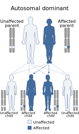
Crouzon syndrome is an autosomal dominant genetic disorder known as a branchial arch syndrome. Specifically, this syndrome affects the first branchial arch, which is the precursor of the maxilla and mandible. Since the branchial arches are important developmental features in a growing embryo, disturbances in their development create lasting and widespread effects.

<span class="mw-page-title-main">Stickler syndrome</span> Genetic connective tissue disorder

Stickler syndrome is a group of rare genetic disorders affecting connective tissue, specifically collagen. Stickler syndrome is a subtype of collagenopathy, types II and XI. Stickler syndrome is characterized by distinctive facial abnormalities, ocular problems, hearing loss, and joint and skeletal problems. It was first studied and characterized by Gunnar B. Stickler in 1965.

Norrie disease is a rare disease and genetic disorder that primarily affects the eyes and almost always leads to blindness. It is caused by mutations in the Norrin cystine knot growth factor (NDP) gene, which is located on the X chromosome. In addition to the congenital ocular symptoms, the majority of patients experience a progressive hearing loss starting mostly in their 2nd decade of life, and some may have learning difficulties among other additional characteristics.

Kniest dysplasia is a rare form of dwarfism caused by a mutation in the COL2A1 gene on chromosome 12. The COL2A1 gene is responsible for producing type II collagen. The mutation of COL2A1 gene leads to abnormal skeletal growth and problems with hearing and vision. What characterizes Kniest dysplasia from other type II osteochondrodysplasia is the level of severity and the dumb-bell shape of shortened long tubular bones.

Albinism-black lock-cell migration disorder is the initialism for the following terms and concepts that describe a condition affecting a person's physical appearance and physiology: (1) A – albinism, (2) B – black lock of hair, (3) C – cell migration disorder of the neurocytes of the gut, and (4) D – sensorineural deafness. The syndrome is caused by mutation in the endothelin B receptor gene (EDNRB).

<span class="mw-page-title-main">SHORT syndrome</span> Medical condition

SHORT syndrome is a medical condition in which affected individuals have multiple birth defects in different organ systems.

<span class="mw-page-title-main">Wagner's disease</span> Medical condition

Wagner's disease is a familial disease of the eye that can cause reduced visual acuity. Wagner's disease was originally described in 1938. This disorder was frequently confused with Stickler syndrome, but lacks the systemic features and high incidence of retinal detachments. Inheritance is autosomal dominant.

<span class="mw-page-title-main">Barakat syndrome</span> Disease characterized by hypoparathyroidism, sensorineural deafness and renal disease

Barakat syndrome is a rare disease characterized by hypoparathyroidism, sensorineural deafness and renal disease, and hence also known as HDR syndrome. It was first described by Amin J. Barakat et al. in 1977.

Abruzzo–Erickson syndrome is an extremely rare disorder characterized by deafness, protruding ears, coloboma, a cleft palate or palatal rugosity, radial synostosis, and short stature. It was first characterized by Abruzzo and Erickson in 1977 as a CHARGE like syndrome as variably expressed among a family of two brothers, their mother, and their maternal uncle. Members of this family exhibited many of the CHARGE symptoms, but notably did not have choanal atresia and the brothers experienced typical genital development. Due to the recent discovery of this disorder, its etiology is not fully known but it is understood that it arises from mutations on the TBX22 gene on the X-chromosome. The disorder is inherited in an X-linked recessive manner. There is currently no known cure but its symptoms can be treated.

<span class="mw-page-title-main">Pierre Robin sequence</span> Medical condition

Pierre Robin sequence is a congenital defect observed in humans which is characterized by facial abnormalities. The three main features are micrognathia, which causes glossoptosis, which in turn causes breathing problems due to obstruction of the upper airway. A wide, U-shaped cleft palate is commonly also present. PRS is not merely a syndrome, but rather it is a sequence—a series of specific developmental malformations which can be attributed to a single cause.

Vici syndrome, also called immunodeficiency with cleft lip/palate, cataract, hypopigmentation and absent corpus callosum, is a rare autosomal recessive congenital disorder characterized by albinism, agenesis of the corpus callosum, cataracts, cardiomyopathy, severe psychomotor retardation, seizures, immunodeficiency and recurrent severe infections. To date, about 50 cases have been reported.

<span class="mw-page-title-main">Collagen, type XI, alpha 1</span> Mammalian protein found in Homo sapiens

Collagen alpha-1(XI) chain is a protein that in humans is encoded by the COL11A1 gene.

<span class="mw-page-title-main">Donnai–Barrow syndrome</span> Medical condition

Donnai–Barrow syndrome is a genetic disorder first described by Dian Donnai and Margaret Barrow in 1993. It is associated with LRP2. It is an inherited (genetic) disorder that affects many parts of the body.

Nasodigitoacoustic syndrome, also called Keipert syndrome, is a rare congenital syndrome first described by J.A. Keipert and colleagues in 1973. The syndrome is characterized by a misshaped nose, broad thumbs and halluces, brachydactyly, sensorineural hearing loss, facial features such as hypertelorism, and developmental delay.

<span class="mw-page-title-main">Distal 18q-</span> Human disease

Distal 18q- is a genetic condition caused by a deletion of genetic material within one of the two copies of chromosome 18. The deletion involves the distal section of 18q and typically extends to the tip of the long arm of chromosome 18.

<span class="mw-page-title-main">Oculofaciocardiodental syndrome</span> Medical condition

Oculofaciocardiodental syndrome is a rare X-linked dominant genetic disorder.

<span class="mw-page-title-main">Otodental syndrome</span> Medical condition

Otodental syndrome, also known as otodental dysplasia, is an exceptionally rare disease that is distinguished by a specific phenotype known as globodontia, that in rare cases can be associated with eye coloboma and high frequency hearing loss. Globodontia is an abnormal condition that can occur in both the primary and secondary dentition, except for the incisors which are normal in shape and size. This is demonstrated by significant enlargement of the canine and molar teeth. The premolars are either reduced in size or are absent. In some cases, the defects affecting the teeth, eye and ear can be either individual or combined. When these conditions are combined with eye coloboma, the condition is also known as oculo-otodental syndrome. The first known case of otodental syndrome was found in Hungary in a mother and her son by Denes and Csiba in 1969. Prevalence is less than 1 out of every 1 million individuals. The cause of otodental syndrome is considered to be genetic. It is an autosomal dominant inheritance and is variable in its expressivity. Haploinsufficiency in the fibroblast growth factor 3 (FGF3) gene (11q13) has been reported in patients with otodental syndrome and is thought to cause the phenotype. Both males and females are equally affected. Individuals diagnosed with otodental syndrome can be of any age; age is not a relevant factor. Currently there are no specific genetic treatments for otodental syndrome. Dental and orthodontic management are the recommended course of action.

<span class="mw-page-title-main">H syndrome</span> Medical condition

H syndrome, also known as Histiocytosis-lymphadenopathy plus syndrome or PHID, is a rare genetic condition caused by mutations in the SLC29A3 gene which encode the human equilibrative nucleoside transporter (hENT3) protein.

References

  1. "Marshall syndrome | Genetic and Rare Diseases Information Center (GARD) – an NCATS Program". rarediseases.info.nih.gov. Retrieved 20 June 2019.
  2. James, William; Berger, Timothy; Elston, Dirk (2005). Andrews' Diseases of the Skin: Clinical Dermatology (10th ed.). Saunders. pp.  146–7. ISBN   0-7216-2921-0.
  3. Annunen S, Körkkö J, Czarny M, et al. (October 1999). "Splicing mutations of 54-bp exons in the COL11A1 gene cause Marshall syndrome, but other mutations cause overlapping Marshall/Stickler phenotypes". Am. J. Hum. Genet. 65 (4): 974–83. doi:10.1086/302585. PMC   1288268 . PMID   10486316.
  4. Griffith AJ, Sprunger LK, Sirko-Osadsa DA, Tiller GE, Meisler MH, Warman ML (April 1998). "Marshall syndrome associated with a splicing defect at the COL11A1 locus". Am. J. Hum. Genet. 62 (4): 816–23. doi:10.1086/301789. PMC   1377029 . PMID   9529347.
  5. "Entry - #154780 - MARSHALL SYNDROME; MRSHS - OMIM". Online Mendelian Inheritance in Man . Retrieved 2023-09-15.
  6. Annunen S, Korkko J, Czarny M, Warman ML, Brunner HG, Kaariainen H, Mulliken JB, Tranebjaerg L, Brooks DG, Cox GF, Cruysberg JR, Curtis MA, Davenport SL, Friedrich CA, Kaitila I, Krawczynski MR, Latos-Bielenska A, Mukai S, Olsen BR, Shinno N, Somer M, Vikkula M, Zlotogora J, Prockop DJ, Ala-Kokko L (1999). "Splicing mutations of 54-bp exons in the COL11A1 gene cause Marshall syndrome, but other mutations cause overlapping Marshall/Stickler phenotypes". Am J Hum Genet. 65 (4): 974–83. doi:10.1086/302585. PMC   1288268 . PMID   10486316.
  7. Majava, Marja (2007). Molecular genetics of Stickler and Marshall syndromes, and the role of collagen II and other candidate proteins in high myopia and impaired hearing. Oulu: Oulun yliopisto. ISBN   978-951-42-8362-8.