Masud Husain

Last updated
Husain in 2023 Masud cropped.jpg
Husain in 2023

Masud Husain FMedSci [1] is a clinical neurologist and neuroscientist working in the UK. He is Professor of Neurology & Cognitive Neuroscience at the Nuffield Department of Clinical Neurosciences [2] and Departmental of Experimental Psychology, [3] University of Oxford, a Professorial Fellow at New College, Oxford, [4] and Editor-in-Chief of the journal Brain [5] . He was born in East Pakistan (now Bangladesh).

Contents

Education

Husain was educated at King Edward VI Camp Hill School for Boys, Birmingham, and studied Physiological Sciences (Medicine) at New College, Oxford, [4] before completing his Doctor of Philosophy degree at Christ Church, Oxford, where he was a Senior Scholar. He held a Harkness Fellowship while a postdoctoral fellow in the laboratory of Richard A. Andersen at MIT. [6] Husain completed his clinical and neurological training at hospitals in Oxford and London. [2]

Research and career

Husain's research focuses on cognitive functions in people with neurological diseases and healthy people. [7] [8] [9] [10] [11] [12] [13] [14] [15]

Attention and inattention

His work on people with hemispatial neglect following stroke demonstrated several novel components of this syndrome. Using the attentional blink paradigm he showed that there were non-spatial, selective attention deficits in these patients, [16] in addition to their well-established directional attentional bias. Subsequent behavioural studies revealed that some people with hemispatial neglect can also suffer from impaired spatial working memory, [17] often revisiting locations without being aware that they have fixated them before. [18] [19] Some may have poor sustained attention as measured on vigilance tasks, [20] or even directional motor deficits as indexed by paradigms where the spatial location of a visual target is dissociated from direction of movement required to reach it. [21] [22] These findings provided further evidence for neglect being a multi-component syndrome, with different patients having different deficits, depending upon the extent of their lesion. [23] [24] Critical brain regions associated with neglect that were identified by this work, included the right inferior posterior parietal and frontal regions. [25] [26] Experimental medicine studies by Husain's group using the dopamine agonist rotigotine [27] and the noradrenergic agonist guanfacine [28] [29] demonstrated that these drugs can ameliorate hemispatial neglect to some extent by improving attention.

Visual short-term or working memory

By using new methods to measure the precision of recall in healthy people, work in Husain's lab challenged the view that capacity of visual short-term memory or working memory is limited to a fixed number of items. [30] [31] Instead, this research revealed that although short-term memory is a highly limited resource, it can be flexibly deployed depending upon task demands. [32] This work led to the application of new methods to measure short-term memory in patients with Alzheimer's disease, [33] [34] Parkinson's disease [33] [34] [35] and individuals at risk of developing these conditions. [33] [34] [35] [36] The techniques that have been developed can provide more sensitive ways to measure short-term memory than traditional methods. [37] They have also revealed how different mechanisms might underlie short-term memory disorders in different neurological conditions. [34]

Motivation, apathy and initiation of action

Work from Husain's lab showed that lesions to ventral basal ganglia leads to a condition of profound apathy, manifest as a lack of motivation to initiate action and specifically attributable to a deficit in reward sensitivity. [38] Using the dopamine receptor agonist ropinirole, it was possible to improve reward sensitivity, restore motivational vigour and reverse apathy. [38] These observations stimulated larger-scale studies in Parkinson's disease, a condition associated with basal ganglia pathology and often debilitating apathy. The syndrome of pathological apathy in Parkinson's disease [39] [40] and small vessel cerebrovascular disease [41] [42] was found to be characterised by reduced sensitivity to rewards, a deficit that could be ameliorated by dopaminergic drugs in Parkinson's disease. [39] [40] This has led to a theoretical framework to understand mechanisms underlying apathy across brain disorders which incorporates concepts from cost-benefit decision making to formalise how people differ in their willingness to engage in effort in order to obtain potential rewards. [43]

The basal ganglia are considered to be essential for linking motivation to action systems. [44] Outputs of the basal ganglia are strongly connected to medial frontal cortex. Husain's group identified a mechanism that resolves competition between conflicting action plans, in medial frontal brain regions, including the supplementary eye field, [45] supplementary motor area and pre-supplementary motor area. [46] [47] A key component of voluntary control paradoxically appears to involve inhibition of unwanted actions that are primed automatically by seeing objects around us. [48] This control is lost following supplementary motor area and pre-supplementary motor area lesions. Findings from lesion, stimulation and physiological studies were incorporated to provide a new theoretical framework for the role of the supplementary motor area and pre-supplementary motor area complex. [49]

Awards and honours

Husain held a Wellcome Trust Senior Research Fellowship at Imperial College London (2000-2007) and University College London, UCL (2007–12). He was awarded a Wellcome Trust Principal Research Fellowship (2012-2023) and elected Fellow of the UK Academy of Medical Sciences (2008). [1] Husain won the Royal College of Physicians' (London) Graham Bull Prize in Clinical Science (2006), British Neuropsychological Society's Elizabeth Warrington Prize (2006), [50] and the European Academy of Neurology Investigator Award (2016). [51] He is Fellow of the American Academy of Neurology (2018) and Fellow of the European Academy of Neurology (2018), [52] and is co-lead of the NIHR Oxford Health Biomedical Research Centre Dementia theme (2022 -) [53] and Dementia Research Oxford at the University of Oxford [54]

Related Research Articles

<span class="mw-page-title-main">Cerebral cortex</span> Outer layer of the cerebrum of the mammalian brain

The cerebral cortex, also known as the cerebral mantle, is the outer layer of neural tissue of the cerebrum of the brain in humans and other mammals. It is the largest site of neural integration in the central nervous system. and plays a key role in attention, perception, awareness, thought, memory, language, and consciousness. The cerebral cortex is part of the brain responsible for cognition.

<span class="mw-page-title-main">Basal ganglia</span> Group of subcortical nuclei involved in the motor and reward systems

The basal ganglia (BG) or basal nuclei are a group of subcortical nuclei found in the brains of vertebrates. In humans and other primates, differences exist, primarily in the division of the globus pallidus into external and internal regions, and in the division of the striatum. Positioned at the base of the forebrain and the top of the midbrain, they have strong connections with the cerebral cortex, thalamus, brainstem and other brain areas. The basal ganglia are associated with a variety of functions, including regulating voluntary motor movements, procedural learning, habit formation, conditional learning, eye movements, cognition, and emotion.

<span class="mw-page-title-main">Apathy</span> State of indifference, or the suppression of emotions

Apathy is a lack of feeling, emotion, interest, or concern about something. It is a state of indifference, or the suppression of emotions such as concern, excitement, motivation, or passion. An apathetic individual has an absence of interest in or concern about emotional, social, spiritual, philosophical, virtual, or physical life and the world. Apathy can also be defined as a person's lack of goal orientation. Apathy falls in the less extreme spectrum of diminished motivation, with abulia in the middle and akinetic mutism being more extreme than both apathy and abulia.

<span class="mw-page-title-main">Caudate nucleus</span> Structure of the striatum in the basal ganglia of the brain

The caudate nucleus is one of the structures that make up the corpus striatum, which is a component of the basal ganglia in the human brain. While the caudate nucleus has long been associated with motor processes due to its role in Parkinson's disease, it plays important roles in various other nonmotor functions as well, including procedural learning, associative learning and inhibitory control of action, among other functions. The caudate is also one of the brain structures which compose the reward system and functions as part of the cortico–basal ganglia–thalamic loop.

<span class="mw-page-title-main">Parietal lobe</span> Part of the brain responsible for sensory input and some language processing

The parietal lobe is one of the four major lobes of the cerebral cortex in the brain of mammals. The parietal lobe is positioned above the temporal lobe and behind the frontal lobe and central sulcus.

<span class="mw-page-title-main">Temporal lobe</span> One of the four lobes of the mammalian brain

The temporal lobe is one of the four major lobes of the cerebral cortex in the brain of mammals. The temporal lobe is located beneath the lateral fissure on both cerebral hemispheres of the mammalian brain.

Anosognosia is a condition in which a person with a disability is cognitively unaware of having it due to an underlying physical condition. Anosognosia results from physiological damage to brain structures, typically to the parietal lobe or a diffuse lesion on the fronto-temporal-parietal area in the right hemisphere, and is thus a neuropsychiatric disorder. A deficit of self-awareness, the term was first coined by the neurologist Joseph Babinski in 1914, in order to describe the unawareness of hemiplegia.

<span class="mw-page-title-main">Frontotemporal dementia</span> Types of dementia involving the frontal or temporal lobes

Frontotemporal dementia (FTD), frontotemporal degeneration disease, or frontotemporal neurocognitive disorder encompasses several types of dementia involving the progressive degeneration of the brain's frontal and temporal lobes. FTDs broadly present as behavioral or language disorders with gradual onsets.

<span class="mw-page-title-main">Hemispatial neglect</span> Medical condition

Hemispatial neglect is a neuropsychological condition in which, after damage to one hemisphere of the brain, a deficit in attention and awareness towards the side of space opposite brain damage is observed. It is defined by the inability of a person to process and perceive stimuli towards the contralesional side of the body or environment. Hemispatial neglect is very commonly contralateral to the damaged hemisphere, but instances of ipsilesional neglect have been reported.

<span class="mw-page-title-main">Hypokinesia</span> Decreased movement due to basal ganglia dysfunction

Hypokinesia is one of the classifications of movement disorders, and refers to decreased bodily movement. Hypokinesia is characterized by a partial or complete loss of muscle movement due to a disruption in the basal ganglia. Hypokinesia is a symptom of Parkinson's disease shown as muscle rigidity and an inability to produce movement. It is also associated with mental health disorders and prolonged inactivity due to illness, amongst other diseases.

<span class="mw-page-title-main">Frontal lobe disorder</span> Brain disorder

Frontal lobe disorder, also frontal lobe syndrome, is an impairment of the frontal lobe of the brain due to disease or frontal lobe injury. The frontal lobe plays a key role in executive functions such as motivation, planning, social behaviour, and speech production. Frontal lobe syndrome can be caused by a range of conditions including head trauma, tumours, neurodegenerative diseases, neurodevelopmental disorders, neurosurgery and cerebrovascular disease. Frontal lobe impairment can be detected by recognition of typical signs and symptoms, use of simple screening tests, and specialist neurological testing.

<span class="mw-page-title-main">Posterior cingulate cortex</span> Caudal part of the cingulate cortex of the brain

The posterior cingulate cortex (PCC) is the caudal part of the cingulate cortex, located posterior to the anterior cingulate cortex. This is the upper part of the "limbic lobe". The cingulate cortex is made up of an area around the midline of the brain. Surrounding areas include the retrosplenial cortex and the precuneus.

Frontostriatal circuits are neural pathways that connect frontal lobe regions with the striatum and mediate motor, cognitive, and behavioural functions within the brain. They receive inputs from dopaminergic, serotonergic, noradrenergic, and cholinergic cell groups that modulate information processing. Frontostriatal circuits are part of the executive functions. Executive functions include the following: selection and perception of important information, manipulation of information in working memory, planning and organization, behavioral control, adaptation to changes, and decision making. These circuits are involved in neurodegenerative disorders such as Alzheimer's disease and Parkinson's disease as well as neuropsychiatric disorders including schizophrenia, depression, obsessive compulsive disorder (OCD), and in neurodevelopmental disorder such as attention-deficit hyperactivity disorder (ADHD).

<span class="mw-page-title-main">Posterior parietal cortex</span>

The posterior parietal cortex plays an important role in planned movements, spatial reasoning, and attention.

The neuroanatomy of memory encompasses a wide variety of anatomical structures in the brain.

Constructional apraxia is a neurological disorder in which people are unable to perform tasks or movements even though they understand the task, are willing to complete it, and have the physical ability to perform the movements. It is characterized by an inability or difficulty to build, assemble, or draw objects. Constructional apraxia may be caused by lesions in the parietal lobe following stroke or it may serve as an indicator for Alzheimer's disease.

The temporal dynamics of music and language describes how the brain coordinates its different regions to process musical and vocal sounds. Both music and language feature rhythmic and melodic structure. Both employ a finite set of basic elements that are combined in ordered ways to create complete musical or lingual ideas.

The neuroscience of aging is the study of the changes in the nervous system that occur with ageing. Aging is associated with many changes in the central nervous system, such as mild atrophy of the cortex that is considered non-pathological. Aging is also associated with many neurological and neurodegenerative disease such as amyotrophic lateral sclerosis, dementia, mild cognitive impairment, Parkinson's disease, and Creutzfeldt–Jakob disease.

<span class="mw-page-title-main">Cortico-basal ganglia-thalamo-cortical loop</span> System of neural circuits in the brain

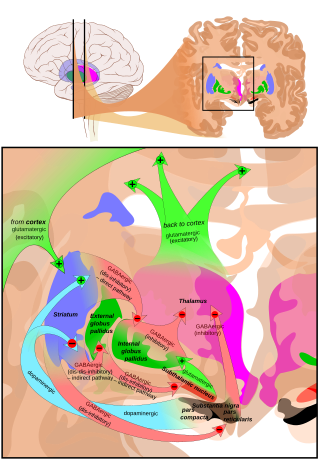
The cortico-basal ganglia-thalamo-cortical loop is a system of neural circuits in the brain. The loop involves connections between the cortex, the basal ganglia, the thalamus, and back to the cortex. It is of particular relevance to hyperkinetic and hypokinetic movement disorders, such as Parkinson's disease and Huntington's disease, as well as to mental disorders of control, such as attention deficit hyperactivity disorder (ADHD), obsessive–compulsive disorder (OCD), and Tourette syndrome.

Dyschiria, also known as dyschiric syndrome, is a neurological disorder where one-half of an individual's body or space cannot be recognized or respond to sensations. The term dyschiria is rarely used in modern scientific research and literature. Dyschiria has been often referred to as unilateral neglect, visuo-spatial neglect, or hemispatial neglect from the 20th century onwards. Psychologists formerly characterized dyschiric patients to be unable to discriminate or report external stimuli. This left the patients incapable of orienting sensory responses in their extrapersonal and personal space. Patients with dyschiria are unable to distinguish one side of their body in general, or specific segments of the body. There are three stages to dyschiria: achiria, allochiria, and synchiria, in which manifestations of dyschiria evolve in varying degrees.

References

  1. 1 2 "Professor Masud Husain | The Academy of Medical Sciences". acmedsci.ac.uk. Retrieved 2023-06-23.
  2. 1 2 "Masud Husain". www.ndcn.ox.ac.uk. Retrieved 2023-11-10.
  3. "Masud Husain". www.psy.ox.ac.uk. Retrieved 2023-11-10.
  4. 1 2 "Masud Husain | New College". www.new.ox.ac.uk. Retrieved 2023-11-10.
  5. "Editorial_Board". Oxford Academic. Retrieved 2023-11-10.
  6. "Alumni". Andersen Lab. Retrieved 2023-11-10.
  7. "Masud Husain". www.ndcn.ox.ac.uk. Retrieved 2023-06-23.
  8. "Masud Husain". www.psy.ox.ac.uk. Retrieved 2023-06-23.
  9. "Brain structure may be root of apathy | University of Oxford". www.ox.ac.uk. 2015-11-13. Retrieved 2024-03-11.
  10. "High blood pressure and diabetes impair brain function, study suggests". The Guardian. ISSN   0261-3077 . Retrieved 2024-03-11.
  11. "Covid-19: Infection raises brain conditions risk". BBC News. Retrieved 2024-03-11.
  12. "Canongate acquires neurologist's debut book on brain disorders and identity". The Bookseller. Retrieved 2024-03-11.
  13. "ORCID". orcid.org. Retrieved 2024-03-11.
  14. Prior, Ryan (2021-04-07). "A third of Covid-19 survivors suffer 'brain disease,' study shows". CNN. Retrieved 2024-03-11.
  15. "Brain disorders affect 1 in 3 Covid survivors, large UK study shows". www.ft.com. Retrieved 2024-03-11.
  16. Husain, Masud; Shapiro, Kimron; Martin, Jesse; Kennard, Christopher (1997). "Abnormal temporal dynamics of visual attention in spatial neglect patients". Nature. 385 (6612): 154–156. Bibcode:1997Natur.385..154H. doi:10.1038/385154a0. ISSN   0028-0836. PMID   8990117. S2CID   4281607.
  17. Malhotra, Paresh; Jager, H. Rolf; Parton, Andrew; Greenwood, Richard; Playford, E. Diane; Brown, Martin M.; Driver, John; Husain, Masud (2004-12-22). "Spatial working memory capacity in unilateral neglect". Brain. 128 (2): 424–435. doi: 10.1093/brain/awh372 . ISSN   1460-2156. PMID   15644422.
  18. Husain, M. (2001-05-01). "Impaired spatial working memory across saccades contributes to abnormal search in parietal neglect". Brain. 124 (5): 941–952. doi: 10.1093/brain/124.5.941 . PMID   11335696.
  19. Mannan, Sabira K.; Mort, Dominic J.; Hodgson, Tim L.; Driver, Jon; Kennard, Christopher; Husain, Masud (2005-02-01). "Revisiting Previously Searched Locations in Visual Neglect: Role of Right Parietal and Frontal Lesions in Misjudging Old Locations as New". Journal of Cognitive Neuroscience. 17 (2): 340–354. doi:10.1162/0898929053124983. ISSN   0898-929X. PMID   15811244. S2CID   9434730.
  20. Malhotra, Paresh; Coulthard, Elizabeth J.; Husain, Masud (2009). "Role of right posterior parietal cortex in maintaining attention to spatial locations over time". Brain. 132 (3): 645–660. doi:10.1093/brain/awn350. ISSN   1460-2156. PMC   2664449 . PMID   19158107.
  21. Mattingley, Jason B.; Husain, Masud; Rorden, Chris; Kennard, Christopher; Driver, Jon (1998). "Motor role of human inferior parietal lobe revealed in unilateral neglect patients". Nature. 392 (6672): 179–182. Bibcode:1998Natur.392..179M. doi:10.1038/32413. ISSN   0028-0836. PMID   9515962. S2CID   205001866.
  22. Husain, M.; Mattingley, Jason B.; Rorden, Chris; Kennard, Christopher; Driver, John (2000-08-01). "Distinguishing sensory and motor biases in parietal and frontal neglect". Brain. 123 (8): 1643–1659. doi: 10.1093/brain/123.8.1643 . PMID   10908194.
  23. Husain, Masud; Rorden, Chris (2003). "Non-spatially lateralized mechanisms in hemispatial neglect". Nature Reviews Neuroscience. 4 (1): 26–36. doi:10.1038/nrn1005. ISSN   1471-003X. PMID   12511859. S2CID   11450338.
  24. Husain, Masud (2019). "Visual Attention: What Inattention Reveals about the Brain". Current Biology. 29 (7): R262–R264. doi: 10.1016/j.cub.2019.02.026 . PMID   30939313. S2CID   89617355.
  25. Mort, Dominic J.; Malhotra, Paresh; Mannan, Sabira K; Rorden, Chris; Pambakian, Alidz; Kennard, Chris; Husain, Masud (2003-09-01). "The anatomy of visual neglect". Brain. 126 (9): 1986–1997. doi: 10.1093/brain/awg200 . ISSN   1460-2156. PMID   12821519.
  26. Husain, Masud; Kennard, Christopher (1996-09-01). "Visual neglect associated with frontal lobe infarction". Journal of Neurology. 243 (9): 652–657. doi:10.1007/BF00878662. ISSN   1432-1459. PMID   8892067. S2CID   11280313.
  27. Gorgoraptis, Nikos; Mah, Yee-Haur; Machner, Bjoern; Singh-Curry, Victoria; Malhotra, Paresh; Hadji-Michael, Maria; Cohen, David; Simister, Robert; Nair, Ajoy; Kulinskaya, Elena; Ward, Nick; Greenwood, Richard; Husain, Masud (2012). "The effects of the dopamine agonist rotigotine on hemispatial neglect following stroke". Brain. 135 (8): 2478–2491. doi:10.1093/brain/aws154. ISSN   1460-2156. PMC   3407421 . PMID   22761293.
  28. Malhotra, Paresh A.; Parton, Andrew D.; Greenwood, Richard; Husain, Masud (2006). "Noradrenergic modulation of space exploration in visual neglect". Annals of Neurology. 59 (1): 186–190. doi:10.1002/ana.20701. ISSN   0364-5134. PMID   16261567. S2CID   41900093.
  29. Dalmaijer, Edwin S.; Li, Korina M. S.; Gorgoraptis, Nikos; Leff, Alexander P.; Cohen, David L.; Parton, Andrew D.; Husain, Masud; Malhotra, Paresh A. (2018-06-01). "Randomised, double-blind, placebo-controlled crossover study of single-dose guanfacine in unilateral neglect following stroke". Journal of Neurology, Neurosurgery & Psychiatry. 89 (6): 593–598. doi:10.1136/jnnp-2017-317338. ISSN   0022-3050. PMC   6031270 . PMID   29436486.
  30. Bays, Paul M.; Husain, Masud (2008-08-08). "Dynamic Shifts of Limited Working Memory Resources in Human Vision". Science. 321 (5890): 851–854. Bibcode:2008Sci...321..851B. doi:10.1126/science.1158023. ISSN   0036-8075. PMC   2532743 . PMID   18687968.
  31. Bays, P. M.; Catalao, R. F. G.; Husain, M. (2009-09-01). "The precision of visual working memory is set by allocation of a shared resource". Journal of Vision. 9 (10): 7.1–11. doi:10.1167/9.10.7. ISSN   1534-7362. PMC   3118422 . PMID   19810788.
  32. Ma, Wei Ji; Husain, Masud; Bays, Paul M. (2014). "Changing concepts of working memory". Nature Neuroscience. 17 (3): 347–356. doi:10.1038/nn.3655. ISSN   1546-1726. PMC   4159388 . PMID   24569831.
  33. 1 2 3 Zokaei, Nahid; Sillence, Annie; Kienast, Annika; Drew, Daniel; Plant, Olivia; Slavkova, Ellie; Manohar, Sanjay G.; Husain, Masud (2020-11-01). "Different patterns of short-term memory deficit in Alzheimer's disease, Parkinson's disease and subjective cognitive impairment". Cortex. 132: 41–50. doi:10.1016/j.cortex.2020.06.016. ISSN   0010-9452. PMC   7651994 . PMID   32919108.
  34. 1 2 3 4 Zokaei, Nahid; Husain, Masud (2019), Hodgson, Timothy (ed.), "Working Memory in Alzheimer's Disease and Parkinson's Disease", Processes of Visuospatial Attention and Working Memory, Current Topics in Behavioral Neurosciences, vol. 41, Cham: Springer International Publishing, pp. 325–344, doi:10.1007/7854_2019_103, ISBN   978-3-030-31026-4, PMID   31347008, S2CID   198912072 , retrieved 2023-10-30
  35. 1 2 Rolinski, Michal; Zokaei, Nahid; Baig, Fahd; Giehl, Kathrin; Quinnell, Timothy; Zaiwalla, Zenobia; Mackay, Clare E.; Husain, Masud; Hu, Michele T. M. (2016). "Visual short-term memory deficits in REM sleep behaviour disorder mirror those in Parkinson's disease". Brain. 139 (1): 47–53. doi:10.1093/brain/awv334. ISSN   0006-8950. PMC   4949392 . PMID   26582557.
  36. Liang, Yuying; Pertzov, Yoni; Nicholas, Jennifer M.; Henley, Susie M.D.; Crutch, Sebastian; Woodward, Felix; Leung, Kelvin; Fox, Nick C.; Husain, Masud (2016). "Visual short-term memory binding deficit in familial Alzheimer's disease". Cortex. 78: 150–164. doi:10.1016/j.cortex.2016.01.015. PMC   4865502 . PMID   27085491.
  37. Zokaei, Nahid; Burnett Heyes, Stephanie; Gorgoraptis, Nikos; Budhdeo, Sanjay; Husain, Masud (2015). "Working memory recall precision is a more sensitive index than span". Journal of Neuropsychology. 9 (2): 319–329. doi:10.1111/jnp.12052. ISSN   1748-6645. PMID   25208525. S2CID   23417389.
  38. 1 2 Adam, Robert; Leff, Alexander; Sinha, Nihal; Turner, Christopher; Bays, Paul; Draganski, Bogdan; Husain, Masud (2013). "Dopamine reverses reward insensitivity in apathy following globus pallidus lesions". Cortex. 49 (5): 1292–1303. doi:10.1016/j.cortex.2012.04.013. PMC   3639369 . PMID   22721958.
  39. 1 2 Muhammed, Kinan; Manohar, Sanjay; Ben Yehuda, Michael; Chong, Trevor T.-J.; Tofaris, George; Lennox, Graham; Bogdanovic, Marko; Hu, Michele; Husain, Masud (2016). "Reward sensitivity deficits modulated by dopamine are associated with apathy in Parkinson's disease". Brain. 139 (10): 2706–2721. doi:10.1093/brain/aww188. ISSN   0006-8950. PMC   5035817 . PMID   27452600.
  40. 1 2 Le Heron, Campbell; Plant, Olivia; Manohar, Sanjay; Ang, Yuen-Siang; Jackson, Matthew; Lennox, Graham; Hu, Michele T; Husain, Masud (2018-05-01). "Distinct effects of apathy and dopamine on effort-based decision-making in Parkinson's disease". Brain. 141 (5): 1455–1469. doi:10.1093/brain/awy110. ISSN   0006-8950. PMC   5917786 . PMID   29672668.
  41. Le Heron, Campbell; Manohar, Sanjay; Plant, Olivia; Muhammed, Kinan; Griffanti, Ludovica; Nemeth, Andrea; Douaud, Gwenaëlle; Markus, Hugh S; Husain, Masud (2018-10-20). "Dysfunctional effort-based decision-making underlies apathy in genetic cerebral small vessel disease". Brain. 141 (11): 3193–3210. doi:10.1093/brain/awy257. ISSN   0006-8950. PMC   6202575 . PMID   30346491.
  42. Saleh, Youssuf; Le Heron, Campbell; Petitet, Pierre; Veldsman, Michele; Drew, Daniel; Plant, Olivia; Schulz, Ursula; Sen, Arjune; Rothwell, Peter M; Manohar, Sanjay; Husain, Masud (2021-05-07). "Apathy in small vessel cerebrovascular disease is associated with deficits in effort-based decision making". Brain. 144 (4): 1247–1262. doi:10.1093/brain/awab013. ISSN   0006-8950. PMC   8240747 . PMID   33734344.
  43. Husain, Masud; Roiser, Jonathan P. (2018). "Neuroscience of apathy and anhedonia: a transdiagnostic approach". Nature Reviews Neuroscience. 19 (8): 470–484. doi:10.1038/s41583-018-0029-9. ISSN   1471-0048. PMID   29946157. S2CID   49428707.
  44. Mogenson, G; Jones, D; Yim, C (1980). "From motivation to action: Functional interface between the limbic system and the motor system". Progress in Neurobiology. 14 (2–3): 69–97. doi:10.1016/0301-0082(80)90018-0.
  45. Husain, Masud; Parton, Andrew; Hodgson, Timothy L.; Mort, Dominic; Rees, Geraint (2003). "Self-control during response conflict by human supplementary eye field". Nature Neuroscience. 6 (2): 117–118. doi:10.1038/nn1005. ISSN   1097-6256. PMID   12536212. S2CID   11770770.
  46. Nachev, Parashkev; Rees, Geraint; Parton, Andrew; Kennard, Christopher; Husain, Masud (2005). "Volition and Conflict in Human Medial Frontal Cortex". Current Biology. 15 (2): 122–128. doi:10.1016/j.cub.2005.01.006. PMC   2648721 . PMID   15668167.
  47. Nachev, Parashkev; Wydell, Henrietta; O’Neill, Kevin; Husain, Masud; Kennard, Christopher (2007). "The role of the pre-supplementary motor area in the control of action". NeuroImage. 36 (3): T155–T163. doi:10.1016/j.neuroimage.2007.03.034. PMC   2648723 . PMID   17499162.
  48. Sumner, Petroc; Nachev, Parashkev; Morris, Peter; Peters, Andrew M.; Jackson, Stephen R.; Kennard, Christopher; Husain, Masud (2007). "Human Medial Frontal Cortex Mediates Unconscious Inhibition of Voluntary Action". Neuron. 54 (5): 697–711. doi:10.1016/j.neuron.2007.05.016. PMC   1890004 . PMID   17553420.
  49. Nachev, Parashkev; Kennard, Christopher; Husain, Masud (2009). "Cognition and the supplementary motor complex". Nature Reviews Neuroscience. 10 (1): 78. doi: 10.1038/nrn2478-c2 . ISSN   1471-003X. S2CID   26352556.
  50. "Prizes & Bursaries". www.the-bns.org. Retrieved 2023-06-23.
  51. "Investigator Awards". ean.org. Retrieved 2023-06-23.
  52. "Fellows of EAN". ean.org. Retrieved 2023-06-23.
  53. "Dementia – NIHR Oxford Health Biomedical Research Centre". oxfordhealthbrc.nihr.ac.uk. Retrieved 2023-06-23.
  54. "Dementia Research Oxford — Oxford Neuroscience". www.neuroscience.ox.ac.uk. Retrieved 2023-06-23.