
Microscopy is the technical field of using microscopes to view objects and areas of objects that cannot be seen with the naked eye. There are three well-known branches of microscopy: optical, electron, and scanning probe microscopy, along with the emerging field of X-ray microscopy.

Optical engineering is the field of science and engineering encompassing the physical phenomena and technologies associated with the generation, transmission, manipulation, detection, and utilization of light. Optical engineers use optics to solve problems and to design and build devices that make light do something useful. They design and operate optical equipment that uses the properties of light using physics and chemistry, such as lenses, microscopes, telescopes, lasers, sensors, fiber-optic communication systems and optical disc systems.

Optical coherence tomography (OCT) is an imaging technique that uses interferometry with short-coherence-length light to obtain micrometer-level depth resolution and uses transverse scanning of the light beam to form two- and three-dimensional images from light reflected from within biological tissue or other scattering media. Short-coherence-length light can be obtained using a superluminescent diode (SLD) with a broad spectral bandwidth or a broadly tunable laser with narrow linewidth. The first demonstration of OCT imaging was published by a team from MIT and Harvard Medical School in a 1991 article in the journal Science. The article introduced the term "OCT" to credit its derivation from optical coherence-domain reflectometry, in which the axial resolution is based on temporal coherence. The first demonstrations of in vivo OCT imaging quickly followed.
SPIE is an international not-for-profit professional society for optics and photonics technology, founded in 1955. It organizes technical conferences, trade exhibitions, and continuing education programs for researchers and developers in the light-based fields of physics, including: optics, photonics, and imaging engineering. The society publishes peer-reviewed scientific journals, conference proceedings, monographs, tutorial texts, field guides, and reference volumes in print and online. SPIE is especially well-known for Photonics West, one of the laser and photonics industry's largest combined conferences and tradeshows which is held annually in San Francisco. SPIE also participates as partners in leading educational initiatives, and in 2020, for example, provided more than $5.8 million in support of optics education and outreach programs around the world.

A profilometer is a measuring instrument used to measure a surface's profile, in order to quantify its roughness. Critical dimensions as step, curvature, flatness are computed from the surface topography.

Bruce J. Tromberg is an American photochemist and a leading researcher in the field of biophotonics. He is the director of the National Institute of Biomedical Imaging and Bioengineering (NIBIB) within the National Institutes of Health (NIH). Before joining NIH, he was Professor of Biomedical Engineering at The Henry Samueli School of Engineering and of Surgery at the School of Medicine, University of California, Irvine. He was the principal investigator of the Laser Microbeam and Medical Program (LAMMP), and the Director of the Beckman Laser Institute and Medical Clinic at Irvine. He was a co-leader of the Onco-imaging and Biotechnology Program of the NCI Chao Family Comprehensive Cancer Center at Irvine.
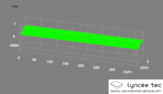
Digital holographic microscopy (DHM) is digital holography applied to microscopy. Digital holographic microscopy distinguishes itself from other microscopy methods by not recording the projected image of the object. Instead, the light wave front information originating from the object is digitally recorded as a hologram, from which a computer calculates the object image by using a numerical reconstruction algorithm. The image forming lens in traditional microscopy is thus replaced by a computer algorithm. Other closely related microscopy methods to digital holographic microscopy are interferometric microscopy, optical coherence tomography and diffraction phase microscopy. Common to all methods is the use of a reference wave front to obtain amplitude (intensity) and phase information. The information is recorded on a digital image sensor or by a photodetector from which an image of the object is created (reconstructed) by a computer. In traditional microscopy, which do not use a reference wave front, only intensity information is recorded and essential information about the object is lost.
Novacam Technologies Inc. specializes in designing and manufacturing advanced metrology and imaging systems for industrial and bio-medical applications. Novacam's fiber-based optical profilometers and Optical Coherence Tomography (OCT) systems are based on low coherence interferometry. The fiber-based nature of Novacam's detector probes is unique in the optical metrology industry.
Through-Focus Scanning Optical Microscopy (TSOM) is an imaging method that produces nanometer-scale three-dimensional measurement sensitivity using a conventional bright-field optical microscope. TSOM has been introduced and maintained by Ravikiran Attota at NIST. It was given an R&D 100 Award in 2010. In the TSOM method a target is scanned through the focus of an optical microscope, acquiring conventional optical images at different focal positions. The TSOM images are constructed using the through-focus optical images. A TSOM image is unique under given experimental conditions and is sensitive to changes in the dimensions of a target in a distinct way, which is very well applicable in nanoscale dimensional metrology. The TSOM method is alleged to have several nanometrology applications ranging from nanoparticles to through-silicon-vias (TSV).

Coherence scanning interferometry (CSI) is any of a class of optical surface measurement methods wherein the localization of interference fringes during a scan of optical path length provides a means to determine surface characteristics such as topography, transparent film structure, and optical properties. CSI is currently the most common interference microscopy technique for areal surface topography measurement. The term "CSI" was adopted by the International Organization for Standardization (ISO).

Organic photonics includes the generation, emission, transmission, modulation, signal processing, switching, amplification, and detection/sensing of light, using organic optical materials.

Din Ping Tsai is a physicist known for his work in the fields of photonics. He is currently a Distinguished Professor at the National Taiwan University and Director of the Research Center for Applied Sciences, Academia Sinica. He has been President of Taiwan Information Storage Association (TISA) since 2015.

Anatoly V. Zayats is a British experimental physicist of Ukrainian origin known for his work in nanophotonics, plasmonics, metamaterials and applied nanotechnology. He is currently a Chair in Experimental Physics and the head of the Photonics & Nanotechnology Group at King's College London. He is a co-director of the London Centre for Nanotechnology and the London Institute for Advanced Light Technologies

Stephen A. Boppart is a principal investigator at the Beckman Institute for Advanced Science and Technology at the University of Illinois at Urbana-Champaign, where he holds an Abel Bliss Professorship in engineering. He is a faculty member in the departments of electrical and computer engineering, bioengineering, and internal medicine. His research focus is biophotonics, where he has pioneered new optical imaging technologies in the fields of optical coherence tomography, multi-photon microscopy, and computational imaging.
Elizabeth M. C. Hillman is a British-born academic who is Professor of Biomedical Engineering and Radiology at Columbia University. She was awarded the 2011 Adolph Lomb Medal from The Optical Society and the 2018 SPIE Biophotonics Technology Innovator Award.

Anita Mahadevan-Jansen is a Professor of Biomedical Engineering and holds the Orrin H. Ingram Chair in Biomedical Engineering at Vanderbilt University. Her research considers the development of optical techniques for clinical diagnosis and surgical guidance, particularly using Raman and fluorescence spectroscopy. She serves on the Board of Directors of SPIE, and is a Fellow of SPIE, The Optical Society, Society for Applied Spectroscopy, and the American Society for Lasers in Medicine and Surgery. She was elected to serve as the 2020 Vice President of SPIE. With her election, Mahadevan-Jansen joined the SPIE presidential chain and served as President-Elect in 2021 and the Society's President in 2022.
Jannick Rolland is the Brian J. Thompson Professor of Optical Engineering at the Institute of Optics at the University of Rochester. She is also the co-founder and CTO of LighTopTech, a women-owner business founded in 2013 to create medical imaging technologies with biomimetic noninvasive imaging technology. At the University of Rochester, she is the Director of the NSF I/UCRC Center for Freeform Optics (CeFO). She is also the Director of the R.E. Hopkins Center for Optical Design and Engineering that engages undergraduates in optical design, fabrication, and metrology.

Jürgen W. Czarske is a German electrical engineer and a measurement system technician. He is the director of the TU Dresden Biomedical Computational Laser Systems competence center and a co-opted professor of physics.
Julie Lynn Bentley is an American optical physicist who is a professor at the University of Rochester. She is a Fellow of the Optical Society of America and the Society of Photo-Optical Instrumentation Engineers (SPIE).

Wibool Piyawattanametha is the head of Advanced Imaging Research (AIR) Center, King Mongkut's Institute of Technology Ladkrabang, Thailand.