Membrane fluidity

Last updated

In biology, membrane fluidity refers to the viscosity of the lipid bilayer of a cell membrane or a synthetic lipid membrane. Lipid packing can influence the fluidity of the membrane. Viscosity of the membrane can affect the rotation and diffusion of proteins and other bio-molecules within the membrane, there-by affecting the functions of these things. [1]

Contents

Membrane fluidity is affected by fatty acids. More specifically, whether the fatty acids are saturated or unsaturated has an effect on membrane fluidity. Saturated fatty acids have no double bonds in the hydrocarbon chain, and the maximum amount of hydrogen. The absence of double bonds increases fluidity. Unsaturated fatty acids have at least one double bond, creating a "kink" in the chain. The double bond decreases fluidity. While the addition of one double bond raises the melting temperature, research conducted by Xiaoguang Yang et. al. supports that four or more double bonds has a direct correlation to membrane fluidity. Membrane fluidity is also affected by cholesterol. [2] Cholesterol can make the cell membrane fluid as well as rigid.

Factors determining membrane fluidity

Membrane fluidity can be affected by a number of factors. [1] The main factors affecting membrane fluidity are environmental (ie. temperature), and compositionally. [3] One way to increase membrane fluidity is to heat up the membrane. Lipids acquire thermal energy when they are heated up; energetic lipids move around more, arranging and rearranging randomly, making the membrane more fluid. At low temperatures, the lipids are laterally ordered and organized in the membrane, and the lipid chains are mostly in the all-trans configuration and pack well together.

The melting temperature of a membrane is defined as the temperature across which the membrane transitions from a crystal-like to a fluid-like organization, or vice versa. This phase transition is not an actual state transition, but the two levels of organizations are very similar to a solid and liquid state.

The composition of a membrane can also affect its fluidity. The membrane phospholipids incorporate fatty acyl chains of varying length and saturation. Lipids with shorter chains are less stiff and less viscous because they are more susceptible to changes in kinetic energy due to their smaller molecular size and they have less surface area to undergo stabilizing London forces with neighboring hydrophobic chains. Molecules with carbon-carbon double bonds (unsaturated) are more rigid than those that are saturated with hydrogens, as double bonds cannot freely turn. As a result, the presence of fatty acyl chains with unsaturated double bonds makes it harder for the lipids to pack together by putting kinks into the otherwise straightened hydrocarbon chain. While unsaturated lipids may have more rigid individual bonds, membranes made with such lipids are more fluid because the individual lipids cannot pack as tightly as saturated lipids and thus have lower melting points: less thermal energy is required to achieve the same level of fluidity as membranes made with lipids with saturated hydrocarbon chains. [1] Incorporation of particular lipids, such as sphingomyelin, into synthetic lipid membranes is known to stiffen a membrane. Such membranes can be described as "a glass state, i.e., rigid but without crystalline order". [4]

Cholesterol acts as a bidirectional regulator of membrane fluidity because at high temperatures, it stabilizes the membrane and raises its melting point, whereas at low temperatures it intercalates between the phospholipids and prevents them from clustering together and stiffening. Some drugs, e.g. Losartan, are also known to alter membrane viscosity. [4] Another way to change membrane fluidity is to change the pressure. [1] In the laboratory, supported lipid bilayers and monolayers can be made artificially. In such cases, one can still speak of membrane fluidity. These membranes are supported by a flat surface, e.g. the bottom of a box. The fluidity of these membranes can be controlled by the lateral pressure applied, e.g. by the side walls of a box.

Heterogeneity in membrane physical property

Discrete lipid domains with differing composition, and thus membrane fluidity, can coexist in model lipid membranes; this can be observed using fluorescence microscopy. [4] The biological analogue, 'lipid raft', is hypothesized to exist in cell membranes and perform biological functions. [5] Also, a narrow annular lipid shell of membrane lipids in contact with integral membrane proteins have low fluidity compared to bulk lipids in biological membranes, as these lipid molecules stay stuck to surface of the protein macromolecules.

Measurement methods

Membrane fluidity can be measured with electron spin resonance, fluorescence, atomic force microscopy-based force spectroscopy, or deuterium nuclear magnetic resonance spectroscopy. Electron spin resonance measurements involve observing spin probe behaviour in the membrane. Fluorescence experiments involve observing fluorescent probes incorporated into the membrane. Atomic force microscopy experiments can measure fluidity on synthetic [6] or isolated patches of native membranes. [7] Solid state deuterium nuclear magnetic resonance spectroscopy involves observing deuterated lipids. [1] The techniques are complementary in that they operate on different timescales.

Membrane fluidity can be described by two different types of motion: rotational and lateral. In electron spin resonance, rotational correlation time of spin probes is used to characterize how much restriction is imposed on the probe by the membrane. In fluorescence, steady-state anisotropy of the probe can be used, in addition to the rotation correlation time of the fluorescent probe. [1] Fluorescent probes show varying degree of preference for being in an environment of restricted motion. In heterogeneous membranes, some probes will only be found in regions of higher membrane fluidity, while others are only found in regions of lower membrane fluidity. [8] Partitioning preference of probes can also be a gauge of membrane fluidity. In deuterium nuclear magnetic resonance spectroscopy, the average carbon-deuterium bond orientation of the deuterated lipid gives rise to specific spectroscopic features. All three of techniques can give some measure of the time-averaged orientation of the relevant (probe) molecule, which is indicative of the rotational dynamics of the molecule. [1]

Lateral motion of molecules within the membrane can be measured by a number of fluorescence techniques: fluorescence recovery after photobleaching involves photobleaching a uniformly labelled membrane with an intense laser beam and measuring how long it takes for fluorescent probes to diffuse back into the photobleached spot. [1] Fluorescence correlation spectroscopy monitors the fluctuations in fluorescence intensity measured from a small number of probes in a small space. These fluctuations are affected by the mode of lateral diffusion of the probe. Single particle tracking involves following the trajectory of fluorescent molecules or gold particles attached to a biomolecule and applying statistical analysis to extract information about the lateral diffusion of the tracked particle. [9]

Phospholipid-deficient bio-membranes

A study of central linewidths of electron spin resonance spectra of thylakoid membranes and aqueous dispersions of their total extracted lipids, labeled with stearic acid spin label (having spin or doxyl moiety at 5,7,9,12,13,14 and 16th carbons, with reference to carbonyl group), reveals a fluidity gradient. Decreasing linewidth from 5th to 16th carbons represents increasing degree of motional freedom (fluidity gradient) from headgroup-side to methyl terminal in both native membranes and their aqueous lipid extract (a multilamellar liposomal structure, typical of lipid bilayer organization). This pattern points at similarity of lipid bilayer organization in both native membranes and liposomes. This observation is critical, as thylakoid membranes comprising largely galactolipids, contain only 10% phospholipid, unlike other biological membranes consisting largely of phospholipids. Proteins in chloroplast thylakoid membranes, apparently, restrict lipid fatty acyl chain segmental mobility from 9th to 16th carbons vis a vis their liposomal counterparts. Surprisingly, liposomal fatty acyl chains are more restricted at 5th and 7th carbon positions as compared at these positions in thylakoid membranes. This is explainable as due to motional restricting effect at these positions, because of steric hindrance by large chlorophyll headgroups, specially so, in liposomes. However, in native thylakoid membranes, chlorophylls are mainly complexed with proteins as light-harvesting complexes and may not largely be free to restrain lipid fluidity, as such. [10]

Diffusion coefficients

Diffusion coefficients of fluorescent lipid analogues are about 10−8cm2/s in fluid lipid membranes. In gel lipid membranes and natural biomembranes, the diffusion coefficients are about 10−11cm2/s to 10−9cm2/s. [1]

Charged lipid membranes

The melting of charged lipid membranes, such as 1,2-dimyristoyl-sn-glycero-3-phosphoglycerol, can take place over a wide range of temperature. Within this range of temperatures, these membranes become very viscous. [4]

Biological relevance

Microorganisms subjected to thermal stress are known to alter the lipid composition of their cell membrane (see homeoviscous adaptation). This is one way they can adjust the fluidity of their membrane in response to their environment. [1] Membrane fluidity is known to affect the function of biomolecules residing within or associated with the membrane structure. For example, the binding of some peripheral proteins is dependent on membrane fluidity. [11] Lateral diffusion (within the membrane matrix) of membrane-related enzymes can affect reaction rates. [1] Consequently, membrane-dependent functions, such as phagocytosis and cell signalling, can be regulated by the fluidity of the cell-membrane. [12]

See also

Related Research Articles

<span class="mw-page-title-main">Biological membrane</span> Enclosing or separating membrane in organisms acting as selective semi-permeable barrier

A biological membrane, biomembrane or cell membrane is a selectively permeable membrane that separates the interior of a cell from the external environment or creates intracellular compartments by serving as a boundary between one part of the cell and another. Biological membranes, in the form of eukaryotic cell membranes, consist of a phospholipid bilayer with embedded, integral and peripheral proteins used in communication and transportation of chemicals and ions. The bulk of lipids in a cell membrane provides a fluid matrix for proteins to rotate and laterally diffuse for physiological functioning. Proteins are adapted to high membrane fluidity environment of the lipid bilayer with the presence of an annular lipid shell, consisting of lipid molecules bound tightly to the surface of integral membrane proteins. The cell membranes are different from the isolating tissues formed by layers of cells, such as mucous membranes, basement membranes, and serous membranes.

<span class="mw-page-title-main">Fatty acid</span> Carboxylic acid

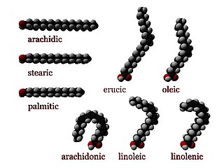
In chemistry, particularly in biochemistry, a fatty acid is a carboxylic acid with an aliphatic chain, which is either saturated or unsaturated. Most naturally occurring fatty acids have an unbranched chain of an even number of carbon atoms, from 4 to 28. Fatty acids are a major component of the lipids in some species such as microalgae but in some other organisms are not found in their standalone form, but instead exist as three main classes of esters: triglycerides, phospholipids, and cholesteryl esters. In any of these forms, fatty acids are both important dietary sources of fuel for animals and important structural components for cells.

<span class="mw-page-title-main">Lipid</span> Substance of biological origin that is soluble in nonpolar solvents

Lipids are a broad group of organic compounds which include fats, waxes, sterols, fat-soluble vitamins, monoglycerides, diglycerides, phospholipids, and others. The functions of lipids include storing energy, signaling, and acting as structural components of cell membranes. Lipids have applications in the cosmetic and food industries, and in nanotechnology.

<span class="mw-page-title-main">Lipid bilayer</span> Membrane of two layers of lipid molecules

The lipid bilayer is a thin polar membrane made of two layers of lipid molecules. These membranes are flat sheets that form a continuous barrier around all cells. The cell membranes of almost all organisms and many viruses are made of a lipid bilayer, as are the nuclear membrane surrounding the cell nucleus, and membranes of the membrane-bound organelles in the cell. The lipid bilayer is the barrier that keeps ions, proteins and other molecules where they are needed and prevents them from diffusing into areas where they should not be. Lipid bilayers are ideally suited to this role, even though they are only a few nanometers in width, because they are impermeable to most water-soluble (hydrophilic) molecules. Bilayers are particularly impermeable to ions, which allows cells to regulate salt concentrations and pH by transporting ions across their membranes using proteins called ion pumps.

An unsaturated fat is a fat or fatty acid in which there is at least one double bond within the fatty acid chain. A fatty acid chain is monounsaturated if it contains one double bond, and polyunsaturated if it contains more than one double bond.

<span class="mw-page-title-main">Fluid mosaic model</span> Describe the fluid mosaic model of plasma membrane

The fluid mosaic model explains various characteristics regarding the structure of functional cell membranes. According to this biological model, there is a lipid bilayer in which protein molecules are embedded. The phospholipid bilayer gives fluidity and elasticity to the membrane. Small amounts of carbohydrates are also found in the cell membrane. The biological model, which was devised by Seymour Jonathan Singer and Garth L. Nicolson in 1972, describes the cell membrane as a two-dimensional liquid that restricts the lateral diffusion of membrane components. Such domains are defined by the existence of regions within the membrane with special lipid and protein cocoon that promote the formation of lipid rafts or protein and glycoprotein complexes. Another way to define membrane domains is the association of the lipid membrane with the cytoskeleton filaments and the extracellular matrix through membrane proteins. The current model describes important features relevant to many cellular processes, including: cell-cell signaling, apoptosis, cell division, membrane budding, and cell fusion. The fluid mosaic model is the most acceptable model of the plasma membrane. In this definition of the cell membrane, its main function is to act as a barrier between the contents inside the cell and the extracellular environment.

<span class="mw-page-title-main">Glycerophospholipid</span> Class of lipids

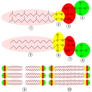
Glycerophospholipids or phosphoglycerides are glycerol-based phospholipids. They are the main component of biological membranes in eukaryotic cells. They are a type of lipid, of which its composition affects membrane structure and properties. Two major classes are known: those for bacteria and eukaryotes and a separate family for archaea.

Homeoviscous adaptation is the adaptation of the cell membrane lipid composition to keep the adequate membrane fluidity.

<span class="mw-page-title-main">Surfactin</span> Chemical compound

Surfactin is a cyclic lipopeptide, commonly used as an antibiotic for its capacity as a surfactant. It is an amphiphile capable of withstanding hydrophilic and hydrophobic environments. The Gram-positive bacterial species Bacillus subtilis produces surfactin for its antibiotic effects against competitors. Surfactin showcases antibacterial, antiviral, antifungal, and hemolytic effects.

<span class="mw-page-title-main">Membrane lipid</span> Lipid molecules on cell membrane

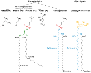
Membrane lipids are a group of compounds which form the lipid bilayer of the cell membrane. The three major classes of membrane lipids are phospholipids, glycolipids, and cholesterol. Lipids are amphiphilic: they have one end that is soluble in water ('polar') and an ending that is soluble in fat ('nonpolar'). By forming a double layer with the polar ends pointing outwards and the nonpolar ends pointing inwards membrane lipids can form a 'lipid bilayer' which keeps the watery interior of the cell separate from the watery exterior. The arrangements of lipids and various proteins, acting as receptors and channel pores in the membrane, control the entry and exit of other molecules and ions as part of the cell's metabolism. In order to perform physiological functions, membrane proteins are facilitated to rotate and diffuse laterally in two dimensional expanse of lipid bilayer by the presence of a shell of lipids closely attached to protein surface, called annular lipid shell.

<span class="mw-page-title-main">Lipid polymorphism</span>

Polymorphism in biophysics is the ability of lipids to aggregate in a variety of ways, giving rise to structures of different shapes, known as "phases". This can be in the form of spheres of lipid molecules (micelles), pairs of layers that face one another, a tubular arrangement (hexagonal), or various cubic phases. More complicated aggregations have also been observed, such as rhombohedral, tetragonal and orthorhombic phases.

α-Parinaric acid Chemical compound

α-Parinaric acid is a conjugated polyunsaturated fatty acid. Discovered by Tsujimoto and Koyanagi in 1933, it contains 18 carbon atoms and 4 conjugated double bonds. The repeating single bond-double bond structure of α-parinaric acid distinguishes it structurally and chemically from the usual "methylene-interrupted" arrangement of polyunsaturated fatty acids that have double-bonds and single bonds separated by a methylene unit (−CH2−). Because of the fluorescent properties conferred by the alternating double bonds, α-parinaric acid is commonly used as a molecular probe in the study of biomembranes.

Lipid microdomains are formed when lipids undergo lateral phase separations yielding stable coexisting lamellar domains. These phase separations can be induced by changes in temperature, pressure, ionic strength or by the addition of divalent cations or proteins. The question of whether such lipid microdomains observed in model lipid systems also exist in biomembranes had motivated considerable research efforts. Lipid domains are not readily isolated and examined as unique species, in contrast to the examples of lateral heterogeneity. One can disrupt the membrane and demonstrate a heterogeneous range of composition in the population of the resulting vesicles or fragments. Electron microscopy can also be used to demonstrate lateral inhomogeneities in biomembranes.

Protein–lipid interaction is the influence of membrane proteins on the lipid physical state or vice versa.

Lipid bilayer characterization is the use of various optical, chemical and physical probing methods to study the properties of lipid bilayers. Many of these techniques are elaborate and require expensive equipment because the fundamental nature of the lipid bilayer makes it a very difficult structure to study. An individual bilayer, since it is only a few nanometers thick, is invisible in traditional light microscopy. The bilayer is also a relatively fragile structure since it is held together entirely by non-covalent bonds and is irreversibly destroyed if removed from water. In spite of these limitations dozens of techniques have been developed over the last seventy years to allow investigations of the structure and function of bilayers. The first general approach was to utilize non-destructive in situ measurements such as x-ray diffraction and electrical resistance which measured bilayer properties but did not actually image the bilayer. Later, protocols were developed to modify the bilayer and allow its direct visualization at first in the electron microscope and, more recently, with fluorescence microscopy. Over the past two decades, a new generation of characterization tools including AFM has allowed the direct probing and imaging of membranes in situ with little to no chemical or physical modification. More recently, dual polarisation interferometry has been used to measure the optical birefringence of lipid bilayers to characterise order and disruption associated with interactions or environmental effects.

One property of a lipid bilayer is the relative mobility (fluidity) of the individual lipid molecules and how this mobility changes with temperature. This response is known as the phase behavior of the bilayer. Broadly, at a given temperature a lipid bilayer can exist in either a liquid or a solid phase. The solid phase is commonly referred to as a “gel” phase. All lipids have a characteristic temperature at which they undergo a transition (melt) from the gel to liquid phase. In both phases the lipid molecules are constrained to the two dimensional plane of the membrane, but in liquid phase bilayers the molecules diffuse freely within this plane. Thus, in a liquid bilayer a given lipid will rapidly exchange locations with its neighbor millions of times a second and will, through the process of a random walk, migrate over long distances.

<span class="mw-page-title-main">Lipid bilayer fusion</span>

In membrane biology, fusion is the process by which two initially distinct lipid bilayers merge their hydrophobic cores, resulting in one interconnected structure. If this fusion proceeds completely through both leaflets of both bilayers, an aqueous bridge is formed and the internal contents of the two structures can mix. Alternatively, if only one leaflet from each bilayer is involved in the fusion process, the bilayers are said to be hemifused. In hemifusion, the lipid constituents of the outer leaflet of the two bilayers can mix, but the inner leaflets remain distinct. The aqueous contents enclosed by each bilayer also remain separated.

<span class="mw-page-title-main">Ethanol-induced non-lamellar phases in phospholipids</span>

The presence of ethanol can lead to the formations of non-lamellar phases also known as non-bilayer phases. Ethanol has been recognized as being an excellent solvent in an aqueous solution for inducing non-lamellar phases in phospholipids. The formation of non-lamellar phases in phospholipids is not completely understood, but it is significant that this amphiphilic molecule is capable of doing so. The formation of non-lamellar phases is significant in biomedical studies which include drug delivery, the transport of polar and non-polar ions using solvents capable of penetrating the biomembrane, increasing the elasticity of the biomembrane when it is being disrupted by unwanted substances and functioning as a channel or transporter of biomaterial.

<span class="mw-page-title-main">Cell membrane</span> Biological membrane that separates the interior of a cell from its outside environment

The cell membrane is a biological membrane that separates and protects the interior of a cell from the outside environment. The cell membrane consists of a lipid bilayer, made up of two layers of phospholipids with cholesterols interspersed between them, maintaining appropriate membrane fluidity at various temperatures. The membrane also contains membrane proteins, including integral proteins that span the membrane and serve as membrane transporters, and peripheral proteins that loosely attach to the outer (peripheral) side of the cell membrane, acting as enzymes to facilitate interaction with the cell's environment. Glycolipids embedded in the outer lipid layer serve a similar purpose. The cell membrane controls the movement of substances in and out of a cell, being selectively permeable to ions and organic molecules. In addition, cell membranes are involved in a variety of cellular processes such as cell adhesion, ion conductivity, and cell signalling and serve as the attachment surface for several extracellular structures, including the cell wall and the carbohydrate layer called the glycocalyx, as well as the intracellular network of protein fibers called the cytoskeleton. In the field of synthetic biology, cell membranes can be artificially reassembled.

<span class="mw-page-title-main">Laurdan</span> Chemical compound

Laurdan is an organic compound which is used as a fluorescent dye when applied to fluorescence microscopy. It is used to investigate membrane qualities of the phospholipid bilayers of cell membranes. One of its most important characteristics is its sensitivity to membrane phase transitions as well as other alterations to membrane fluidity such as the penetration of water.

References

  1. 1 2 3 4 5 6 7 8 9 10 11 Gennis, R. B. (1989) Biomembranes: Molecular Structure and Function. Springer, ISBN   0387967605.
  2. Yang, Xiaoguang; Sheng, Wenwen; Sun, Grace Y.; Lee, James C-M. (February 2011). "Effects of fatty acid unsaturation numbers on membrane fluidity and α-secretase-dependent amyloid precursor protein processing". Neurochemistry International. 58 (3): 321–329. doi:10.1016/j.neuint.2010.12.004. ISSN   0197-0186. PMC   3040984 . PMID   21184792.
  3. Los, Dmitry A.; Murata, Norio (2004-11-03). "Membrane fluidity and its roles in the perception of environmental signals". Biochimica et Biophysica Acta (BBA) - Biomembranes. Lipid-Protein Interactions. 1666 (1): 142–157. doi:10.1016/j.bbamem.2004.08.002. ISSN   0005-2736.
  4. 1 2 3 4 Heimburg, T. (2007) Thermal Biophysics of Membranes. Wiley-VCH, ISBN   3527404716.
  5. Simons K, Vaz WL (2004). "Model systems, lipid rafts, and cell membranes" (PDF). Annual Review of Biophysics and Biomolecular Structure. 33: 269–95. doi:10.1146/annurev.biophys.32.110601.141803. hdl: 10316/11254 . PMID   15139814.
  6. Chiantia, Salvatore (2006). "Combined AFM and Two‐Focus SFCS Study of Raft‐Exhibiting Model Membranes". ChemPhysChem. 7 (11): 2409–2418. doi:10.1002/cphc.200600464. PMID   17051578.
  7. Galvanetto, Nicola (2018). "Single-cell unroofing: probing topology and nanomechanics of native membranes". Biochimica et Biophysica Acta (BBA) - Biomembranes. 1860 (12): 2532–2538. arXiv: 1810.01643 . doi:10.1016/j.bbamem.2018.09.019. PMID   30273580. S2CID   52897823.
  8. Baumgart, Tobias; Hunt, Geoff; Farkas, Elaine R.; Webb, Watt W.; Feigenson, Gerald W. (2007). "Fluorescence probe partitioning between Lo/Ld phases in lipid membranes". Biochimica et Biophysica Acta (BBA) - Biomembranes. 1768 (9): 2182–94. doi:10.1016/j.bbamem.2007.05.012. PMC   2702987 . PMID   17588529.
  9. Almeida, P. and Vaz, W. (1995). "Lateral diffusion in membranes", Ch. 6, pp. 305–357 in: Lipowsky, R. and Sackmann, E. (eds.) Handbook of biological physics. Elsevier Science B.V. doi : 10.1016/S1383-8121(06)80023-0, ISBN   978-0-444-81975-8
  10. YashRoy R C (1990) Magnetic resonance studies of dynamic organisation of lipids in chloroplast membranes. Journal of Biosciences, vol. 15(4), pp. 281-288.https://www.researchgate.net/publication/225688482_Magnetic_resonance_studies_of_dynamic_organisation_of_lipids_in_chloroplast_membranes?ev=prf_pub
  11. Heimburg, Thomas & Marsh, Derek (1996). "Thermodynamics of the Interaction of Proteins with Lipid Membranes". In Kenneth M. Merz Jr. & Benoît Roux (eds.). Biological Membranes. Boston: Birkhäuser. pp. 405–462. doi:10.1007/978-1-4684-8580-6_13. ISBN   978-1-4684-8580-6.
  12. Helmreich EJ (2003). "Environmental influences on signal transduction through membranes: A retrospective mini-review". Biophysical Chemistry. 100 (1–3): 519–34. doi:10.1016/S0301-4622(02)00303-4. PMID   12646388.