Memory T cell

Last updated

Memory T cells are a subset of T lymphocytes that might have some of the same functions as memory B cells. Their lineage is unclear.

Contents

Function

Antigen-specific memory T cells specific to viruses or other microbial molecules can be found in both central memory T cells(TCM) and effector memory T cells(TEM) subsets. Although most information is currently based on observations in the cytotoxic T cells (CD8-positive) subset, similar populations appear to exist for both the helper T cells (CD4-positive) and the cytotoxic T cells. Primary function of memory cells is augmented immune response after reactivation of those cells by reintroduction of relevant pathogen into the body. It is important to note that this field is intensively studied and some information may not be available as of yet.

Homeostatic maintenance

Clones of memory T cells expressing a specific T cell receptor can persist for decades in our body. Since memory T cells have shorter half-lives than naïve T cells do, continuous replication and replacement of old cells are likely involved in the maintenance process. [3] Currently, the mechanism behind memory T cell maintenance is not fully understood. Activation through the T cell receptor may play a role. [3] It is found that memory T cells can sometimes react to novel antigens, potentially caused by intrinsic the diversity and breadth of the T cell receptor binding targets. [3] These T cells could cross-react to environmental or resident antigens in our bodies (like bacteria in our gut) and proliferate. These events would help maintain the memory T cell population. [3] The cross-reactivity mechanism may be important for memory T cells in the mucosal tissues since these sites have higher antigen density. [3] For those resident in blood, bone marrow, lymphoid tissues, and spleen, homeostatic cytokines (including IL-17 and IL-15) or major histocompatibility complex II (MHCII) signaling may be more important. [3]

Lifetime overview

Memory T cells undergo different changes and play different roles in different life stages for humans. At birth and early childhood, T cells in the peripheral blood are mainly naïve T cells. [10] Through frequent antigen exposure, the population of memory T cells accumulates. This is the memory generation stage, which lasts from birth to about 20–25 years old when our immune system encounters the greatest number of new antigens. [3] [10] During the memory homeostasis stage that comes next, the number of memory T cells plateaus and is stabilized by homeostatic maintenance. [10] At this stage, the immune response shifts more towards maintaining homeostasis since few new antigens are encountered. [10] Tumor surveillance also becomes important at this stage. [10] At later stages of life, at about 65–70 years of age, immunosenescence stage comes, in which stage immune dysregulation, decline in T cell function and increased susceptibility to pathogens are observed. [3] [10]

Lineage debate

On-Off-On model:
After the naive T cell (N) encounters an antigen it becomes activated and begins to proliferate (divide) into many clones or daughter cells.
Some of the T cell clones will differentiate into effector T cells (E) that will perform the function of that cell (e.g. produce cytokines in the case of helper T cells or invoke cell killing in the case of cytotoxic T cells).
Some of the cells will form memory T cells (M) that will survive in an inactive state in the host for a long period of time until they re-encounter the same antigen and reactivate. T cell prolif.jpg
On-Off-On model:
  1. After the naive T cell (N) encounters an antigen it becomes activated and begins to proliferate (divide) into many clones or daughter cells.
  2. Some of the T cell clones will differentiate into effector T cells (E) that will perform the function of that cell (e.g. produce cytokines in the case of helper T cells or invoke cell killing in the case of cytotoxic T cells).
  3. Some of the cells will form memory T cells (M) that will survive in an inactive state in the host for a long period of time until they re-encounter the same antigen and reactivate.

As of April 2020, the lineage relationship between effector and memory T cells is unclear. [11] [12] [13] Two competing models exist. One is called the On-Off-On model. [12] When naive T cells are activated by T cell receptor (TCR) binding to antigen and its downstream signaling pathway, they actively proliferate and form a large clone of effector cells. Effector cells undergo active cytokine secretion and other effector activities. [11] After antigen clearance, some of these effector cells form memory T cells, either in a randomly determined manner or are selected based on their superior specificity. [11] These cells would reverse from the active effector role to a state more similar to naive T cells and would be "turned on" again upon the next antigen exposure. [13] This model predicts that effector T cells can transit into memory T cells and survive, retaining the ability to proliferate. [11] It also predicts that certain gene expression profiles would follow the on-off-on pattern during naive, effector, and memory stages. [13] Evidence supporting this model includes the finding of genes related to survival and homing that follow the on-off-on expression pattern, including interleukin-7 receptor alpha (IL-7Rα), Bcl-2, CD26L, and others. [13]

Developmental differentiation model:
In this model, memory T cells generate effector T cells, not the other way around. A picture for the developmental differentiation model for memory T cell lineage.png
Developmental differentiation model:
In this model, memory T cells generate effector T cells, not the other way around.

The other model is the developmental differentiation model. [12] This model argues that effector cells produced by the highly activated naive T cells would all undergo apoptosis after antigen clearance. [11] Memory T cells are instead produced by naive T cells that are activated but never entered with full strength into the effector stage. [11] The progeny of memory T cells are not fully activated because they are not as specific to the antigen as the expanding effector T cells. Studies looking at cell division history found that the length of telomere and activity of telomerase were reduced in effector T cells compared to memory T cells, which suggests that memory T cells did not undergo as much cell division as effector T cells, which is inconsistent with the On-Off-On model. [11] Repeated or chronic antigenic stimulation of T cells, like HIV infection, would induce elevated effector functions but reduce memory. [12] It was also found that massively proliferated T cells are more likely to generate short-lived effector cells, while minimally proliferated T cells would form more long-lived cells. [11]

Epigenetic modifications

Epigenetic modifications are involved in the change from naive T-cells. [14] For example, in CD4 + memory T cells, positive histone modifications mark key cytokine genes that are up-regulated during the secondary immune response, including IFNγ, IL4, and IL17A. [14] Some of these modifications persisted after antigen clearance, establishing an epigenetic memory that allows a faster activation upon re-encounter with the antigen. [14] For CD8 + memory T cells, certain effector genes, such as IFNγ, would not be expressed but they are transcriptionally poised for fast expression upon activation. [14] Additionally, the enhancement of expression for certain genes also depends on the strength of the initial TCR signaling for the progeny of memory T cells, which is correlated to the regulatory element activation that directly changes gene expression level. [14]

Sub-populations

Historically, memory T cells were thought to belong to either the effector (TEM cells) or central memory (TCM cells) subtypes, each with its own distinguishing set of cell surface markers (see below). [15] Subsequently, numerous additional populations of memory T cells were discovered including tissue-resident memory T (TRM) cells, stem memory TSCM cells, and virtual memory T cells. The single unifying theme for all memory T cell subtypes is that they are long-lived and can quickly expand to large numbers of effector T cells upon re-exposure to their cognate antigen. By this mechanism, they provide the immune system with "memory" against previously encountered pathogens. Memory T cells may be either CD4 + or CD8 + and usually express CD45RO and at the same time lack CD45RA. [16]

Memory T cell subtypes

There have been numerous other subpopulations of memory T cells suggested. Investigators have studied Stem memory TSCM cells. Like naive T cells, TSCM cells are CD45RO−, CCR7+, CD45RA+, CD62L+ (L-selectin), CD27+, CD28+, and IL-7Rα+, but they also express large amounts of CD95, IL-2Rβ, CXCR3, and LFA-1, and show numerous functional attributes distinctive of memory cells. [6]

TCR-independent (bystander) activation

T cells possess the ability to be activated independently of their cognate antigen stimulation, i.e. without TCR stimulation. At early stages of infection, T cells specific for unrelated antigen are activated only by the presence of inflammation. This happens in the inflammatory milieu resulting from microbial infection, cancer or autoimmunity in both mice and humans and occurs locally as well as systematically [25] [26] [27] [28] [29]  . Moreover, bystander activated T cells can migrate to the site of infection, due to increased CCR5 expression. [26]

This phenomenon was observed predominantly in memory CD8+ T cells, which have lower sensitivity to cytokine stimulation, compared to their naive counterparts and get activated in this manner more easily. [25] Virtual memory CD8+ T cells also display heightened sensitivity to cytokine-induced activation in mouse models, but this was not directly demonstrated in humans. [26]  Conversely, TCR-independent activation of naive CD8+ T cells remains controversial. [26] [28]

Apart from infections, bystander activation also plays an important role in antitumor immunity. [30] In human cancerous tissues, a high number of virus-specific, not tumor-specific, CD8+ T cells was detected. [30] This type of activation is considered to be beneficial for the host in terms of cancer clearance efficiency. [26]

Drivers of bystander activation

The major drivers of bystander activation are cytokines, such as IL-15, IL-18, IL-12 or type I IFNs, often working synergistically. [25] [26] [28] [29] IL-15 is responsible for cytotoxic activity of bystander-activated T cells. It induces the NKG2D (a receptor typically expressed on NK cells) expression on memory CD8+ T cells, leading to innate-like cytotoxicity, i.e. recognition of NKG2D ligands as indicators of infection, cell stress and cell transformation as well as destruction of altered cells in an NK-like manner. [25] [26] [28] [29] TCR activation was shown to abrogate IL-15 mediated NKG2D expression on T cells. [28] [29] Additionally, IL-15 induces expression of cytolytic molecules, cell expansion and enhances the cell response to IL-18. [25] [26] [29] IL-18 is another cytokine involved in this process, typically acting in synergy with IL-12, enhancing the differentiation of memory T cells into effector cells, i.e. it induces IFN-γ production and cell proliferation. [25] [26] [29] Toll-like receptors (TLRs), especially TLR2, have been linked to TCR-independent activation of CD8+ T cells upon bacterial infection as well. [25] [29]

Bystander activation of CD4+ T cells

Despite TCR-independent activation being studied more extensively in CD8+ T cells, there's a clear evidence of this phenomenon occurring in CD4+ T cells. However, it's considered to be less efficient, presumably due to lower CD122 (also known as IL2RB or IL15RB) expression. [31] [32] Similarly to their CD8+ counterparts, memory and effector CD4+ T cells exhibit increased sensitivity to TCR-independent activation. [26] [32] IL-1β, synergistically with IL-12 and IL-23, stimulates memory CD4+ T cells and drives Th17 response. [32] Moreover, IL-18, IL-12 and IL-27 induce cytokine expression in effector and memory CD4+ T cells [32]  and IL-2 is considered to be a strong activation inducer of CD4+ T cells that can replace TCR stimulation even in naive cells. [32] TLR2 was also reported to be present on memory CD4+ T cells, which respond to their agonist by IFNγ production, even without TCR stimulation. [32]

Role in pathogenicity

Bystander activation plays role in the elimination of the spread of infection in its early stages and helps in tumor clearance. However, this type of activation can also have deleterious outcome, especially in chronic infections and autoimmune diseases. [26] [27] [28] [29] Liver injury during chronic Hepatitis B virus infection is a result of non-HBV-specific CD8+ T cell infiltration into the tissue. [26] A similar situation occurs during the acute Hepatitis A virus infection [26] and activated virus unrelated CD4+ T cells contribute to ocular lesions in Herpes Simplex Virus infections. [26] [32]

Increased IL-15 expression and subsequent excessive NKG2D expression was linked to exacerbation of some autoimmune disorders, such as, type I diabetes, multiple sclerosis and inflammatory bowel diseases, for instance Crohn's disease and celiac disease. [25] Furthermore, enhanced TLR2 expression was observed in joints, cartilage and bones of rheumatoid arthritis patients and the presence of its ligand, peptidoglycan, was detected in their synovial fluid. [25]

Related Research Articles

<span class="mw-page-title-main">T cell</span> White blood cells of the immune system

T cells are one of the important types of white blood cells of the immune system and play a central role in the adaptive immune response. T cells can be distinguished from other lymphocytes by the presence of a T-cell receptor (TCR) on their cell surface.

<span class="mw-page-title-main">Cytotoxic T cell</span> T cell that kills infected, damaged or cancerous cells

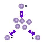
A cytotoxic T cell (also known as TC, cytotoxic T lymphocyte, CTL, T-killer cell, cytolytic T cell, CD8+ T-cell or killer T cell) is a T lymphocyte (a type of white blood cell) that kills cancer cells, cells that are infected by intracellular pathogens (such as viruses or bacteria), or cells that are damaged in other ways.

<span class="mw-page-title-main">T helper cell</span> Type of immune cell

The T helper cells (Th cells), also known as CD4+ cells or CD4-positive cells, are a type of T cell that play an important role in the adaptive immune system. They aid the activity of other immune cells by releasing cytokines. They are considered essential in B cell antibody class switching, breaking cross-tolerance in dendritic cells, in the activation and growth of cytotoxic T cells, and in maximizing bactericidal activity of phagocytes such as macrophages and neutrophils. CD4+ cells are mature Th cells that express the surface protein CD4. Genetic variation in regulatory elements expressed by CD4+ cells determines susceptibility to a broad class of autoimmune diseases.

<span class="mw-page-title-main">Cell-mediated immunity</span> Immune response that does not involve antibodies

Cellular immunity, also known as cell-mediated immunity, is an immune response that does not rely on the production of antibodies. Rather, cell-mediated immunity is the activation of phagocytes, antigen-specific cytotoxic T-lymphocytes, and the release of various cytokines in response to an antigen.

The regulatory T cells (Tregs or Treg cells), formerly known as suppressor T cells, are a subpopulation of T cells that modulate the immune system, maintain tolerance to self-antigens, and prevent autoimmune disease. Treg cells are immunosuppressive and generally suppress or downregulate induction and proliferation of effector T cells. Treg cells express the biomarkers CD4, FOXP3, and CD25 and are thought to be derived from the same lineage as naïve CD4+ cells. Because effector T cells also express CD4 and CD25, Treg cells are very difficult to effectively discern from effector CD4+, making them difficult to study. Research has found that the cytokine transforming growth factor beta (TGF-β) is essential for Treg cells to differentiate from naïve CD4+ cells and is important in maintaining Treg cell homeostasis.

<span class="mw-page-title-main">Adaptive immune system</span> Subsystem of the immune system

The adaptive immune system, also known as the acquired immune system, or specific immune system is a subsystem of the immune system that is composed of specialized, systemic cells and processes that eliminate pathogens or prevent their growth. The acquired immune system is one of the two main immunity strategies found in vertebrates.

Alloimmunity is an immune response to nonself antigens from members of the same species, which are called alloantigens or isoantigens. Two major types of alloantigens are blood group antigens and histocompatibility antigens. In alloimmunity, the body creates antibodies against the alloantigens, attacking transfused blood, allotransplanted tissue, and even the fetus in some cases. Alloimmune (isoimmune) response results in graft rejection, which is manifested as deterioration or complete loss of graft function. In contrast, autoimmunity is an immune response to the self's own antigens. Alloimmunization (isoimmunization) is the process of becoming alloimmune, that is, developing the relevant antibodies for the first time.

In immunology, a naive T cell (Th0 cell) is a T cell that has differentiated in the thymus, and successfully undergone the positive and negative processes of central selection in the thymus. Among these are the naive forms of helper T cells (CD4+) and cytotoxic T cells (CD8+). Any naive T cell is considered immature and, unlike activated or memory T cells, has not encountered its cognate antigen within the periphery. After this encounter, the naive T cell is considered a mature T cell.

<span class="mw-page-title-main">Interleukin 21</span> Mammalian protein found in humans

Interleukin 21 (IL-21) is a protein that in humans is encoded by the IL21 gene.

<span class="mw-page-title-main">Intraepithelial lymphocyte</span>

Intraepithelial lymphocytes (IEL) are lymphocytes found in the epithelial layer of mammalian mucosal linings, such as the gastrointestinal (GI) tract and reproductive tract. However, unlike other T cells, IELs do not need priming. Upon encountering antigens, they immediately release cytokines and cause killing of infected target cells. In the GI tract, they are components of gut-associated lymphoid tissue (GALT).

In immunology, peripheral tolerance is the second branch of immunological tolerance, after central tolerance. It takes place in the immune periphery. Its main purpose is to ensure that self-reactive T and B cells which escaped central tolerance do not cause autoimmune disease. Peripheral tolerance prevents immune response to harmless food antigens and allergens, too.

<span class="mw-page-title-main">CD69</span> Human lectin protein

CD69 is a human transmembrane C-Type lectin protein encoded by the CD69 gene. It is an early activation marker that is expressed in hematopoietic stem cells, T cells, and many other cell types in the immune system. It is also implicated in T cell differentiation as well as lymphocyte retention in lymphoid organs.

Gamma delta T cells are T cells that have a γδ T-cell receptor (TCR) on their surface. Most T cells are αβ T cells with TCR composed of two glycoprotein chains called α (alpha) and β (beta) TCR chains. In contrast, γδ T cells have a TCR that is made up of one γ (gamma) chain and one δ (delta) chain. This group of T cells is usually less common than αβ T cells. Their highest abundance is in the gut mucosa, within a population of lymphocytes known as intraepithelial lymphocytes (IELs).

<span class="mw-page-title-main">Lymphocyte-activation gene 3</span>

Lymphocyte-activation gene 3, also known as LAG-3, is a protein which in humans is encoded by the LAG3 gene. LAG3, which was discovered in 1990 and was designated CD223 after the Seventh Human Leucocyte Differentiation Antigen Workshop in 2000, is a cell surface molecule with diverse biological effects on T cell function but overall has an immune inhibitory effect. It is an immune checkpoint receptor and as such is the target of various drug development programs by pharmaceutical companies seeking to develop new treatments for cancer and autoimmune disorders. In soluble form it is also being developed as a cancer drug in its own right.

T helper 3 cells (Th3) are a subset of T lymphocytes with immunoregulary and immunosuppressive functions, that can be induced by administration of foreign oral antigen. Th3 cells act mainly through the secretion of anti-inflammatory cytokine transforming growth factor beta (TGF-β). Th3 have been described both in mice and human as CD4+FOXP3 regulatory T cells. Th3 cells were first described in research focusing on oral tolerance in the experimental autoimmune encephalitis (EAE) mouse model and later described as CD4+CD25FOXP3LAP+ cells, that can be induced in the gut by oral antigen through T cell receptor (TCR) signalling.

Natural killer T (NKT) cells are a heterogeneous group of T cells that share properties of both T cells and natural killer cells. Many of these cells recognize the non-polymorphic CD1d molecule, an antigen-presenting molecule that binds self and foreign lipids and glycolipids. They constitute only approximately 1% of all peripheral blood T cells. Natural killer T cells should neither be confused with natural killer cells nor killer T cells.

Mucosal-associated invariant T cells make up a subset of T cells in the immune system that display innate, effector-like qualities. In humans, MAIT cells are found in the blood, liver, lungs, and mucosa, defending against microbial activity and infection. The MHC class I-like protein, MR1, is responsible for presenting bacterially-produced vitamin B2 and B9 metabolites to MAIT cells. After the presentation of foreign antigen by MR1, MAIT cells secrete pro-inflammatory cytokines and are capable of lysing bacterially-infected cells. MAIT cells can also be activated through MR1-independent signaling. In addition to possessing innate-like functions, this T cell subset supports the adaptive immune response and has a memory-like phenotype. Furthermore, MAIT cells are thought to play a role in autoimmune diseases, such as multiple sclerosis, arthritis and inflammatory bowel disease, although definitive evidence is yet to be published.

Tissue-resident memory T cells or TRM cells represent a subset of a long-lived memory T cells that occupies epithelial, mucosal and other tissues without recirculating. TRM cells are transcriptionally, phenotypically and functionally distinct from central memory (TCM) and effector memory (TEM) T cells which recirculate between blood, the T cell zones of secondary lymphoid organ, lymph and nonlymphoid tissues. Moreover, TRM cells themself represent a diverse populations because of the specializations for the resident tissues. The main role of TRM cells is to provide superior protection against infection in extralymphoid tissues.

Virtual memory T cells (TVM) are a subtype of T lymphocytes. These are cells that have a memory phenotype but have not been exposed to a foreign antigen. They are classified as memory cells but do not have an obvious memory function. They were first observed and described in 2009. The name comes from a computerized "virtual memory" that describes a working memory based on an alternative use of an existing space.

A T memory stem cell (TSCM) is a type of long-lived memory T cell with the ability to reconstitute the full diversity of memory and effector T cell subpopulations as well as to maintain their own pool through self-renewal. TSCM represent an intermediate subset between naïve (Tn) and central memory (Tcm) T cells, expressing both naïve T cells markers, such as CD45RA+, CD45RO-, high levels of CD27, CD28, IL-7Rα (CD127), CD62L, and C-C chemokine receptor 7 (CCR7), as well as markers of memory T cells, such as CD95, CD122 (IL-2Rβ), CXCR3, LFA-1. These cells represent a small fraction of circulating T cells, approximately 2-3%. Like naïve T cells, TSCM cells are found more abundantly in lymph nodes than in the spleen or bone marrow; but in contrast to naïve T cells, TSCM cells are clonally expanded. Similarly to memory T cells, TSCM are able to rapidly proliferate and secrete pro-inflammatory cytokines in response to antigen re-exposure, but show higher proliferation potential compared with Tcm cells; their homeostatic turnover is also dependent on IL-7 and IL-15.

References

  1. 1 2 Wherry EJ, Teichgräber V, Becker TC, Masopust D, Kaech SM, Antia R, et al. (March 2003). "Lineage relationship and protective immunity of memory CD8 T cell subsets". Nature Immunology. 4 (3): 225–34. doi: 10.1038/ni889 . PMID   12563257. S2CID   7209417.
  2. Klebanoff CA, Gattinoni L, Torabi-Parizi P, Kerstann K, Cardones AR, Finkelstein SE, et al. (July 2005). "Central memory self/tumor-reactive CD8+ T cells confer superior antitumor immunity compared with effector memory T cells". Proceedings of the National Academy of Sciences of the United States of America. 102 (27): 9571–6. Bibcode:2005PNAS..102.9571K. doi: 10.1073/pnas.0503726102 . PMC   1172264 . PMID   15980149.
  3. 1 2 3 4 5 6 7 8 9 Farber DL, Yudanin NA, Restifo NP (January 2014). "Human memory T cells: generation, compartmentalization and homeostasis". Nature Reviews. Immunology. 14 (1): 24–35. doi:10.1038/nri3567. PMC   4032067 . PMID   24336101.
  4. Gebhardt T, Wakim LM, Eidsmo L, Reading PC, Heath WR, Carbone FR (May 2009). "Memory T cells in nonlymphoid tissue that provide enhanced local immunity during infection with herpes simplex virus". Nature Immunology. 10 (5): 524–30. doi:10.1038/ni.1718. PMID   19305395. S2CID   24388.
  5. 1 2 Shin H, Iwasaki A (September 2013). "Tissue-resident memory T cells". Immunological Reviews. 255 (1): 165–81. doi:10.1111/imr.12087. PMC   3748618 . PMID   23947354.
  6. 1 2 Gattinoni L, Lugli E, Ji Y, Pos Z, Paulos CM, Quigley MF, et al. (September 2011). "A human memory T cell subset with stem cell-like properties". Nature Medicine. 17 (10): 1290–7. doi:10.1038/nm.2446. PMC   3192229 . PMID   21926977.
  7. White JT, Cross EW, Kedl RM (June 2017). "+ T cells: where they come from and why we need them". Nature Reviews. Immunology. 17 (6): 391–400. doi:10.1038/nri.2017.34. PMC   5569888 . PMID   28480897.
  8. Lee JY, Hamilton SE, Akue AD, Hogquist KA, Jameson SC (August 2013). "Virtual memory CD8 T cells display unique functional properties". Proceedings of the National Academy of Sciences of the United States of America. 110 (33): 13498–503. Bibcode:2013PNAS..11013498L. doi: 10.1073/pnas.1307572110 . PMC   3746847 . PMID   23898211.
  9. Drobek A, Moudra A, Mueller D, Huranova M, Horkova V, Pribikova M, et al. (July 2018). "Strong homeostatic TCR signals induce formation of self-tolerant virtual memory CD8 T cells". The EMBO Journal. 37 (14). doi:10.15252/embj.201798518. PMC   6043851 . PMID   29752423.
  10. 1 2 3 4 5 6 7 8 9 Kumar BV, Connors TJ, Farber DL (February 2018). "Human T Cell Development, Localization, and Function throughout Life". Immunity. 48 (2): 202–213. doi:10.1016/j.immuni.2018.01.007. PMC   5826622 . PMID   29466753.
  11. 1 2 3 4 5 6 7 8 Restifo NP, Gattinoni L (October 2013). "Lineage relationship of effector and memory T cells". Current Opinion in Immunology. Special section: Systems biology and bioinformatics / Immunogenetics and transplantation. 25 (5): 556–63. doi:10.1016/j.coi.2013.09.003. PMC   3858177 . PMID   24148236.
  12. 1 2 3 4 Henning AN, Roychoudhuri R, Restifo NP (May 2018). "+ T cell differentiation". Nature Reviews. Immunology. 18 (5): 340–356. doi:10.1038/nri.2017.146. PMC   6327307 . PMID   29379213.
  13. 1 2 3 4 Youngblood B, Hale JS, Ahmed R (July 2013). "T-cell memory differentiation: insights from transcriptional signatures and epigenetics". Immunology. 139 (3): 277–84. doi:10.1111/imm.12074. PMC   3701173 . PMID   23347146.
  14. 1 2 3 4 5 Schmidl C, Delacher M, Huehn J, Feuerer M (September 2018). "Epigenetic mechanisms regulating T-cell responses". The Journal of Allergy and Clinical Immunology. 142 (3): 728–743. doi: 10.1016/j.jaci.2018.07.014 . PMID   30195378.
  15. Sallusto F, Lenig D, Förster R, Lipp M, Lanzavecchia A (October 1999). "Two subsets of memory T lymphocytes with distinct homing potentials and effector functions". Nature. 401 (6754): 708–12. Bibcode:1999Natur.401..708S. doi:10.1038/44385. PMID   10537110. S2CID   4378970.
  16. Akbar AN, Terry L, Timms A, Beverley PC, Janossy G (April 1988). "Loss of CD45R and gain of UCHL1 reactivity is a feature of primed T cells". Journal of Immunology. 140 (7): 2171–8. doi: 10.4049/jimmunol.140.7.2171 . PMID   2965180. S2CID   22340282.
  17. Willinger T, Freeman T, Hasegawa H, McMichael AJ, Callan MF (November 2005). "Molecular signatures distinguish human central memory from effector memory CD8 T cell subsets". Journal of Immunology. 175 (9): 5895–903. doi: 10.4049/jimmunol.175.9.5895 . hdl: 20.500.11820/f28e936e-a6a7-4f06-bdc9-79a1355c5f02 . PMID   16237082.
  18. Koch S, Larbi A, Derhovanessian E, Ozcelik D, Naumova E, Pawelec G (July 2008). "Multiparameter flow cytometric analysis of CD4 and CD8 T cell subsets in young and old people". Immunity & Ageing. 5 (6): 6. doi: 10.1186/1742-4933-5-6 . PMC   2515281 . PMID   18657274.
  19. Gerlach, Carmen; Moseman, E. Ashley; Loughhead, Scott M.; Alvarez, David; Zwijnenburg, Anthonie J.; Waanders, Lisette; Garg, Rohit; de la Torre, Juan C.; von Andrian, Ulrich H. (December 2016). "The Chemokine Receptor CX3CR1 Defines Three Antigen-Experienced CD8 T Cell Subsets with Distinct Roles in Immune Surveillance and Homeostasis". Immunity. 45 (6): 1270–1284. doi:10.1016/j.immuni.2016.10.018. PMC   5177508 . PMID   27939671.
  20. 1 2 3 Mueller SN, Mackay LK (February 2016). "Tissue-resident memory T cells: local specialists in immune defence". Nature Reviews. Immunology. 16 (2): 79–89. doi:10.1038/nri.2015.3. PMID   26688350. S2CID   3155731.
  21. Steinert EM, Schenkel JM, Fraser KA, Beura LK, Manlove LS, Igyártó BZ, et al. (May 2015). "Quantifying Memory CD8 T Cells Reveals Regionalization of Immunosurveillance". Cell. 161 (4): 737–49. doi:10.1016/j.cell.2015.03.031. PMC   4426972 . PMID   25957682.
  22. 1 2 3 "Study highlights possible Achilles' heel in key immune memory cells".
  23. Lee YJ, Jameson SC, Hogquist KA (February 2011). "Alternative memory in the CD8 T cell lineage". Trends in Immunology. 32 (2): 50–6. doi:10.1016/j.it.2010.12.004. PMC   3039080 . PMID   21288770.
  24. Marusina AI, Ono Y, Merleev AA, Shimoda M, Ogawa H, Wang EA, et al. (February 2017). "+ virtual memory: Antigen-inexperienced T cells reside in the naïve, regulatory, and memory T cell compartments at similar frequencies, implications for autoimmunity". Journal of Autoimmunity. 77: 76–88. doi:10.1016/j.jaut.2016.11.001. PMC   6066671 . PMID   27894837.
  25. 1 2 3 4 5 6 7 8 9 Whiteside, Sarah K.; Snook, Jeremy P.; Williams, Matthew A.; Weis, Janis J. (December 2018). "Bystander T Cells: A Balancing Act of Friends and Foes". Trends in Immunology. 39 (12): 1021–1035. doi:10.1016/j.it.2018.10.003. PMC   6269193 . PMID   30413351.
  26. 1 2 3 4 5 6 7 8 9 10 11 12 13 14 Lee, Hoyoung; Jeong, Seongju; Shin, Eui-Cheol (January 2022). "Significance of bystander T cell activation in microbial infection". Nature Immunology. 23 (1): 13–22. doi:10.1038/s41590-021-00985-3. ISSN   1529-2908. PMID   34354279. S2CID   236933989.
  27. 1 2 Pacheco, Yovana; Acosta-Ampudia, Yeny; Monsalve, Diana M.; Chang, Christopher; Gershwin, M. Eric; Anaya, Juan-Manuel (September 2019). "Bystander activation and autoimmunity". Journal of Autoimmunity. 103: 102301. doi:10.1016/j.jaut.2019.06.012. PMID   31326230. S2CID   198133084.
  28. 1 2 3 4 5 6 Maurice, Nicholas J.; Taber, Alexis K.; Prlic, Martin (2021-02-01). "The Ugly Duckling Turned to Swan: A Change in Perception of Bystander-Activated Memory CD8 T Cells". The Journal of Immunology. 206 (3): 455–462. doi:10.4049/jimmunol.2000937. ISSN   0022-1767. PMC   7839146 . PMID   33468558.
  29. 1 2 3 4 5 6 7 8 Kim, Tae-Shin; Shin, Eui-Cheol (December 2019). "The activation of bystander CD8+ T cells and their roles in viral infection". Experimental & Molecular Medicine. 51 (12): 1–9. doi:10.1038/s12276-019-0316-1. ISSN   1226-3613. PMC   6906361 . PMID   31827070.
  30. 1 2 Borras, DM, Verbandt, S, Ausserhofer, M, et al. (November 2023). "Single cell dynamics of tumor specificity vs bystander activity in CD8+ T cells define the diverse immune landscapes in colorectal cancer". Cell Discovery. 9 (114): 114. doi: 10.1038/s41421-023-00605-4 . PMC   10652011 . PMID   37968259.
  31. Boyman, Onur (April 2010). "Bystander activation of CD4 + T cells: HIGHLIGHTS". European Journal of Immunology. 40 (4): 936–939. doi: 10.1002/eji.201040466 . PMID   20309907. S2CID   7918378.
  32. 1 2 3 4 5 6 7 Lee, Hong-Gyun; Cho, Min-Zi; Choi, Je-Min (August 2020). "Bystander CD4+ T cells: crossroads between innate and adaptive immunity". Experimental & Molecular Medicine. 52 (8): 1255–1263. doi:10.1038/s12276-020-00486-7. ISSN   1226-3613. PMC   8080565 . PMID   32859954.