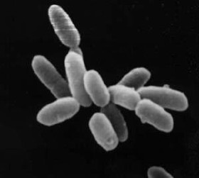
Bacillus subtilis, known also as the hay bacillus or grass bacillus, is a Gram-positive, catalase-positive bacterium, found in soil and the gastrointestinal tract of ruminants, humans and marine sponges. As a member of the genus Bacillus, B. subtilis is rod-shaped, and can form a tough, protective endospore, allowing it to tolerate extreme environmental conditions. B. subtilis has historically been classified as an obligate aerobe, though evidence exists that it is a facultative anaerobe. B. subtilis is considered the best studied Gram-positive bacterium and a model organism to study bacterial chromosome replication and cell differentiation. It is one of the bacterial champions in secreted enzyme production and used on an industrial scale by biotechnology companies.
Heliobacteria are a unique subset of prokaryotic bacteria that process light for energy. Distinguishable from other phototrophic bacteria, they utilize a unique photosynthetic pigment, bacteriochlorophyll g and are the only known Gram-positive phototroph. They are a key player in symbiotic nitrogen fixation alongside plants, and use a type I reaction center like green-sulfur bacteria.
Aquifex pyrophilus is a gram-negative, non-spore forming, rod-shaped bacteria. It is one of a handful of species in the Aquificota phylum, which are a group of thermophilic bacteria that are found near underwater volcanoes or hot springs.
The family Micrococcaceae includes bacterial genera of Gram positive cocci that inhabit the air and skin, such as Micrococcus luteus.
Halobacterium is a genus in the family Halobacteriaceae.

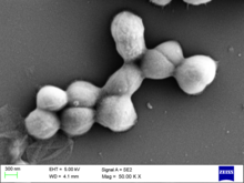
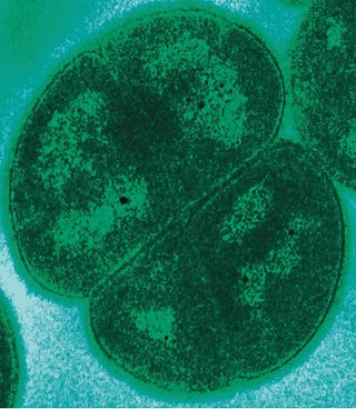
Micrococcus is a genus of bacteria in the Micrococcaceae family. Micrococcus occurs in a wide range of environments, including water, dust, and soil. Micrococci have Gram-positive spherical cells ranging from about 0.5 to 3 micrometers in diameter and typically appear in tetrads. They are catalase positive, oxidase positive, indole negative and citrate negative. Micrococcus has a substantial cell wall, which may comprise as much as 50% of the cell mass. The genome of Micrococcus is rich in guanine and cytosine (GC), typically exhibiting 65 to 75% GC-content. Micrococci often carry plasmids that provide the organism with useful traits.
Chromobacterium violaceum is a Gram-negative, facultative anaerobic, non-sporing coccobacillus. It is motile with the help of a single flagellum which is located at the pole of the coccobacillus. Usually, there are one or two more lateral flagella as well. It is part of the normal flora of water and soil of tropical and sub-tropical regions of the world. It produces a natural antibiotic called violacein, which may be useful for the treatment of colon and other cancers. It grows readily on nutrient agar, producing distinctive smooth low convex colonies with a dark violet metallic sheen. Some strains of the bacteria which do not produce this pigment have also been reported. It has the ability to break down tarballs.

Enterobacter cloacae is a clinically significant Gram-negative, facultatively-anaerobic, rod-shaped bacterium.

Streptomyces griseus is a species of bacteria in the genus Streptomyces commonly found in soil. A few strains have been also reported from deep-sea sediments. It is a Gram-positive bacterium with high GC content. Along with most other streptomycetes, S. griseus strains are well known producers of antibiotics and other such commercially significant secondary metabolites. These strains are known to be producers of 32 different structural types of bioactive compounds. Streptomycin, the first antibiotic ever reported from a bacterium, comes from strains of S. griseus. Recently, the whole genome sequence of one of its strains had been completed.
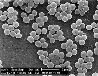
Staphylococcus is a genus of Gram-positive bacteria in the family Staphylococcaceae from the order Bacillales. Under the microscope, they appear spherical (cocci), and form in grape-like clusters. Staphylococcus species are facultative anaerobic organisms.
Deinococcus radiodurans is an extremophilic bacterium and one of the most radiation-resistant organisms known. It can survive cold, dehydration, vacuum, and acid, and therefore is known as a polyextremophile. It has been listed as the world's toughest known bacterium in The Guinness Book Of World Records.
Mycosporine-like amino acids (MAAs) are small secondary metabolites produced by organisms that live in environments with high volumes of sunlight, usually marine environments. The exact number of compounds within this class of natural products is yet to be determined, since they have only relatively recently been discovered and novel molecular species are constantly being discovered; however, to date their number is around 30. They are commonly described as “microbial sunscreens” although their function is believed not to be limited to sun protection. MAAs represent high potential in cosmetics, and biotechnological applications. Indeed, their UV-absorbing properties would allow to create products derived from natural photoprotectors, potentially harmless to the environment and efficient against UV damage.
Rothia kristinae is a Gram positive bacterium. R. kristinae is a common human skin organism, but can cause opportunistic infections in humans.
Micrococcus lylae is a gram positive bacterium. The normal habitat for this Micrococcus species is skin, dust, and water. Its type strain is ATCC 27566. It grows in tetrads, irregular clusters, and cubical packets of eight, and colonies are often brightly pigmented. They are strictly aerobic.
Kocuria is a genus of gram-positive bacteria. Kocuria is named after Miloslav Kocur, a Czech microbiologist. It has been found in the milk of water deer and reindeer. Cells are coccoid, resembling Staphylococcus and Micrococcus, and can group in pairs, chains, tetrads, cubical arrangements of eight, or irregular clusters. They have rigid cell walls and are either aerobic or facultative anaerobic. Kocuria can usually survive in mesophilic temperatures.
Kocuria rhizophila is a soil dwelling Gram positive bacterium in the genus Kocuria. It is used in industry for antimicrobial testing and in food preparation.

Kytococcus sedentarius is a marine dwelling Gram positive bacterium in the genus Kytococcus. It is known for the production of polyketide antibiotics as well as for its role as an opportunistic pathogen. It is strictly aerobic and can only grow when amino acids are provided.
Arthrobacter luteus (ALU) is a species of gram-positive bacteria in the genus Arthrobacter. A. luteus is facultatively anaerobic, pleomorphic, branching, non-motile, non-sporulating, non-acid-fast, catalase-positive, and rod-shaped.
Kocuria rosea is a gram-positive bacteria that is catalase-positive and oxidase-positive. It has a coccus shape that occurs in the tetrad arrangement and is a strict aerobe that grows best from 25 to 37 °C. K. rosea has also been found to cause urinary tract infections in people with weakened immune systems.
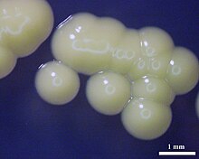
Kocuria varians is a gram-positive species of bacteria in the genus Kocuria. It has been isolated from milk, meat, skin, soil, and beach sand. It is 0.9 to 1.5 micrometers in diameter, and occurs in clusters, which can be up to 4 millimeters in diameter and are yellow. It is known to cause ocular infections, brain abscesses, and endophthalmitis.