Mycobacterium

Last updated

Mycobacterium
Mycobacterium tuberculosis 01.jpg
TEM micrograph of M. tuberculosis
Scientific classification OOjs UI icon edit-ltr.svg
Domain: Bacteria
Phylum: Actinomycetota
Class: Actinomycetia
Order: Mycobacteriales
Family: Mycobacteriaceae
Genus: Mycobacterium
Lehmann & Neumann 1896 [1]
Species

Over 190 species, see LPSN

Synonyms [2]
  • MycolicibacteriumGupta et al. 2018
  • Mycolicibacillus Gupta et al. 2018
  • MycolicibacterGupta et al. 2018
  • MycobacteroidesGupta et al. 2018

Mycobacterium is a genus of over 190 species in the phylum Actinomycetota, assigned its own family, Mycobacteriaceae. This genus includes pathogens known to cause serious diseases in mammals, including tuberculosis ( M. tuberculosis ) and leprosy ( M. leprae ) in humans. The Greek prefix myco- means 'fungus', alluding to this genus' mold-like colony surfaces. [3] Since this genus has cell walls with a waxy lipid-rich outer layer that contains high concentrations of mycolic acid, [4] acid-fast staining is used to emphasize their resistance to acids, compared to other cell types. [5]

Contents

Mycobacterial species are generally aerobic, non-motile, and capable of growing with minimal nutrients. The genus is divided based on each species' pigment production and growth rate. [6] While most Mycobacterium species are non-pathogenic, the genus' characteristic complex cell wall contributes to evasion from host defenses. [7]

Microbiology

Morphology

Model of the Mycobacterium spp. cell envelope with 3-D protein structures Model of the Mycobacterial Cell Envelope.png
Model of the Mycobacterium spp. cell envelope with 3-D protein structures

Mycobacteria are aerobic with 0.2-0.6 μm wide and 1.0-10 μm long rod shapes. They are generally non-motile, except for the species Mycobacterium marinum , which has been shown to be motile within macrophages. [8] Mycobacteria possess capsules and most do not form endospores. M. marinum and perhaps M. bovis have been shown to sporulate; [9] however, this has been contested by further research. [10] The distinguishing characteristic of all Mycobacterium species is a thick, hydrophobic, and mycolic acid-rich cell wall made of peptidoglycan and arabinogalactan, with these unique components offering targets for new tuberculosis drugs. [11]

Physiology

Many Mycobacterium species readily grow with minimal nutrients, using ammonia and/or amino acids as nitrogen sources and glycerol as a carbon source in the presence of mineral salts. Temperatures for optimal growth vary between species and media conditions, ranging from 25 to 45 °C. [6]

Most Mycobacterium species, including most clinically relevant species, can be cultured in blood agar. [12] However, some species grow very slowly due to extremely long reproductive cycles, such as M. leprae requiring 12 days per division cycle compared to 20 minutes for some E. coli strains. [13]

Ecology

Whereas Mycobacterium tuberculosis and M. leprae are pathogenic, most mycobacteria do not cause disease unless they enter skin lesions of those with pulmonary and/or immune dysfunction, despite being widespread across aquatic and terrestrial environments. Through biofilm formation, cell wall resistance to chlorine, and association with amoebas, mycobacteria can survive a variety of environmental stressors. The agar media used for most water testing does not support the growth of mycobacteria, allowing it to go undetected in municipal and hospital systems. [14]

Genomics

Hundreds of Mycobacterium genomes have been completely sequenced. [15]

The genome sizes of mycobacteria range from relatively small ones (e.g. in M. leprae) to quite large ones, such as that as M. vulneris, encoding 6,653 proteins, larger than the ~6000 proteins of eukaryotic yeast. [16]

Protein-Coding Genomic Information
OrganismNumber of Protein Coding Genes
M. intracellulare5,289 [17]
M. colombiense5,084 [18]
M. leprae1,603 [19]
M. tuberculosis3,995 [19]
M. smegmatis6,602 [20]
M. chelonae4,948 [21]

Pathogenicity

Mycobacterium tuberculosis complex

Mycobacterium tuberculosis can remain latent in human hosts for decades after an initial infection, allowing it to continue infecting others. It has been estimated that a third of the world population has latent tuberculosis (TB). [22] M. tuberculosis has many virulence factors, which can be divided across lipid and fatty acid metabolism, cell envelope proteins, macrophage inhibitors, kinase proteins, proteases, metal-transporter proteins, and gene expression regulators. [23] Several lineages such as M. t. var. bovis (bovine TB) were considered separate species in the M, tuberculosis complex until they were finally merged into the main species in 2018. [24]

Leprosy

The development of Leprosy is caused by infection with either Mycobacterium leprae or Mycobacterium lepromatosis , two closely related bacteria. Roughly 200,000 new cases of infection are reported each year, and 80% of new cases are reported in Brazil, India, and Indonesia. [25] M. leprae infection localizes within the skin macrophages and Schwann cells found in peripheral nerve tissue.

Nontuberculosis Mycobacteria

Orthologous proteins found in each species (based on OMA identifiers). Unique proteins for each species are localized in the outer section for each species. Three Pathogenic Mycobacteria.png
Orthologous proteins found in each species (based on OMA identifiers). Unique proteins for each species are localized in the outer section for each species.

Nontuberculosis Mycobacteria (NTM), which exclude M. tuberculosis, M. leprae, and M. lepromatosis, can infect mammalian hosts. These bacteria are referred to as "atypical mycobacteria." Although person-to-person transmission is rare, transmission of M. abscessus has been observed between patients with cystic fibrosis. [26] The four primary diseases observed in humans are chronic pulmonary disease, disseminated disease in immunocompromised patients, skin and soft tissue infections, and superficial lymphadenitis. 80-90% of recorded NTM infections manifest as pulmonary diseases. [27]

M. abscessus is the most virulent rapidly-growing mycobacterium (RGM), as well as the leading cause of RGM based pulmonary infections. Although it has been traditionally viewed as an opportunistic pathogen like other NTMs, analysis of various virulence factors (VFs) have shifted this view to that of a true pathogen. This is due to the presence of known mycobacterial VFs and other non-mycobacterial VFs found in other prokaryotic pathogens. [27]

Virulence factors

Mycobacteria have cell walls with peptidoglycan, arabinogalactan, and mycolic acid; a waxy outer mycomembrane of mycolic acid; and an outermost capsule of glucans and secreted proteins for virulence. It constantly remodels these layers to survive in stressful environments and avoid host immune defenses. This cell wall structure results in colony surfaces resembling fungi, leading to the genus' use of the Greek prefix myco-. [28] This unique structure makes penicillins ineffective, instead requiring a multi-drug antibiotic treatment of isoniazid to inhibit mycolic acid synthesis, rifampicin to interfere with transcription, ethambutol to hinder arabinogalactan synthesis, and pyrazinamide to impede Coenzyme A synthesis. [7]

Mycobacterial Infection Information
OrganismCommon Symptoms of InfectionKnown TreatmentsReported Cases (Region, Year)
M. tuberculosisFatigue, weight loss, fever, hemoptysis, chest pain. [29] isoniazid INH, rifampin, pyrazinamide, ethambutol. [30] 1.6 Million (Global, 2021) [31]
M. leprae

M. lepromatosis

Skin discoloration, nodule development, dry skin, loss of eyebrows and/or eyelashes, numbness, nosebleeds, paralysis, blindness, nerve pain. [32] dapson, rifampicin, clofazimine. [32] 133,802 (Global, 2021) [33]
M. avium complexTender skin, development of boils or pus-filled vesicles, fevers, chills, muscle aches. [34] clarithromycin, azithromycin, amikacin, cefoxitin, imipenem. [35] 3000 (US, Annual estimated) [36]
M. abscessus complexCoughing, hemoptysis, fever, cavitary lesions. [37] clarithromycin, amikacin, cefoxitin, imipenem. [37] Unknown

History

Cladogram of Key Species

Mycobacteria have historically been categorized through phenotypic testing, such as the Runyon classification of analyzing growth rate and production of yellow/orange carotenoid pigments. Group I contains photochromogens (pigment production induced by light), Group II comprises scotochromogens (constitutive pigment production), and the non-chromogens of Groups III and IV have a pale yellow/tan pigment, regardless of light exposure. Group IV species are "rapidly-growing" mycobacteria compared to the "slowly-growing" Group III species because samples grow into visible colonies in less than seven days. [6]

Because the International Code of Nomenclature of Prokaryotes (ICNP) currently recognizes 195 Mycobacterium species, classification and identification systems now rely on DNA sequencing and computational phylogenetics. The major disease-causing groups are the M. tuberculosis complex (tuberculosis), M. avium complex (mycobacterium avium-intracellulare infection), M. leprae and M. lepromatosis (leprosy), and M. abscessus (chronic lung infection). [3]

Microbiologist Enrico Tortoli has constructed a phylogenetic tree of the genus' key species based on the earlier genetic sequencing of Rogall, et al. (1990), alongside new phylogentic trees based on Tortoli's 2017 sequencing of 148 Mycobacterium species: [38]

Phylogenetic tree of slowly-growing members of the Mycobacterium genus Phylogentic Tree of Slowly-Growing Mycobacterium Tortoli 2017.png
Phylogenetic tree of slowly-growing members of the Mycobacterium genus
Phylogenetic tree of rapidly-growing members of the Mycobacterium genus, alongside the M. terrae complex. Phylogentic Tree of Rapidly-Growing Mycobacterium Tortoli 2017.png
Phylogenetic tree of rapidly-growing members of the Mycobacterium genus, alongside the M. terrae complex.

Proposed division of the genus

Gupta et al. have proposed dividing Mycobacterium into five genera, based on an analysis of 150 species in this genus. Due to controversy over complicating clinical diagnoses and treatment, all of the renamed species have retained their original identity in the Mycobacterium genus as a valid taxonomic synonym: [40] [41]

Diagnosis

The two most common methods for visualizing these acid-fast bacilli as bright red against a blue background are the Ziehl-Neelsen stain and modified Kinyoun stain. Fite's stain is used to color M. leprae cells as pink against a blue background. Rapid Modified Auramine O Fluorescent staining has specific binding to slowly-growing mycobacteria for yellow staining against a dark background. Newer methods include Gomori-Methenamine Silver staining and Perioidic Acid Schiff staining to color Mycobacterium avium complex (MAC) cells black and pink, respectively. [5]

While some mycobacteria can take up to eight weeks to grow visible colonies from a cultured sample, most clinically relevant species will grow within the first four weeks, allowing physicians to consider alternative causes if negative readings continue past the first month. [42] Growth media include Löwenstein–Jensen medium and mycobacteria growth indicator tube (MGIT).

Mycobacteriophages

Mycobacteria can be infected by mycobacteriophages, a class of viruses with high specificity for their targets. By hijacking the cellular machinery of mycobacteria to produce additional phages, such viruses can be used in phage therapy for eukaryotic hosts, as they would die alongside the mycobacteria. Since only some mycobacteriophages are capable of penetrating the M. tuberculosis membrane, the viral DNA may be delivered through artificial liposomes because bacteria uptake, transcribe, and translate foreign DNA into proteins. [43]

Mycosides

Mycosides are glycolipids isolated from Mycobacterium species with Mycoside A found in photochromogenic strains, Mycoside B in bovine strains, and Mycoside C in avian strains. [44] Different forms of Mycoside C have varying success as a receptor to inactivate mycobacteriophages. [45] Replacement of the gene encoding mycocerosic acid synthase in M. bovis prevents formation of mycosides. [46]

Notes

  1. From left to right in image of slant tubes of Löwenstein-Jensen medium:
    - Negative control
    - M. tuberculosis : Dry-appearing colonies
    - Mycobacterium avium complex : Wet-appearing colonies
    - M. gordonae : Yellowish colonies

Related Research Articles

<i>Mycobacterium tuberculosis</i> Species of pathogenic bacteria that causes tuberculosis

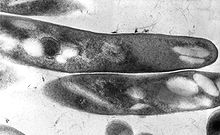
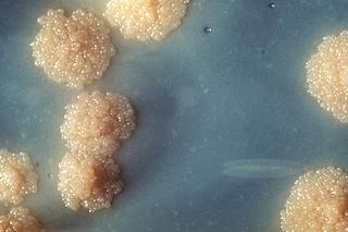
Mycobacterium tuberculosis, also known as Koch's bacillus, is a species of pathogenic bacteria in the family Mycobacteriaceae and the causative agent of tuberculosis. First discovered in 1882 by Robert Koch, M. tuberculosis has an unusual, waxy coating on its cell surface primarily due to the presence of mycolic acid. This coating makes the cells impervious to Gram staining, and as a result, M. tuberculosis can appear weakly Gram-positive. Acid-fast stains such as Ziehl–Neelsen, or fluorescent stains such as auramine are used instead to identify M. tuberculosis with a microscope. The physiology of M. tuberculosis is highly aerobic and requires high levels of oxygen. Primarily a pathogen of the mammalian respiratory system, it infects the lungs. The most frequently used diagnostic methods for tuberculosis are the tuberculin skin test, acid-fast stain, culture, and polymerase chain reaction.

<i>Mycobacterium leprae</i> Bacterium that causes leprosy

Mycobacterium leprae is one of the two species of bacteria that cause Hansen's disease (leprosy), a chronic but curable infectious disease that damages the peripheral nerves and targets the skin, eyes, nose, and muscles.

<span class="mw-page-title-main">Isoniazid</span> Antibiotic for treatment of tuberculosis

Isoniazid, also known as isonicotinic acid hydrazide (INH), is an antibiotic used for the treatment of tuberculosis. For active tuberculosis, it is often used together with rifampicin, pyrazinamide, and either streptomycin or ethambutol. For latent tuberculosis, it is often used alone. It may also be used for atypical types of mycobacteria, such as M. avium, M. kansasii, and M. xenopi. It is usually taken by mouth, but may be used by injection into muscle.

Nontuberculous mycobacteria (NTM), also known as environmental mycobacteria, atypical mycobacteria and mycobacteria other than tuberculosis (MOTT), are mycobacteria which do not cause tuberculosis or leprosy/Hansen's disease. NTM are able to cause pulmonary diseases that resemble tuberculosis. Mycobacteriosis is any of these illnesses, usually meant to exclude tuberculosis. They occur in many animals, including humans and are commonly found in soil and water.

<span class="mw-page-title-main">Ziehl–Neelsen stain</span> Bacteriological technique

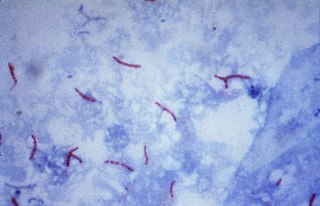
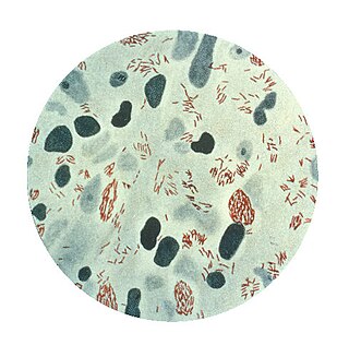
The Ziehl-Neelsen stain, also known as the acid-fast stain, is a bacteriological staining technique used in cytopathology and microbiology to identify acid-fast bacteria under microscopy, particularly members of the Mycobacterium genus. This staining method was initially introduced by Paul Ehrlich (1854–1915) and subsequently modified by the German bacteriologists Franz Ziehl (1859–1926) and Friedrich Neelsen (1854–1898) during the late 19th century.

<i>Mycobacterium smegmatis</i> Species of bacterium

Mycobacterium smegmatis is an acid-fast bacterial species in the phylum Actinomycetota and the genus Mycobacterium. It is 3.0 to 5.0 μm long with a bacillus shape and can be stained by Ziehl–Neelsen method and the auramine-rhodamine fluorescent method. It was first reported in November 1884 by Lustgarten, who found a bacillus with the staining appearance of tubercle bacilli in syphilitic chancres. Subsequent to this, Alvarez and Tavel found organisms similar to that described by Lustgarten also in normal genital secretions (smegma). This organism was later named M. smegmatis.

<i>Mycobacterium avium-intracellulare</i> infection Medical condition

Mycobacterium avium-intracellulare infection (MAI) is an atypical mycobacterial infection, i.e. one with nontuberculous mycobacteria or NTM, caused by Mycobacterium avium complex (MAC), which is made of two Mycobacterium species, M. avium and M. intracellulare. This infection causes respiratory illness in birds, pigs, and humans, especially in immunocompromised people. In the later stages of AIDS, it can be very severe. It usually first presents as a persistent cough. It is typically treated with a series of three antibiotics for a period of at least six months.

<span class="mw-page-title-main">Ethionamide</span> Chemical compound

Ethionamide is an antibiotic used to treat tuberculosis. Specifically it is used, along with other antituberculosis medications, to treat active multidrug-resistant tuberculosis. It is no longer recommended for leprosy. It is taken by mouth.

<span class="mw-page-title-main">Mycobacteriophage</span> Virus infecting mycobacteria

A mycobacteriophage is a member of a group of bacteriophages known to have mycobacteria as host bacterial species. While originally isolated from the bacterial species Mycobacterium smegmatis and Mycobacterium tuberculosis, the causative agent of tuberculosis, more than 4,200 mycobacteriophage have since been isolated from various environmental and clinical sources. 2,042 have been completely sequenced. Mycobacteriophages have served as examples of viral lysogeny and of the divergent morphology and genetic arrangement characteristic of many phage types.

The Timpe and Runyon classification of nontuberculous mycobacteria based on the rate of growth, production of yellow pigment and whether this pigment was produced in the dark or only after exposure to light.

<i>Mycobacteroides abscessus</i> Species of bacterium

Mycobacteroides abscessus is a species of rapidly growing, multidrug-resistant, nontuberculous mycobacteria (NTM) that is a common soil and water contaminant. Although M. abscessus most commonly causes chronic lung infection and skin and soft tissue infection (SSTI), it can also cause infection in almost all human organs, mostly in patients with suppressed immune systems. Amongst NTM species responsible for disease, infection caused by M. abscessus complex are more difficult to treat due to antimicrobial drug resistance.

<i>Mycobacterium fortuitum</i> Species of bacterium

Mycobacterium fortuitum is a nontuberculous species of the phylum Actinomycetota, belonging to the genus Mycobacterium.

Mycobacterium gordonae is a species of Mycobacterium named for Ruth E. Gordon. It is a species of the phylum Actinomycetota, belonging to the genus Mycobacterium.

Mycobacterium avium complex is a group of mycobacteria comprising Mycobacterium intracellulare and Mycobacterium avium that are commonly grouped because they infect humans together; this group, in turn, is part of the group of nontuberculous mycobacteria. These bacteria cause Mycobacterium avium-intracellulare infections or Mycobacterium avium complex infections in humans. These bacteria are common and are found in fresh and salt water, in household dust and in soil. MAC bacteria usually cause infection in those who are immunocompromised or those with severe lung disease.

<i>Mycobacterium kansasii</i> Species of bacterium

Mycobacterium kansasii is a bacterium in the Mycobacterium genus. It is an environmental bacteria that causes opportunistic infections in humans, and is one of the leading mycobacterial causes of human disease after tuberculosis and leprosy.

<span class="mw-page-title-main">Cord factor</span> Chemical compound

Cord factor, or trehalose dimycolate (TDM), is a glycolipid molecule found in the cell wall of Mycobacterium tuberculosis and similar species. It is the primary lipid found on the exterior of M. tuberculosis cells. Cord factor influences the arrangement of M. tuberculosis cells into long and slender formations, giving its name. Cord factor is virulent towards mammalian cells and critical for survival of M. tuberculosis in hosts, but not outside of hosts. Cord factor has been observed to influence immune responses, induce the formation of granulomas, and inhibit tumor growth. The antimycobacterial drug SQ109 is thought to inhibit TDM production levels and in this way disrupts its cell wall assembly.

Mycobacterium scrofulaceum is a species of Mycobacterium.

Mycobacterium lepromatosis is an aerobic, acid-fast bacillus (AFB), and the second known causative agent of Hansen's disease (leprosy). It was discovered in 2008. Analysis of the 16S rRNA gene confirms that the species is distinct from Mycobacterium leprae.

<span class="mw-page-title-main">Lung cavity</span> Medical condition

A lung cavity or pulmonary cavity is an abnormal, thick-walled, air-filled space within the lung. Cavities in the lung can be caused by infections, cancer, autoimmune conditions, trauma, congenital defects, or pulmonary embolism. The most common cause of a single lung cavity is lung cancer. Bacterial, mycobacterial, and fungal infections are common causes of lung cavities. Globally, tuberculosis is likely the most common infectious cause of lung cavities. Less commonly, parasitic infections can cause cavities. Viral infections almost never cause cavities. The terms cavity and cyst are frequently used interchangeably; however, a cavity is thick walled, while a cyst is thin walled. The distinction is important because cystic lesions are unlikely to be cancer, while cavitary lesions are often caused by cancer.

Mycobacteroides franklinii is a species of bacteria from the phylum Actinomycetota belonging to the genus Mycobacteroides. Most of the original strains were isolated from clinical specimens in Pennsylvania, but some have been found in conduit water in the Netherlands. In general, human M. franklinii infections present with symptoms similar to an infection with Mycobacteroides abscessus, but it can also be associated with tattoo infections. M. franklinii is also associated with outbreaks of mycobacteriosis in farmed fish. M. fanklinii is susceptible to cefoxitin and bedaquiline.

References

  1. Lehmann KB, Neumann R (1896). Atlas und Grundriss der Bakteriologie und Lehrbuch der speziellen bakteriologischen Diagnostik[Atlas and Outline of Bacteriology and Textbook of Special Bacteriological Diagnostics] (1st ed.). München: J.F. Lehmann.
  2. Euzéby JP, Parte AC. "Mycobacterium". List of Prokaryotic names with Standing in Nomenclature (LPSN). Retrieved June 16, 2021.[ permanent dead link ]
  3. 1 2 Mycobacteria: Health Advisory (PDF). Environmental Protection Agency (Report). August 1999.
  4. Batt SM, Minnikin DE, Besra GS (May 2020). "The thick waxy coat of mycobacteria, a protective layer against antibiotics and the host's immune system". The Biochemical Journal. 477 (10): 1983–2006. doi:10.1042/BCJ20200194. PMC   7261415 . PMID   32470138.
  5. 1 2 Pennington KM, Vu A, Challener D, Rivera CG, Shweta FN, Zeuli JD, Temesgen Z (August 2021). "Approach to the diagnosis and treatment of non-tuberculous mycobacterial disease". Journal of Clinical Tuberculosis and Other Mycobacterial Diseases. 24: 100244. doi:10.1016/j.jctube.2021.100244. PMC   8135042 . PMID   34036184.
  6. 1 2 3 Forbes BA, Hall GS, Miller MB, Novak SM, Rowlinson MC, Salfinger M, et al. (April 2018). "Practical Guidance for Clinical Microbiology Laboratories: Mycobacteria". Clinical Microbiology Reviews. 31 (2): e00038–17. doi:10.1128/CMR.00038-17. PMC   5967691 . PMID   29386234.
  7. 1 2 Dulberger CL, Rubin EJ, Boutte CC (January 2020). "The mycobacterial cell envelope - a moving target". Nature Reviews. Microbiology. 18 (1): 47–59. doi:10.1038/s41579-019-0273-7. PMID   31728063. S2CID   208020338.
  8. Stamm LM, Morisaki JH, Gao LY, Jeng RL, McDonald KL, Roth R, et al. (November 2003). "Mycobacterium marinum escapes from phagosomes and is propelled by actin-based motility". The Journal of Experimental Medicine. 198 (9): 1361–1368. doi:10.1084/jem.20031072. PMC   2194249 . PMID   14597736.
  9. Ghosh J, Larsson P, Singh B, Pettersson BM, Islam NM, Sarkar SN, et al. (June 2009). "Sporulation in mycobacteria". Proceedings of the National Academy of Sciences of the United States of America. 106 (26): 10781–10786. Bibcode:2009PNAS..10610781G. doi: 10.1073/pnas.0904104106 . PMC   2705590 . PMID   19541637.
  10. Traag BA, Driks A, Stragier P, Bitter W, Broussard G, Hatfull G, et al. (January 2010). "Do mycobacteria produce endospores?". Proceedings of the National Academy of Sciences of the United States of America. 107 (2): 878–881. Bibcode:2010PNAS..107..878T. doi: 10.1073/pnas.0911299107 . PMC   2818926 . PMID   20080769.
  11. Bhamidi S (2009). "Mycobacterial Cell Wall Arabinogalactan". Bacterial Polysaccharides: Current Innovations and Future Trends. Caister Academic Press. ISBN   978-1-904455-45-5.
  12. Lagier JC, Edouard S, Pagnier I, Mediannikov O, Drancourt M, Raoult D (January 2015). "Current and past strategies for bacterial culture in clinical microbiology". Clinical Microbiology Reviews. 28 (1): 208–236. doi:10.1128/CMR.00110-14. PMC   4284306 . PMID   25567228.
  13. Shepard CC, Mcrae DH (February 1965). "Mycobacterium leprae in Mice: Minimal Infectious Dose, Relationship Between Staining Quality and Infectivity, and Effect of Cortisone". Journal of Bacteriology. 89 (2): 365–372. doi:10.1128/jb.89.2.365-372.1965. PMC   305516 . PMID   14255702.
  14. Vaerewijck MJ, Huys G, Palomino JC, Swings J, Portaels F (November 2005). "Mycobacteria in drinking water distribution systems: ecology and significance for human health". FEMS Microbiology Reviews. 29 (5): 911–934. doi: 10.1016/j.femsre.2005.02.001 . PMID   16219512.
  15. "JGI GOLD | Projects". gold.jgi.doe.gov. Retrieved 2023-05-13.
  16. Croce O, Robert C, Raoult D, Drancourt M (May 2014). "Draft Genome Sequence of Mycobacterium vulneris DSM 45247T". Genome Announcements. 2 (3). doi:10.1128/genomeA.00370-14. PMC   4014686 . PMID   24812218.
  17. "UniProt". www.uniprot.org. Retrieved 2023-05-07.
  18. "UniProt". www.uniprot.org. Retrieved 2023-05-07.
  19. 1 2 "UniProt". www.uniprot.org. Retrieved 2023-05-07.
  20. "UniProt". www.uniprot.org. Retrieved 2023-05-07.
  21. "UniProt". www.uniprot.org. Retrieved 2023-05-07.
  22. Getahun H, Matteelli A, Chaisson RE, Raviglione M (May 2015). "Latent Mycobacterium tuberculosis infection". The New England Journal of Medicine. 372 (22): 2127–2135. doi:10.1056/NEJMra1405427. PMID   26017823.
  23. Forrellad MA, Klepp LI, Gioffré A, Sabio y García J, Morbidoni HR, de la Paz Santangelo M, et al. (January 2013). "Virulence factors of the Mycobacterium tuberculosis complex". Virulence. 4 (1): 3–66. doi:10.4161/viru.22329. PMC   3544749 . PMID   23076359.
  24. Riojas, Marco A.; McGough, Katya J.; Rider-Riojas, Cristin J.; Rastogi, Nalin; Hazbón, Manzour Hernando (1 January 2018). "Phylogenomic analysis of the species of the Mycobacterium tuberculosis complex demonstrates that Mycobacterium africanum, Mycobacterium bovis, Mycobacterium caprae, Mycobacterium microti and Mycobacterium pinnipedii are later heterotypic synonyms of Mycobacterium tuberculosis". International Journal of Systematic and Evolutionary Microbiology. 68 (1): 324–332. doi: 10.1099/ijsem.0.002507 . PMID   29205127.
  25. Sugawara-Mikami M, Tanigawa K, Kawashima A, Kiriya M, Nakamura Y, Fujiwara Y, Suzuki K (December 2022). "Pathogenicity and virulence of Mycobacterium leprae". Virulence. 13 (1): 1985–2011. doi:10.1080/21505594.2022.2141987. PMC   9635560 . PMID   36326715.
  26. "Nontuberculous Mycobacteria (NTM) Infections | HAI | CDC". www.cdc.gov. 2019-08-12. Retrieved 2023-04-28.
  27. 1 2 To K, Cao R, Yegiazaryan A, Owens J, Venketaraman V (August 2020). "General Overview of Nontuberculous Mycobacteria Opportunistic Pathogens: Mycobacterium avium and Mycobacterium abscessus". Journal of Clinical Medicine. 9 (8): 2541. doi: 10.3390/jcm9082541 . PMC   7463534 . PMID   32781595.
  28. "Mycobacteria: Health Advisory EPA-822-B-01-007" (PDF). epa.gov. US Environmental Protection Agency (EPA). August 1999. p. 2. Retrieved 10 March 2023.
  29. "Fact Sheets | General | Tuberculosis: General Information | TB | CDC". www.cdc.gov. 2022-08-17. Retrieved 2023-04-25.
  30. "Diagnosing and Treating Tuberculosis". American Lung Association. Retrieved 2023-04-25.
  31. "Global Tuberculosis Report 2022". www.who.int. Retrieved 2023-05-07.
  32. 1 2 "Signs and Symptoms | Hansen's Disease (Leprosy) | CDC". www.cdc.gov. 2018-10-22. Retrieved 2023-04-25.
  33. "Global leprosy (Hansen disease) update, 2021: moving towards interruption of transmission". www.who.int. Retrieved 2023-05-07.
  34. "Mycobacterium abscessus in Healthcare Settings | HAI | CDC". www.cdc.gov. 2021-11-15. Retrieved 2023-04-27.
  35. Weng YW, Huang CK, Sy CL, Wu KS, Tsai HC, Lee SS (June 2020). "Treatment for Mycobacterium abscessus complex-lung disease". Journal of the Formosan Medical Association = Taiwan Yi Zhi. Consensus Statement of Nontuberculous Mycobacterial Lung Disease in Taiwan. 119 (Suppl 1): S58–S66. doi: 10.1016/j.jfma.2020.05.028 . PMID   32527504. S2CID   219604813.
  36. Scholar E (2007-01-01), Enna SJ, Bylund DB (eds.), "Mycobacterium Avium-Intracellulare Infections", xPharm: The Comprehensive Pharmacology Reference, New York: Elsevier, pp. 1–5, doi:10.1016/b978-008055232-3.60891-8, ISBN   978-0-08-055232-3 , retrieved 2023-05-07
  37. 1 2 Huang YC, Liu MF, Shen GH, Lin CF, Kao CC, Liu PY, Shi ZY (October 2010). "Clinical outcome of Mycobacterium abscessus infection and antimicrobial susceptibility testing". Journal of Microbiology, Immunology, and Infection = Wei Mian Yu Gan Ran Za Zhi. 43 (5): 401–406. doi:10.1016/S1684-1182(10)60063-1. PMID   21075707.
  38. Tortoli E (2019-01-10). "Chapter 1 - The Taxonomy of the Genus Mycobacterium". In Velayati AA, Farnia P (eds.). Nontuberculous Mycobacteria (NTM). Academic Press. pp. 1–10. doi:10.1016/B978-0-12-814692-7.00001-2. ISBN   978-0-12-814692-7. S2CID   92810288.
  39. Tortoli E, Fedrizzi T, Meehan CJ, Trovato A, Grottola A, Giacobazzi E, et al. (December 2017). "The new phylogeny of the genus Mycobacterium: The old and the news". Infection, Genetics and Evolution. 56: 19–25. doi:10.1016/j.meegid.2017.10.013. PMID   29030295.
  40. Gupta RS, Lo B, Son J (2018). "Phylogenomics and Comparative Genomic Studies Robustly Support Division of the Genus Mycobacterium into an Emended Genus Mycobacterium and Four Novel Genera". Frontiers in Microbiology. 9: 67. doi: 10.3389/fmicb.2018.00067 . PMC   5819568 . PMID   29497402.
  41. Tortoli E, Brown-Elliott BA, Chalmers JD, Cirillo DM, Daley CL, Emler S, et al. (July 2019). "Same meat, different gravy: ignore the new names of mycobacteria". The European Respiratory Journal. 54 (1): 1900795. doi: 10.1183/13993003.00795-2019 . PMID   31296783.
  42. Ogwang S, Mubiri P, Bark CM, Joloba ML, Boom WH, Johnson JL (October 2015). "Incubation time of Mycobacterium tuberculosis complex sputum cultures in BACTEC MGIT 960: 4weeks of negative culture is enough for physicians to consider alternative diagnoses". Diagnostic Microbiology and Infectious Disease. 83 (2): 162–164. doi:10.1016/j.diagmicrobio.2015.07.002. PMC   4573350 . PMID   26239846.
  43. Azimi T, Mosadegh M, Nasiri MJ, Sabour S, Karimaei S, Nasser A (2019-09-17). "Phage therapy as a renewed therapeutic approach to mycobacterial infections: a comprehensive review". Infection and Drug Resistance. 12: 2943–2959. doi: 10.2147/IDR.S218638 . PMC   6756577 . PMID   31571947.
  44. Smith DW, Randall HM, Maclennan AP, Lederer E (June 1960). "Mycosides: a new class of type-specific glycolipids of Mycobacteria". Nature. 186 (4728): 887–888. Bibcode:1960Natur.186..887S. doi:10.1038/186887a0. PMID   13831939. S2CID   4149360.
  45. Goren MB, McClatchy JK, Martens B, Brokl O (June 1972). "Mycosides C: behavior as receptor site substance for mycobacteriophage D4". Journal of Virology. 9 (6): 999–1003. doi:10.1128/jvi.9.6.999-1003.1972. PMC   356406 . PMID   4113889.
  46. Azad AK, Sirakova TD, Rogers LM, Kolattukudy PE (May 1996). "Targeted replacement of the mycocerosic acid synthase gene in Mycobacterium bovis BCG produces a mutant that lacks mycosides". Proceedings of the National Academy of Sciences of the United States of America. 93 (10): 4787–4792. Bibcode:1996PNAS...93.4787A. doi: 10.1073/pnas.93.10.4787 . PMC   39357 . PMID   8643481.