Neoscytalidium dimidiatum

Last updated

Neoscytalidium dimidiatum
Neoscytalidium dimidiatum microscopic.jpg
Scientific classification
Kingdom:
Division:
Class:
Order:
Family:
Genus:
Species:
N. dimidiatum
Binomial name
Neoscytalidium dimidiatum
(Penz.) Crous & Slippers (2006) [1] [2]
Synonyms
  • Torula dimidiataPenz. (1882) [1]
  • Hendersonula toruloideaNattrass (1933) [1]
  • Scytalidium dimidiatum(Penz.) B. Sutton & Dyko (1989) [1]
  • Fusicoccum dimidiatum(Penz.) D.F. Farr (2005) [1]

Neoscytalidium dimidiatum was first described in 1933 as Hendersonula toruloidea from diseased orchard trees in Egypt. [3] Decades later, it was determined to be a causative agent of human dermatomycosis-like infections and foot infections predominantly in tropical areas; however the fungus is considered to be widespread. [3] A newer name, Scytalidium dimidiatum, was applied to a synanamorph of Nattrassia mangiferae, [4] otherwise known as Neofusicoccum mangiferae. Substantial confusion has arisen in the literature on this fungus resulting from the use of multiple different names including Torula dimidiata, Fusicoccum dimidiatum, [1] Scytalidium dimidiatum, and Hendersonula toruloidea. [5] Additionally, Scytalidium lignicola and Scytalidium lignicolum are often considered earlier names of N. dimidiatum. [6]

Contents

History and taxonomy

In 1933, British mycologist Dr. Rolland Marshall Nattrass described an arthroconidial asexual fungus that he named H. toruloidea that was responsible for causing die-back disease of plum, apricot and apple trees in Egypt. [3] At the time, he recognized that single spore cultures of the fungus yielded two "forms" in culture – a mycelial form resembling members of the genus Torula that produced fragmenting chains of arthroconidia, and a pycnidial form characterized by the production of greenish, ellipsoidal spores that oozed from tiny sacs. [7] The name H. toruloidea applied to the latter pycnidial form. [7] Others likened the Torula form to Torula dimidiata described by Otto Penzig in 1882.[ citation needed ] Despite that the fungus was known by this name for over 50 years by one or the other of these names, increased scrutiny of the species and its close relatives using molecular genetic methods spawned significant controversy in its taxonomy and naming. [3] [5]

In 1989 Sutton and Dyko created the genus Nattrassia to accommodate H. toruloidea and applied the name Scytalidium dimidiatum to the mycelial synanamorph. [3] They also included in the new genus Nattrassia a fungus described by father-son mycologists Paul and Hans Sydow as Dothiorella mangiferae, which became Nattrassia mangiferae, thought to be very closely related to and perhaps indistinguishable from Nattrass's original pycnidial form. Farr and coworkers recognized that both states were asexual forms affiliated with the genus Fusicoccum, an anamorph of the plant pathogenic ascomycete genus, Botryosphaeria. [3] They proposed the transfer of Scytalidium dimidiatum to the genus Fusicoccum as F. dimidiatum. [3] A reappraisal of the family Botryosphaeriaceae by Crous and coworkers in 2006 concluded that the genus Fusicoccum was polyphyletic, and they created a new genus, Neoscytalidium to accommodate Nattrass's fungus. Separately they erected the genus Neofusicoccum to accommodate Nattrassia mangiferae. [3] [5] Crous and colleagues concluded it inappropriate to collapse the entirety of Scytalidium with Fusicoccum because they demonstrated N. dimidiatum to be phylogenetically distinct from Neofusicoccum mangiferae; thus, they interpreted N. dimidiatum to be the correct name for Nattrass's fungus. [3]

Growth and morphology

UAMH 10614 Neoscytalidium dimidiatum on CER agar incubated at 35 C for 29 days. Neoscytalidium dimidiatum colony.jpg
UAMH 10614 Neoscytalidium dimidiatum on CER agar incubated at 35 C for 29 days.

This filamentous fungus [3] produces sinuous and irregular hyphae and is characterized by rapidly growing colonies that are deeply tufted with dense, darkly coloured, ropy aerial mycelium. [5] [8] [9] Cultures are rapidly growing, initially light in colour and becoming dark brown and then black with age. [8] Both arthroconidia and pycnidia may be produced in the same culture. [5] Cultures of the fungus derived from human skin tend to be black in colouration. [5]

Habitat and ecology

Neoscytalidium dimidiatum is mainly found in tropical to subtropical environments, [10] such as in South America, Southeast Asia, India and Africa. [3] [5] In addition to these regions, this fungus is endemic to parts of west and central Africa, the Caribbean and Asian, [5] but more cases are being seen in temperate countries, possibly as a consequence of immigration from tropical regions. [11] The fungus occurs in nature in soil and on decaying wood. [10]

Human infection

Neoscytalidium dimidiatum has been describe as an agent to cause infections referred to as dermatomycosis, onychomycosis, [9] ringworm or tinea, [11] affecting human nails, toe webs and feet, and skin, [3] [10] [12] forming hyphomycete, [12] and also sometimes infecting the palms of hands but this is a rare occurrence. [3] To cause these infections in humans, infections occurs through contact with contaminated soil or plant materials, or nail or skin tissue from an infected person, [5] causing superficial skin infections similar to dermatophytosis called Scytalidiosis. [3] Although established to cause dermatomycosis and onychomycosis, invasive infection by N. dimidiatum is rare, resulting in limited case reports and limited information available for clinical progression and treatment. [9] as well as there is no currently know oral or topical treatment for infection with this fungus. [11] These result in the invasion of tissue and organs causing systemic diseases. [13] Infection can occur in both immunocompetent and immunosuppressed patients, [13] but deep infections occur mainly in immunosuppressed/ immunocompromised individuals with a 50% case mortality. [3] Melanin is characteristically produced by the fungus in vivo in diseased human tissue where its presence has been interpreted as an important pathogenic factor. [14] Although limited in case reports, there have been some reported cases of N. dimidiatum causing dermatomycosis or onychomycosis in places such as Jamaica, Brazil, Algeria, Canada, the United States and the United Kingdom. [15] In addition, there have been some papers reporting infections besides dermatomycosis and onychomycosis, such as discovery of this fungus confirmed by DNA analysis to cause a fatal case of lung disease. [9] Similarly, a case of lung infection has been reported in a dolphin. [16] A case of rhinosinusitis, a disease where it is believed that fungi play a role in the disease process, was reported to have been caused by N. dimidiatum. [17]

Plant disease

Neoscytalidium dimidiatum has been mainly described as an opportunistic [18] plant pathogen, [14] causing pit canker and spot on the stem of plants or fruits, as well as internal black rot of fruits, [18] but a case of internal brown rot of pitahaya was reported from China. [18] Similarly in Malaysia was a report of the fungus causing stem canker of red-fleshed dragon fruit, [19] and a similar report of stem canker on grapevine in California. [20]

Related Research Articles

<i>Fusarium</i> Genus of fungi

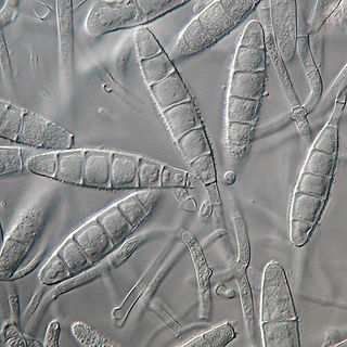
Fusarium is a large genus of filamentous fungi, part of a group often referred to as hyphomycetes, widely distributed in soil and associated with plants. Most species are harmless saprobes, and are relatively abundant members of the soil microbial community. Some species produce mycotoxins in cereal crops that can affect human and animal health if they enter the food chain. The main toxins produced by these Fusarium species are fumonisins and trichothecenes. Despite most species apparently being harmless, some Fusarium species and subspecific groups are among the most important fungal pathogens of plants and animals.

<span class="mw-page-title-main">Onychomycosis</span> Medical condition

Onychomycosis, also known as tinea unguium, is a fungal infection of the nail. Symptoms may include white or yellow nail discoloration, thickening of the nail, and separation of the nail from the nail bed. Fingernails may be affected, but it is more common for toenails. Complications may include cellulitis of the lower leg. A number of different types of fungus can cause onychomycosis, including dermatophytes and Fusarium. Risk factors include athlete's foot, other nail diseases, exposure to someone with the condition, peripheral vascular disease, and poor immune function. The diagnosis is generally suspected based on the appearance and confirmed by laboratory testing.

<i>Basidiobolus ranarum</i> Species of fungus

Basidiobolus ranarum is a filamentous fungus with worldwide distribution. The fungus was first isolated by Eidam in 1886. It can saprophytically live in the intestines of mainly cold-blooded vertebrates and on decaying fruits and soil. The fungus prefers glucose as a carbon source and grows rapidly at room temperature. Basidiobolus ranarum is also known as a cause of subcutaneous zygomycosis, usually causing granulomatous infections on a host's limbs. Infections are generally geographically limited to tropical and subtropical regions such as East and West Africa. Subcutaneous zygomycosis caused by B. ranarum is a rare disease and predominantly affects children and males. Common subcutaneous zygomycosis shows characteristic features and is relatively easy to be diagnosed; while, certain rare cases might show non-specific clinical features that might pose a difficulty on its identification. Although disease caused by this fungus is known to resolve spontaneously on its own, there are a number of treatments available.

<i>Trichophyton rubrum</i> Species of fungus

Trichophyton rubrum is a dermatophytic fungus in the phylum Ascomycota. It is an exclusively clonal, anthropophilic saprotroph that colonizes the upper layers of dead skin, and is the most common cause of athlete's foot, fungal infection of nail, jock itch, and ringworm worldwide. Trichophyton rubrum was first described by Malmsten in 1845 and is currently considered to be a complex of species that comprises multiple, geographically patterned morphotypes, several of which have been formally described as distinct taxa, including T. raubitschekii, T. gourvilii, T. megninii and T. soudanense.

<i>Botryosphaeria dothidea</i> Species of fungus

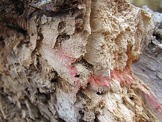
Botryosphaeria dothidea is a plant pathogen that causes the formation of cankers on a wide variety of tree and shrub species. It has been reported on several hundred plant hosts and on all continents except Antarctica. B. dothidea was redefined in 2004, and some reports of its host range from prior to that time likely include species that have since been placed in another genus. Even so, B. dothidea has since been identified on a number of woody plants—including grape, mango, olive, eucalyptus, maple, and oak, among others—and is still expected to have a broad geographical distribution. While it is best known as a pathogen, the species has also been identified as an endophyte, existing in association with plant tissues on which disease symptoms were not observed. It can colonize some fruits, in addition to woody tissues.

Exophiala jeanselmei is a saprotrophic fungus in the family Herpotrichiellaceae. Four varieties have been discovered: Exophiala jeanselmei var. heteromorpha, E. jeanselmei var. lecanii-corni, E. jeanselmei var. jeanselmei, and E. jeanselmei var. castellanii. Other species in the genus Exophiala such as E. dermatitidis and E. spinifera have been reported to have similar annellidic conidiogenesis and may therefore be difficult to differentiate.

Pathogenic fungi are fungi that cause disease in humans or other organisms. Although fungi are eukaryotic, many pathogenic fungi are microorganisms. Approximately 300 fungi are known to be pathogenic to humans; their study is called "medical mycology". Fungal infections kill more people than either tuberculosis or malaria—about 2 million people per year.

<i>Microsporum gypseum</i> Species of fungus

Microsporum gypseum is a soil-associated dermatophyte that occasionally is known to colonise and infect the upper dead layers of the skin of mammals. The name refers to an asexual "form-taxon" that has been associated with four related biological species of fungi: the pathogenic taxa Arthroderma incurvatum, A. gypsea, A. fulva and the non-pathogenic saprotroph A. corniculata. More recent studies have restricted M. gypseum to two teleomorphic species A. gypseum and A. incurvatum. The conidial states of A. fulva and A. corniculata have been assigned to M. fulvum and M. boullardii. Because the anamorphic states of these fungi are so similar, they can be identified reliably only by mating. Two mating strains have been discovered, "+" and "–". The classification of this species has been based on the characteristically rough-walled, blunt, club-shaped, multicelled macroconidia. Synonyms include Achorion gypseum, Microsporum flavescens, M. scorteum, and M. xanthodes. There has been past nomenclatural confusion in the usage of the generic names Microsporum and Microsporon.

<i>Scytalidium</i> Genus of fungi

Scytalidium is a genus of fungi in the Helotiales order. The relationship of this taxon to other taxa within the order is unknown, and it has not yet been placed with certainty into any family. This genus of anamorphic fungi has a widespread distribution and contains 18 species. Scytalidium dimidiatum causes onychomycosis in tea leaf pluckers.

<i>Cladophialophora bantiana</i> Species of fungus

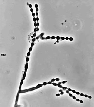
Cladophialophora bantiana is a melanin producing mold known to cause brain abscesses in humans. It is one of the most common causes of systemic phaeohyphomycosis in mammals. Cladophialophora bantiana is a member of the ascomycota and has been isolated from soil samples from around the world.

<i>Onychocola canadensis</i> Species of fungus

Onychocola canadensis is a relative of the dermatophyte and an occasionally causes onychomycosis. It was described in 1990 from 3 clinical reports in Canada.

Coniochaeta hoffmannii, also known as Lecythophora hoffmannii, is an ascomycete fungus that grows commonly in soil. It has also been categorized as a soft-rot fungus capable of bringing the surface layer of timber into a state of decay, even when safeguarded with preservatives. Additionally, it has pathogenic properties, although it causes serious infection only in rare cases. A plant pathogen lacking a known sexual state, C. hoffmannii has been classified as a "dematiaceous fungus" despite its contradictory lack of pigmentation; both in vivo and in vitro, there is no correlation between its appearance and its classification.

Lasiodiplodia citricola is an endophytic fungus. It was first isolated in northern Iran, and is named after its first known host, citrus plants. It has since been isolated in other plants in other continents, and is considered a plant pathogen. L. citricola is phylogenetically related to L. parva, but conidia of the former are longer and wider.

<i>Rhinocladiella mackenziei</i> Species of fungus

Rhinocladiella mackenziei is a deeply pigmented mold that is a common cause of human cerebral phaeohyphomycosis. Rhinocladiella mackenziei was believed to be endemic solely to the Middle East, due to the first cases of infection being limited to the region. However, cases of R. mackenziei infection are increasingly reported from regions outside the Middle East. This pathogen is unique in that the majority of cases have been reported from immunologically normal people.

<i>Cladophialophora carrionii</i> Species of fungus

Cladophialophora carrionii is a melanized fungus in the genus Cladophialophora that is associated with decaying plant material like cacti and wood. It is one of the most frequent species of Cladophialophora implicated in human disease. Cladophialophora carrionii is a causative agent of chromoblastomycosis, a subcutaneous infection that occurs in sub-tropical areas such as Madagascar, Australia and northwestern Venezuela. Transmission occurs through traumatic implantation of plant material colonized by C. carrionii, mainly infecting rural workers. When C. carrionii infects its host, it transforms from a mycelial state to a muriform state to better tolerate the extreme conditions in the host's body.

<i>Epidermophyton floccosum</i> Species of fungus

Epidermophyton floccosum is a filamentous fungus that causes skin and nail infections in humans. This anthropophilic dermatophyte can lead to diseases such as tinea pedis, tinea cruris, tinea corporis and onychomycosis. Diagnostic approaches of the fungal infection include physical examination, culture testing, and molecular detection. Topical antifungal treatment, such as the use of terbinafine, itraconazole, voriconazole, and ketoconazole, is often effective.

Sarocladium kiliense is a saprobic fungus that is occasionally encountered as a opportunistic pathogen of humans, particularly immunocompromised and individuals. The fungus is frequently found in soil and has been linked with skin and systemic infections. This species is also known to cause disease in the green alga, Cladophora glomerata as well as various fruit and vegetable crops grown in warmer climates.

Arthrographis kalrae is an ascomycetous fungus responsible for human nail infections described in 1938 by Cochet as A. langeronii. A. kalrae is considered a weak pathogen of animals including human restricted to the outermost keratinized layers of tissue. Infections caused by this species are normally responsive to commonly used antifungal drugs with only very rare exceptions.

Neofusicoccum arbuti is a fungus species in the genus Neofusicoccum. It was first described by D.F. Farr & M. Elliott, and given its current name by Crous, Slippers & A.J.L. Phillips in 2006. Neofusicoccum arbuti is included in the genus Neofusicoccum and the family Botryosphaeriaceae. This species is known as madrone canker. N. arbuti is a potentially lethal canker disease of Pacific madrone, Arbutus menziesii.

Scytalidium hyalinum is an ascomycete fungus currently in the genus Scytalidium. It causes dermatomycosis and systemic infections in humans and it is widespread throughout the world.

References

  1. 1 2 3 4 5 6 "MycoBank Database". www.mycobank.org. Retrieved 9 October 2016.
  2. "Index Fungorum - Names Record". www.indexfungorum.org.
  3. 1 2 3 4 5 6 7 8 9 10 11 12 13 14 15 Machouart, M.; Menir, P.; Helenon, R.; Quist, D.; Desbois, N. (March 2013). "Scytalidium and scytalidiosis: What's new in 2012?". Journal de Mycologie Médicale / Journal of Medical Mycology. 23 (1): 40–46. doi:10.1016/j.mycmed.2013.01.002. PMID   23416171.
  4. Howard, Dexter H. (2003). Pathogenic fungi in humans and animals (2nd ed.). New York [u.a.]: Dekker. ISBN   978-0-8247-0683-8.
  5. 1 2 3 4 5 6 7 8 9 Tan, Darrell H. S.; Sigler, Lynne; Gibas, Connie F. C.; Fong, Ignatius W. (January 2008). "Disseminated fungal infection in a renal transplant recipient involving Macrophomina phaseolina and Scytalidium dimidiatum: case report and review of taxonomic changes among medically important members of the Botryosphaeriaceae". Medical Mycology. 46 (3): 285–292. doi: 10.1080/13693780701759658 . PMID   18404556.
  6. Onychomycosis caused by Scytalidium dimidiatum. Report of two cases. Review of the taxonomy of the synanamorph and anamorph forms of this coelomycete
  7. 1 2 Nattrass, Roland Marshall (1933). "A new species of Hendersonula (H. Toruloidea) on deciduous trees in Egypt". Transactions of the British Mycological Society. 18 (3): 189–198. doi:10.1016/S0007-1536(33)80030-1.
  8. 1 2 Madrid, H; Ruíz-Cendoya, M; Cano, J; Stchigel, A; Orofino, R; Guarro, J (October 2009). "Genotyping and in vitro antifungal susceptibility of Neoscytalidium dimidiatum isolates from different origins". International Journal of Antimicrobial Agents. 34 (4): 351–4. doi:10.1016/j.ijantimicag.2009.05.006. PMID   19570656.
  9. 1 2 3 4 Dionne, Brandon; Neff, Luke; Lee, Samuel A.; Sutton, Deanna A.; Wiederhold, Nathan P.; Lindner, Jonathan; Fan, Hongxin; Jakeman, Bernadette; Warnock, D. W. (July 2015). "Pulmonary Fungal Infection Caused by Neoscytalidium dimidiatum". Journal of Clinical Microbiology. 53 (7): 2381–2384. doi:10.1128/JCM.00206-15. PMC   4473236 . PMID   25948605.
  10. 1 2 3 Kwon-Chung, K.J; Bennett, J.E. (1992). Medical mycology. Philadelphia: Lea & Febiger. ISBN   978-0812114638.
  11. 1 2 3 Alshawa, Kinda; Beretti, Jean-Luc; Lacroix, Claire; Feuilhade, Martine; Dauphin, Brunhilde; Quesne, Gilles; Hassouni, Noura; Nassif, Xavier; Bougnoux, Marie-Elisabeth (1 July 2012). "Successful identification of clinical dermatophyte and Neoscytalidium species by matrix-assisted laser desorption ionization-time of flight mass spectrometry". Journal of Clinical Microbiology. 50 (7): 2277–2281. doi:10.1128/JCM.06634-11. ISSN   1098-660X. PMC   3405581 . PMID   22535981.
  12. 1 2 Rippon, John Willard (1982). Medical Mycology The Pathogenic Fungi and The Pathogenic Actinomycetes (Second ed.). Philadelphia, PA: W. B. Saunders Company. p.  241. ISBN   9780721675862.
  13. 1 2 da Silva, Roberta Teixeira; Guimarães, Dávson A.; Camargo, Zoilo P.; Rodrigues, Anderson M.; Maceira, Juan P.; Bernardes-Engemann, Andréa R.; Orofino-Costa, Rosane (1 November 2016). "Cutaneous murine model of infection caused by Neoscytalidium dimidiatum: a preliminary study of an emerging human pathogen". Medical Mycology. 54 (8): 890–898. doi: 10.1093/mmy/myw034 . ISSN   1460-2709. PMID   27250925.
  14. 1 2 Ruíz-Cendoya, M; Madrid, H; Pastor, FJ; Mayayo, E; Mariné, M; Guarro, J (August 2010). "Development of murine models of disseminated infection by Neoscytalidium dimidiatum". Medical Mycology. 48 (5): 681–6. doi: 10.3109/13693780903452917 . PMID   20055737.
  15. "UAMH Centre for Global Microfungal Biodiversity". www.uamh.ca.
  16. Morick, Danny; Alvarez, Mariano Domingo; Elad, Daniel (1 August 2011). "Pulmonary fungal infection caused by Neoscytalidium dimidiatum in a Risso's dolphin (Grampus griseus)--update". Medical Mycology. 49 (6): 672. doi: 10.3109/13693786.2011.567303 . ISSN   1460-2709. PMID   21410320.
  17. Bakhshizadeh, Mandana; Hashemian, Hamid Reza; Najafzadeh, Mohammad Javad; Dolatabadi, Somayeh; Zarrinfar, Hossein (1 July 2014). "First report of rhinosinusitis caused by Neoscytalidium dimidiatum in Iran". Journal of Medical Microbiology. 63 (Pt 7): 1017–1019. doi: 10.1099/jmm.0.065292-0 . ISSN   1473-5644. PMID   24850881.
  18. 1 2 3 Yi, Run Hua; Lin, Qiao ling; Mo, Jun Jie; Wu, Feng Fa; Chen, Jing (8 April 2015). "Fruit internal brown rot caused by Neoscytalidium dimidiatum on pitahaya in Guangdong province, China". Australasian Plant Disease Notes. 10 (1): 13. doi: 10.1007/s13314-015-0166-1 . ISSN   1833-928X.
  19. Mohd, Masratul Hawa; Salleh, Baharuddin; Zakaria, Latiffah (1 December 2013). "Identification and Molecular Characterizations of Neoscytalidium dimidiatum Causing Stem Canker of Red-fleshed Dragon Fruit (Hylocereus polyrhizus) in Malaysia". Journal of Phytopathology. 161 (11–12): 841–849. doi:10.1111/jph.12146. ISSN   1439-0434.
  20. Rolshausen, P. E.; Akgül, D. S.; Perez, R.; Eskalen, A.; Gispert, C. (12 June 2013). "First Report of Wood Canker Caused by Neoscytalidium dimidiatum on Grapevine in California". Plant Disease. 97 (11): 1511. doi:10.1094/PDIS-04-13-0451-PDN. ISSN   0191-2917. PMID   30708472.