Neuroferritinopathy

Last updated
Neuroferritinopathy
Other namesAdult-onset basal ganglia disease
Gray745.png
Cerebellum and basal ganglia
Specialty Neurology   OOjs UI icon edit-ltr-progressive.svg

Neuroferritinopathy is a genetic neurodegenerative disorder characterized by the accumulation of iron in the basal ganglia, cerebellum, and motor cortex of the human brain. Symptoms, which are extrapyramidal in nature, progress slowly and generally do not become apparent until adulthood. [1] These symptoms include chorea, dystonia, and cognitive deficits which worsen with age. [2] [3]

Contents

This disorder is autosomal dominant [4] and is caused by mutations in the gene encoding the light chain subunit of the ferritin protein. Wild type ferritin functions as a buffer for iron, sequestering it and controlling its release. Thus, mutations in the light chain of ferritin result in the accumulation of iron in the brain which can be imaged using MRI. [2] Currently, neuroferritinopathy is the only neurodegenerative disease with an iron accumulation in the brain classified as an autosomal dominant syndrome. [4]

Treatment of neuroferritinopathy is focused on managing symptoms associated with chorea and dystonia using standard medications for each. [2] The disorder is progressive and symptoms become worse with age. Fewer than 100 cases of neuroferritinopathy have been reported since its identification in 2001. [2] Its incidence has been largely localized to Northwest England, significantly in the Cumbria region [4] suggesting a founder effect. [2] Due to its genetic nature, current research is focused on therapeutic management of the symptoms caused by the disorder. [4]

Signs and symptoms

Neuroferritinopathy has several distinguishing signs and symptoms. These fall into two categories: diagnostic findings and physically visible symptoms.

Diagnostic findings

Symptoms categorized as medically tested and diagnosed include iron accumulation in the brain, basal ganglia cavitation, and neurodegeneration. [4] Patients who are diagnosed with neuroferritinopathy have abnormal iron accumulation in the brain within the neurons and glia of the striatum and cerebellar cortices. [3] Along with the accumulation of iron in the brain, neuroferritinopathy typically causes severe neuronal loss as well. [3] Secondary symptoms may also arise. It is possible that the initial iron accumulation will cause additional neuronal damage and neuronal death. [3] The damaged neurons may be replaced by other cells in an effort to reverse the neurodegeneration. These cells often have a higher iron content. The breakdown of the blood brain barrier may also occur due to the loss of neurons and will subsequently allow more iron to access the brain and accumulate over time. [3]

Neuroferritinopathy is mainly seen in those who have reached late adulthood and is generally seen to slowly progress throughout many decades in a lifetime with the mean age of onset being 39 years old. [1] A loss of cognition is generally only seen with late stages of the disease. [1] Diagnosed patients are seen to retain most of their cognitive functioning until the most progressive stages of the illness sets in. [1]

Physical symptoms

Symptoms categorized as physically visible symptoms include chorea, dystonia, spasticity, and rigidity, all physical symptoms of the body associated with movement disorders. [3] The symptoms accompanying neuroferritinopathy affecting movement are also progressive, becoming more generalized with time. [4] Usually during the first ten years of onset of the disease only one or two limbs are directly affected. [4] Distinctive symptoms of neuroferritinopathy are chorea, found in 50% of diagnosed patients, dystonia, found in 43% of patients, and parkinsonism, found in 7.5% of patients. [1] Full control of upper limbs on the body generally remains until late onset of the disease. [1] Over time, symptoms seen in a patient can change from one side of the body to the opposite side of the body, jumping from left to right or vice versa. [1] Another route that the physically visible symptoms have been observed to take is the appearance, disappearance, and then reappearance once more of specific symptoms. [1]

While these symptoms are the classic indicators of neuroferritinopathy, symptoms will vary from patient to patient. [1]

Causes

Neuroferritinopathy results from abnormal brain iron accumulation. This iron accumulation is due to mutations in the FTL polypeptide, which is responsible for encoding proteins involved in iron metabolism. Neuroferritinopathy is most commonly caused by a single insertion of the nucleotide adenine into the gene for L-chain ferritin which in turn, alters the carboxyl end of the entire protein chain. [3] However, exact location of the insertion in the exon varies by family. [4] Neuroferritinopathy may also be caused by the insertion of two extra nucleotide bases. The insertion of bases into the L-chain ferritin gene causes the chain to lengthen and alter the sequence of the amino acids found in the gene, also known as a frameshift mutation. [3]

These mutations result in decreased iron-binding ability. [1] The oxidative damage caused by increased iron leads to apoptosis, or programmed cell death. [1] Accumulation of iron in the brain is extremely dangerous as excess iron catalyzes the formation of free radicals, which have damaging effects to the brain. [1] The iron accumulation characteristic of neuroferritinopathy particularly affects the cerebellum, basal ganglia, and motor cortex regions of the brain. [1]

Mechanism

The protein ferritin functions to sequester and release iron, acting as an iron buffering system in cells. Iron is essential to brain function in oxygen transport and cellular metabolism for example. However, careful control of iron is important as increased brain iron levels catalyze the formation of free radicals that create oxidative molecules via the Fenton Reaction. [5] These oxidative molecules can cause oxidative brain damage. Iron that is bound to ferritin in nonreactive. [1]

Fenton Reaction

(1) Fe2+ + H2O2 → Fe3+ + HO• + OH

(2) Fe3+ + H2O2 → Fe2+ + HOO• + H+

The ferritin protein is made up of heavy chain (H) and light chain (L) subunits. In neuroferritinopathy, the gene encoding the light chain is mutated. Several different mutation variations have led to diagnosis as neuroferritinopathy; all of these mutations occur in the light chain. A mutated light chain is believed to inhibit ferritin's ability to effectively sequester and hold iron. Without control of iron, it is free to cause oxidative brain damage as described above. [6]

The concentration of iron in a healthy brain varies greatly from region to region. The specific regions of the brain that are associated with motor functions appear to have larger accumulations of iron than non-motor-related regions. This observation of varying iron concentrations is a possible explanation for the correlation between movement disorders and the iron imbalance within the central nervous system. [3]

Diagnosis

Neuroferritinopathy is primarily diagnosed in older adults, specifically in adults affected by Alzheimer's disease or Parkinson's disease, as iron accumulates in the brain over long periods of time. [3] Neuroferritinopathy is diagnosed using either neuroimaging techniques, physiological tests, or genetic testing.

Classification

Neuroferritinopathy was originally described with hallmark features of neurodegeneration and iron accumulation in the brain, leading it to be classified with other neurodegeneration with brain iron accumulation (NBIA) disorders which share similar symptoms and imaging findings. Over time single-gene causes have been found for many NBIA disorders, like neuroferritinopathy. Before the availability of genetic testing, all such disorders were considered together and known as Hallervorden-Spatz syndrome, a term which is no longer used due to the Nazi party ties of the namesakes. [1] Brain iron disorders are now divided into three categories: genetic neurodegeneration with brain iron accumulation, genetic systemic iron accumulation with neurologic features, and acquired diseases associated with iron excess or iron deficiency. Neuroferritinopathy is classified under the first category. [7] Neuroferritinopathy is classified as a late-onset basal ganglia disease and is a dominantly inherited neurodegenerative disease. [3] Four different alleles are responsible for neuroferritinopathy. Three arise from nucleotide insertions in the ferritin light chain (FTL) polypeptide gene while the fourth arises from a missense mutation in the FTL gene. [4]

Neuroimaging

MRI NPH MRI 274.gif
MRI

Neuroferritinopathy is most commonly diagnosed using MRI and other neuroimaging techniques. [1] MRIs help identify the iron deposits in the cerebellum, basal ganglia, and motor cortex common to neuroferritinopathy. [8] MRIs of affected individuals also show mild cerebellar and cerebral atrophy, or tissue breakdown, and gas cavity formation in the putamen. [8] Most importantly, the MRIs show misfolded ferritin proteins and iron deposits in the glial cells of the caudate, putamen, globus pallidus, cerebral cortex, thalamus, and purkinje cells, causing neuronal death in these areas. [8]

Physiological testing

Blood tests usually come back normal in affected individuals so they do not serve as a reliable means of diagnosis. [1] Blood tests can show low serum ferritin levels. However this is unreliable as method of diagnosis since some patients show typical serum ferritin levels even at the latest stages of neuroferritinopathy. [1] Cerebral spinal fluid tests also are typically normal. [8] Ferritin aggregates found in the skin, liver, kidney and muscle tissues may help in diagnosing neuroferritinopathy. [8] More cytochrome c oxidase-negative fibers are also often found in the muscle biopsies of affected individuals. [8]

Genetic testing

Genetic testing can confirm a neuroferritinopathy diagnosis. A diagnosis can be made by analyzing the protein sequences of affected individuals and comparing them to known neuroferritinopathy sequences. [4]

Treatment

Due to neuroferritinopathy's genetic etiology, the disorder is not currently curable. Furthermore, progression of the disorder cannot be effectively halted. [1] Therefore current treatment focuses on managing symptoms of the disorder. No medication is available to treat all symptoms. [1] Botox has been shown to help with focal dystonia. [1] The dopamine depleter Tetrabenazine shown to help with involuntary movements. [1] Symptoms affecting movement (dystonia) have also been treated with L-Dopa, orphenadrine, benzhexol, sulpiride, diazepam, clonazepam, and deanol. [4] Parkinsonian symptoms were not decreased by L-Dopa. [1] Iron supplements should be avoided. [4]

Epidemiology

Map of England English regions 2009 (named).svg
Map of England

Neuroferritinopathy was first discovered in 2001, with its first case being reported in Cumbria from Northern England. [1] The discovery of neuroferritinopathy was mediated by a study done on a large family suffering from a dominantly inherited basal ganglia disease. [1] It was reported that the disease was instigated by a mutation on the ferritin light chain polypeptide (FTL1) and was found to cause iron accumulation in the brain and neurodegeneration. [1] Following the location of the first case of Neuroferritinopathy, the majority of patients diagnosed with the disease have also been found in Northern and Northeast England. [1] The localization of the majority of cases to Northern and Northeast England suggests that a common ancestor may be responsible for many or possibly all cases. [4] Despite there being fewer than 100 cases reported and the disease's general location of Northern and Northeast England, many more cases of neuroferritinopathy have been diagnosed around the rest of the world in recent years. [4]

Research

New potential treatment options being researched are Venesection (removing red blood cells), Iron chelation with deferiprone, and Coenzyme Q10 (ubiquinone). [4]

Related Research Articles

<span class="mw-page-title-main">Huntington's disease</span> Inherited neurodegenerative disorder

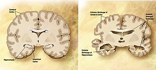
Huntington's disease (HD), also known as Huntington's chorea, is an incurable neurodegenerative disease that is mostly inherited. The earliest symptoms are often subtle problems with mood or mental/psychiatric abilities. A general lack of coordination and an unsteady gait often follow. It is also a basal ganglia disease causing a hyperkinetic movement disorder known as chorea. As the disease advances, uncoordinated, involuntary body movements of chorea become more apparent. Physical abilities gradually worsen until coordinated movement becomes difficult and the person is unable to talk. Mental abilities generally decline into dementia, depression, apathy, and impulsivity at times. The specific symptoms vary somewhat between people. Symptoms usually begin between 30 and 50 years of age, and can start at any age but are usually seen around the age of 40. The disease may develop earlier in each successive generation. About eight percent of cases start before the age of 20 years, and are known as juvenile HD, which typically present with the slow movement symptoms of Parkinson's disease rather than those of chorea.

<span class="mw-page-title-main">Wilson's disease</span> Genetic multisystem copper-transport disease

Wilson's disease is a genetic disorder in which excess copper builds up in the body. Symptoms are typically related to the brain and liver. Liver-related symptoms include vomiting, weakness, fluid build-up in the abdomen, swelling of the legs, yellowish skin, and itchiness. Brain-related symptoms include tremors, muscle stiffness, trouble in speaking, personality changes, anxiety, and psychosis.

<span class="mw-page-title-main">Leigh syndrome</span> Mitochondrial metabolism disease characterized by progressive loss of mental and movement abilities

Leigh syndrome is an inherited neurometabolic disorder that affects the central nervous system. It is named after Archibald Denis Leigh, a British neuropsychiatrist who first described the condition in 1951. Normal levels of thiamine, thiamine monophosphate, and thiamine diphosphate are commonly found, but there is a reduced or absent level of thiamine triphosphate. This is thought to be caused by a blockage in the enzyme thiamine-diphosphate kinase, and therefore treatment in some patients would be to take thiamine triphosphate daily. While the majority of patients typically exhibit symptoms between the ages of 3 and 12 months, instances of adult onset have also been documented.

<span class="mw-page-title-main">Chorea-acanthocytosis</span> Rare autosomal recessive genetic condition

Chorea-acanthocytosis is a rare hereditary disease caused by a mutation in a gene that directs structural proteins in red blood cells. It belongs to a group of four diseases characterized under the name neuroacanthocytosis. When a patient's blood is viewed under a microscope, some of the red blood cells appear thorny. These thorny cells are called acanthocytes.

Pantothenate kinase-associated neurodegeneration (PKAN), formerly called Hallervorden–Spatz syndrome, is a genetic degenerative disease of the brain that can lead to parkinsonism, dystonia, dementia, and ultimately death. Neurodegeneration in PKAN is accompanied by an excess of iron that progressively builds up in the brain.

<span class="mw-page-title-main">Aceruloplasminemia</span> Medical condition

Aceruloplasminemia is a rare autosomal recessive disorder in which the liver can not synthesize the protein ceruloplasmin properly, which is needed to transport copper around the blood. Copper deficiency in the brain results in neurological problems that generally appear in adulthood and worsen over time. .

Neuroacanthocytosis is a label applied to several genetic neurological conditions in which the blood contains misshapen, spiculated red blood cells called acanthocytes.

<span class="mw-page-title-main">Neurodegenerative disease</span> Central nervous system disease

A neurodegenerative disease is caused by the progressive loss of structure or function of neurons, in the process known as neurodegeneration. Such neuronal damage may ultimately involve cell death. Neurodegenerative diseases include amyotrophic lateral sclerosis, multiple sclerosis, Parkinson's disease, Alzheimer's disease, Huntington's disease, multiple system atrophy, tauopathies, and prion diseases. Neurodegeneration can be found in the brain at many different levels of neuronal circuitry, ranging from molecular to systemic. Because there is no known way to reverse the progressive degeneration of neurons, these diseases are considered to be incurable; however research has shown that the two major contributing factors to neurodegeneration are oxidative stress and inflammation. Biomedical research has revealed many similarities between these diseases at the subcellular level, including atypical protein assemblies and induced cell death. These similarities suggest that therapeutic advances against one neurodegenerative disease might ameliorate other diseases as well.

<span class="mw-page-title-main">Infantile neuroaxonal dystrophy</span> Medical condition

Infantile neuroaxonal dystrophy (INAD) is a rare pervasive developmental disorder that primarily affects the nervous system. Individuals with infantile neuroaxonal dystrophy typically do not have any symptoms at birth, but between the ages of about 6 and 18 months they begin to experience delays in acquiring new motor and intellectual skills, such as crawling or beginning to speak. Eventually they lose previously acquired skills.

<span class="mw-page-title-main">PLA2G6</span> Protein-coding gene in the species Homo sapiens

85 kDa calcium-independent phospholipase A2, also known as 85/88 kDa calcium-independent phospholipase A2, Group VI phospholipase A2, Intracellular membrane-associated calcium-independent phospholipase A2 beta, or Patatin-like phospholipase domain-containing protein 9 is an enzyme that in humans is encoded by the PLA2G6 gene.

Leukoencephalopathy with neuroaxonal spheroids (LENAS) is an extremely rare kind of leukoencephalopathy and is classified as a neurodegenerative disease. LENAS is a cause of severe and subacute dementia that results from damage to certain areas of the brain. This damage is to a type of brain tissue called white matter and axon damage due to swellings which are termed spheroids.

Nervous system diseases, also known as nervous system or neurological disorders, refers to a small class of medical conditions affecting the nervous system. This category encompasses over 600 different conditions, including genetic disorders, infections, cancer, seizure disorders, conditions with a cardiovascular origin, congenital and developmental disorders, and degenerative disorders.

Myoclonic dystonia or Myoclonus dystonia syndrome is a rare movement disorder that induces spontaneous muscle contraction causing abnormal posture. The prevalence of myoclonus dystonia has not been reported, however, this disorder falls under the umbrella of movement disorders which affect thousands worldwide. Myoclonus dystonia results from mutations in the SGCE gene coding for an integral membrane protein found in both neurons and muscle fibers. Those suffering from this disease exhibit symptoms of rapid, jerky movements of the upper limbs (myoclonus), as well as distortion of the body's orientation due to simultaneous activation of agonist and antagonist muscles (dystonia).

<span class="mw-page-title-main">Basal ganglia disease</span> Group of physical problems resulting from basal ganglia dysfunction

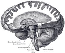
Basal ganglia disease is a group of physical problems that occur when the group of nuclei in the brain known as the basal ganglia fail to properly suppress unwanted movements or to properly prime upper motor neuron circuits to initiate motor function. Research indicates that increased output of the basal ganglia inhibits thalamocortical projection neurons. Proper activation or deactivation of these neurons is an integral component for proper movement. If something causes too much basal ganglia output, then the ventral anterior (VA) and ventral lateral (VL) thalamocortical projection neurons become too inhibited, and one cannot initiate voluntary movement. These disorders are known as hypokinetic disorders. However, a disorder leading to abnormally low output of the basal ganglia leads to reduced inhibition, and thus excitation, of the thalamocortical projection neurons which synapse onto the cortex. This situation leads to an inability to suppress unwanted movements. These disorders are known as hyperkinetic disorders.

<span class="mw-page-title-main">Kufor–Rakeb syndrome</span> Medical condition

Kufor–Rakeb syndrome (KRS) is an autosomal recessive disorder of juvenile onset also known as Parkinson disease-9 (PARK9). It is named after Kufr Rakeb in Irbid, Jordan. Kufor–Rakeb syndrome was first identified in this region in Jordan with a Jordanian couple's 5 children who had rigidity, mask-like face, and bradykinesia. The disease was first described in 1994 by Najim Al-Din et al. The OMIM number is 606693.

Neurodegeneration with brain iron accumulation is a heterogenous group of inherited neurodegenerative diseases, still under research, in which iron accumulates in the basal ganglia, either resulting in progressive dystonia, parkinsonism, spasticity, optic atrophy, retinal degeneration, neuropsychiatric, or diverse neurologic abnormalities. Some of the NBIA disorders have also been associated with several genes in synapse and lipid metabolism related pathways. NBIA is not one disease but an entire group of disorders, characterized by an accumulation of brain iron, sometimes in the presence of axonal spheroids in the central nervous system.

Benign hereditary chorea (BHC), also known as benign familial chorea, is a rare autosomal dominant neurogenetic syndrome. It typically presents itself in childhood with isolated chorea, with average to below average intelligence. Unlike other neurogenetic causes of chorea such as Huntington's disease, BHC is not progressive, and not associated with cognitive decline or psychiatric problems in the vast majority of cases.

<span class="mw-page-title-main">HFE H63D gene mutation</span>

The HFE H63D is a single-nucleotide polymorphism in the HFE gene, which results in the substitution of a histidine for an aspartic acid at amino acid position 63 of the HFE protein (p.His63Asp). HFE participates in the regulation of iron absorption.

Mitochondrial membrane protein-associated neurodegeneration (MPAN) is a genetic neurodegenerative disease that causes dystonia, parkinsonism, and iron accumulation in the brain. It is caused by mutations to the gene C19orf12, which has unknown function. This was originally discovered as an autosomal recessive disorder, caused by individuals having two mutations to the gene C19orf12, but autosomal dominant disease caused by a single mutation in the same gene has also been rarely described. Due to the common features of neurodegeneration, brain iron accumulation, and movement disorder it is classified as a neurodegeneration with brain iron accumulation (NBIA) disorder and another name for the condition is neurodegeneration with brain iron accumulation 4 (NBIA4).

Biotin-thiamine-responsive basal ganglia disease (BTBGD) is a rare disease that affects the nervous system, particularly the basal ganglia in the brain. It is a treatable neurometabolic disorder with autosomal recessive inheritance. First described in 1998 and then genetically distinguished in 2005, the disease is characterized by progressive brain damage that, if left untreated, can lead to coma and/or death. Commonly observed in individuals with BTBGD is recurring subacute encephalopathy along with confusion, seizures, and disordered movement (hypokinesia).

References

  1. 1 2 3 4 5 6 7 8 9 10 11 12 13 14 15 16 17 18 19 20 21 22 23 24 25 26 27 Lehn, A; Boyle, R; Brown, H; Airey, C; Mellick, G (September 2012). "Neuroferritinopathy". Parkinsonism & Related Disorders. 18 (8): 909–15. doi:10.1016/j.parkreldis.2012.06.021. PMID   22818529.
  2. 1 2 3 4 5 Chinnery, PF; Pagon, RA; Adam, MP; Ardinger, HH; Bird, TD; Dolan, CR; Fong, CT; Smith, RJH; Stephens, K (2010). "Neuroferritinopathy". PMID   20301320.{{cite journal}}: Cite journal requires |journal= (help)
  3. 1 2 3 4 5 6 7 8 9 10 11 Zecca, L; Youdim, MB; Riederer, P; Connor, JR; Crichton, RR (November 2004). "Iron, brain ageing and neurodegenerative disorders". Nature Reviews Neuroscience. 5 (11): 863–73. doi:10.1038/nrn1537. PMID   15496864. S2CID   205500060.
  4. 1 2 3 4 5 6 7 8 9 10 11 12 13 14 15 Keogh, MJ; Morris, CM; Chinnery, PF (2013). "Neuroferritinopathy". International Review of Neurobiology. 110: 91–123. doi:10.1016/B978-0-12-410502-7.00006-5. ISBN   9780124105027. PMID   24209436.
  5. Batista-Nascimento, Liliana; Pimentel, Catarina; Andrade Menezes, Regina; Rodrigues-Pousada, Claudina (2012). "Iron and Neurodegeneration: From Cellular Homeostasis to Disease". Oxidative Medicine and Cellular Longevity. 2012: 128647. doi: 10.1155/2012/128647 . PMC   3369498 . PMID   22701145.
  6. Rouault, Tracey A. (3 July 2013). "Iron metabolism in the CNS: implications for neurodegenerative diseases". Nature Reviews Neuroscience. 14 (8): 551–564. doi:10.1038/nrn3453. PMID   23820773. S2CID   21302204.
  7. Woimant, F; Trocello, JM (2014). "Disorders of heavy metals". Handbook of Clinical Neurology. 120: 851–64. doi:10.1016/B978-0-7020-4087-0.00057-7. ISBN   9780702040870. PMID   24365357.
  8. 1 2 3 4 5 6 Levi, Sonia; Finazzi, Dario (7 May 2014). "Neurodegeneration with brain iron accumulation: update on pathogenic mechanisms". Frontiers in Pharmacology. 5: 99. doi: 10.3389/fphar.2014.00099 . PMC   4019866 . PMID   24847269.