Ocular prosthesis

Last updated
Human ocular prosthesis of brown color. Eye-prosthesis-brown.jpg
Human ocular prosthesis of brown color.
Cat with an ocular prosthesis. CatWithOcularProsthetic.jpeg
Cat with an ocular prosthesis.

An ocular prosthesis, artificial eye or glass eye is a type of craniofacial prosthesis that replaces an absent natural eye following an enucleation, evisceration, or orbital exenteration. The prosthesis fits over an orbital implant and under the eyelids. Though often referred to as a glass eye, the ocular prosthesis roughly takes the shape of a convex shell and is made of medical grade plastic acrylic. A few ocular prostheses today are made of cryolite glass. A variant of the ocular prosthesis is a very thin hard shell known as a scleral shell which can be worn over a damaged or eviscerated eye. Makers of ocular prosthetics are known as ocularists. An ocular prosthesis does not provide vision; this would be a visual prosthesis. Someone with an ocular prosthesis is altogether blind on the affected side and has monocular (one sided) vision.

Contents

History

Prosthetic eye and glasses made for an injured World War I soldier by pioneering plastic surgeon Johannes Esser. Prothese met klein oog en bril, item 1.jpg
Prosthetic eye and glasses made for an injured World War I soldier by pioneering plastic surgeon Johannes Esser.
"Making glass eye", c. 1915-1920. Making glass eye (LOC) (15601805189).jpg
"Making glass eye", c.1915–1920.
Glass eye being moulded under heat, 1938. V1-FL9683817.jpg
Glass eye being moulded under heat, 1938.

The earliest known evidence of the use of ocular prosthesis is that of a woman found in Shahr-I Sokhta, Iran [1] dating back to 2900–2800 BC. [2] It has a hemispherical form and a diameter of just over 2.5 cm (1 inch). It consists of very light material, probably bitumen paste. The surface of the artificial eye is covered with a thin layer of gold, engraved with a central circle (representing the iris) and gold lines patterned like sun rays. On both sides of the eye are drilled tiny holes, through which a golden thread could hold the eyeball in place. Since microscopic research has shown that the eye socket showed clear imprints of the golden thread, the eyeball must have been worn during her lifetime. In addition to this, an early Hebrew text references a woman who wore an artificial eye made of gold. [3] Roman and Egyptian priests are known to have produced artificial eyes as early as the fifth century BC constructed from painted clay attached to cloth and worn outside the socket. [4]

The first in-socket artificial eyes were made of gold with colored enamel, later evolving into the use of glass (thus the name "glass eye") by the Venetians in the later part of the sixteenth century. These were crude, uncomfortable, and fragile and the production methodology remained known only to Venetians until the end of the 18th century, when Parisians took over as the center for artificial eye-making. But the center shifted again, this time to Germany because of their superior glass blowing techniques. Shortly following the introduction of the art of glass eye-making to the United States, German goods became unavailable because of World War II. As a result, the US instead made artificial eyes from acrylic plastic. [4]

Production of modern ocular prosthetics has expanded from simply using glass into many different types of materials. In the United States, most custom ocular prostheses are fabricated using PMMA (polymethyl methacrylate), or acrylic. In some countries, Germany especially, prostheses are still most commonly made from glass. [4]

Limits of realism

Ocularist surgeons have always worked together to make artificial eyes look more realistic. For decades, all efforts and investments to improve the appearance of artificial eyes have been dampened by the immobility of the pupil. One solution to this problem has been demonstrated recently in a device based on an LCD which simulates the pupil size as a function of the ambient light. [5]

Implant types and chemical construction

There are many different types of implants, classification ranging from shape (spherical vs egg (oval) shaped), stock vs custom, [4] porous vs nonporous, specific chemical make-up, and the presence of a peg or motility post. The most basic simplification can be to divide implant types into two main groups: non-integrated (non-porous) and integrated (porous). [6]

Nonintegrated implants

Though there is evidence that ocular implants have been around for thousands of years, [2] modern nonintegrated spherical intraconal implants came into existence around 1976 (not just glass eyes). [4] [7] Nonintegrated implants contain no unique apparatus for attachments to the extraocular muscles and do not allow in-growth of organic tissue into their inorganic substance. Such implants have no direct attachment to the ocular prosthesis. [6] Usually, these implants are covered with a material that permits fixation of the extraocular recti muscles, such as donor sclera or polyester gauze which improves implant motility, but does not allow for direct mechanical coupling between the implant and the artificial eye. [7] Non-integrated implants include the acrylic (PMMA), [6] glass, and silicone spheres. [8]

Polymethyl methacrylate (PMMA) (acrylic)

Polymethyl methacrylate (PMMA), [6] commonly known as acrylic, is a transparent thermoplastic available for use as ocular prosthesis, replacement intraocular lenses when the original lens has been removed in the treatment of cataracts and has historically been used as hard contact lenses.

PMMA has a good degree of compatibility with human tissue, much more so than glass. Although various materials have been used to make nonintegrated implants in the past, polymethyl methacrylate is one of the implants of choice. [6]

Integrated implants (porous)

The porous nature of integrated implants allows fibrovascular ingrowth throughout the implant and thus also insertion of pegs or posts. [9] Because direct mechanical coupling is thought to improve artificial eye motility, attempts have been made to develop so-called 'integrated implants' that are directly connected to the artificial eye. [7] Historically, implants that directly attached to the prosthesis were unsuccessful because of chronic inflammation or infection arising from the exposed nonporous implant material. [9] This led to the development of quasi-integrated implants with a specially designed anterior surface that allegedly better transferred implant motility to the artificial eye through the closed conjunctiva and Tenon's capsule. [7] In 1985, the problems associated with integrated implants were thought to be largely solved with the introduction of spherical implants made of porous calcium hydroxyapatite. This material allows for fibrovascular ingrowth within several months. [7] Porous enucleation implants currently are fabricated from a variety of materials including natural and synthetic hydroxyapatite, aluminium oxide, and polyethylene.

The surgeon can alter the contour of porous implants before insertion, and it is also possible to modify the contour in situ, although this is sometimes difficult. [9]

Hydroxyapatite (HA)

Hydroxyapatite implants are spherical and made in a variety of sizes and different materials (coralline/synthetic). [8] [9]

Since their introduction in 1989 when an implant made from hydroxyapatite received Food and Drug Administration approval, spherical hydroxyapatite implants have gained widespread popularity as an enucleation implant [7] [9] and was at one point the most commonly used orbital implant in the United States. [10] [11] The porous nature of this material allows fibrovascular ingrowth throughout the implant and permits insertion of a coupling device (PEG) with reduced risk of inflammation or infection associated with earlier types of exposed integrated implants. [9]

Hydroxyapatite is limited to preformed (stock) [4] spheres (for enucleation) or granules (for building up defects). [12]

One main disadvantage of HA is that it needs to be covered with exogenous material, such as sclera, polyethylene terephthalate, or vicryl mesh (which has the disadvantage of creating a rough implant tissue interface that can lead to technical difficulties in implantation and subsequent erosion of overlying tissue with the end stage being extrusion), as direct suturing is not possible for muscle attachment. Scleral covering carries with it the risk of transmission of infection, inflammation, and rejection. [10]

A 2008 study showed that HA has a more rapid rate of fibrovascularization than MEDPOR, [10] a high-density porous polyethylene [9] implant manufactured from linear high-density polyethylene. [13]

Porous polyethylene (PP)

Development in polymer chemistry has allowed introduction of newer biocompatible material such as porous polyethylene (PP) to be introduced into the field of orbital implant surgery. [10] Porous polyethylene enucleation implants have been used since at least 1989. [9] It is available in dozens of prefabricated spherical and non-spherical shapes and in different sizes or plain blocks for individualized intraoperative customizing. [9] The material is firm but malleable and allows direct suturing of muscles to implant without wrapping or extra steps. Additionally, the smooth surface is less abrasive and irritating than other materials used for similar purposes. [12] Polyethylene also becomes vascularized, allowing placement of a titanium motility post that joins the implant to the prosthesis in the same way that the peg is used for hydroxyapatite implants. [9]

PP has been shown to have a good outcome, and in 2004, it was the most commonly used orbital implant in the United States. [10] [14] Porous polyethylene fulfills several criteria for a successful implant, including little propensity to migrate and restoration of defect in an anatomic fashion; it is readily available, cost-effective, and can be easily modified or custom-fit for each defect. [12] The PP implant does not require to be covered and therefore avoids some of the problems associated with hydroxyapatite implants. [10]

Bioceramic

Bioceramic prosthetics are made of aluminium oxide (Al
2
O
3
). Aluminium oxide is a ceramic biomaterial that has been used for more than 35 years in the orthopedic and dental fields for a variety of prosthetic applications because of its low friction, durability, stability, and inertness. [15] Aluminium oxide ocular implants can be obtained in spherical and non-spherical (egg-shaped) shapes and in different sizes [9] for use in the anophthalmic socket. It received US Food and Drug Administration approval in April 2000 and was approved by Health and Welfare, Canada, in February 2001. [15]

Aluminium oxide has previously been shown to be more biocompatible than HA in cell culture studies and has been suggested as the standard reference material when biocompatibility studies are required to investigate new products. The rate of exposure previously associated with the bioceramic implant (2%) was less than most reports on the HA or porous polyethylene implant (0% to 50%). [15]

Conical orbital implant (COI) and multipurpose conical orbital implant (MCOI)

The safe and effective sphere (still popular and easy to use) was supplemented with the pyramid or COI implant. [12] The COI has unique design elements that have been incorporated into an overall conical shape, including a flat anterior surface, superior projection and preformed channels for the rectus muscles. 5-0 Vicryl suture needles can be passed with slight difficulty straight through the implant to be tied on the anterior surface. In addition, this implant features a slightly recessed slot for the superior rectus and a protrusion to fill the superior fornix. [12]

The newest model[ needs update ] is the multipurpose conical orbital implant (MCOI), which was designed to address the issues of the postoperative anophthalmic orbit being at risk for the development of socket abnormalities including enophthalmos, retraction of the upper eyelid, deepening of the superior sulcus, backward tilt of the prothesis, and stretching of the lower eyelid after evisceration or enucleation. These problems are generally thought to be secondary to orbital volume deficiencies which is also addressed by MCOIs. The conical shape of the MCOI more closely matches the anatomic shape of the orbit than a spherical implant. The wider anterior portion, combined with the narrower and longer posterior portion, allows for a more complete and natural replacement of the lost orbital volume. This shape reduces the risk of superior sulcus deformity and puts more volume within the muscle cone. [16] Muscles can be placed at any location the surgeon desires with these implants. This is advantageous for cases of damaged or lost muscles after trauma, and the remaining muscles are transposed to improve postoperative motility. In anticipation of future peg placement there is a 6 mm (0.24 in) diameter flattened surface, which eliminates the need to shave a flat anterior surface prior to peg placement. [12]

Both implants (COI and MCOI) are composed of interconnecting channels that allow ingrowth of host connective tissue. Complete implant vascularization reduces the risk of infection, extrusion, and other complications associated with nonintegrated implants. Additionally, both implants produce superior motility and postoperative cosmesis. [12]

Pegged (motility post) implants

In hydroxyapatite implants, a secondary procedure can insert an externalized, round-headed peg or screw into the implant. The prosthesis is modified to accommodate the peg, creating a ball-and-socket joint. [9] After fibrovascular ingrowth is completed, a small hole can be drilled into the anterior surface of the implant. After conjunctivalization of this hole, it can be fitted with a peg with a rounded top that fits into a corresponding dimple at the posterior surface of the artificial eye. This peg thus directly transfers implant motility to the artificial eye. [7] However, the motility peg is mounted in a minority of patients. This may partially be due to problems associated with peg placement, whereas hydroxyapatite implants are assumed to yield superior artificial eye motility even without the peg. [7]

Polyethylene also becomes vascularized, allowing placement of a titanium motility post that joins the implant to the prosthesis in the same way that the peg is used for hydroxyapatite implants. [9]

Implant movement

Implant and prosthesis movement are important aspects of the overall cosmetic appearance after enucleation, and are essential to the objective of crafting a lifelike eye similar in all aspects to the normal fellow eye. [6] [17] There are several theories of improved eye movement, such as using integrating prosthetic material, pegging the implant, covering the implant (e.g. with scleral tissue), or suturing the eye muscles directly to the prosthetic implant. The efficiency of transmitting movement from the implant to the prosthesis determines the degree of prosthetic motility. Movement is transmitted from traditional nonporous spherical implants through the surface tension at the conjunctival–prosthetic interface and movement of the fornices. Quasi-integrated implants have irregularly shaped surfaces that create an indirect coupling mechanism between the implant and prosthesis that imparts greater movement to the prosthesis. Directly integrating the implant to the prosthesis through an externalized coupling mechanism would be expected to improve motility further. [9]

Despite the reasoning stating that hydroxyapatite orbital implants without a motility peg would yield a superior artificial eye motility, [18] when similar surgical techniques are used, unpegged porous (hydroxyapatite) enucleation implants and donor sclera-covered nonporous (acrylic) spherical enucleation implants yield comparable artificial eye motility. [7] [9] In two studies, [7] [19] there were no differences in maximum amplitude between hydroxyapatite and acrylic or silicone spherical enucleation implants, [7] thus indicating that the implant material itself may not have a bearing on implant movement as long as the muscles are attached directly or indirectly to the implant and the implant is not pegged. [6] The motility of a nonintegrated artificial eye may be caused by at least two forces:

  1. The rubbing force between the posterior surface of the artificial eye and the conjunctiva that covers the implant may cause the artificial eye to move. Because this force is likely to be approximately equal in all directions, it would cause comparable horizontal and vertical artificial eye amplitudes.[ citation needed ]
  2. An artificial eye usually fits snugly in the conjunctival space (possibly not in the superior fornix). Therefore, any movement of the conjunctival fornices will cause a similar movement of the artificial eye, whereas lack of movement of the fornices will restrict its motility. [7]

Imbrication of the rectus muscles over a nonintegrated implant traditionally was thought to impart movement to the implant and prosthesis. Like a ball-and-socket joint, when the implant moves, the prosthesis moves. However, because the so-called ball and socket are separated by layers of Tenon's capsule, imbricated muscles, and conjunctiva, the mechanical efficiency of transmission of movement from the implant to the prosthesis is suboptimal. Moreover, the concern is that imbrication of the recti over nonintegrated implants actually can result in implant migration. [20] The recent myoconjuctival technique of enucleation is an alternative to muscle imbrication. [6] [19] [21]

Although it is generally accepted that integrating the prosthesis to a porous implant with peg insertion enhances prosthetic movement, there is little available evidence in the literature that documents the degree of improvement. [9] In addition to this, although the porous implants have been reported to offer improved implant movement, [22] these are more expensive and intrusive, require wrapping and subsequent imaging to determine vascularization and pegging to provide for better transmission of implant movement to the prosthesis, and are prone to implant exposure. [6]

Age and size of the implant may also affect the motility, since in a study comparing patients with hydroxyapatite implants and patients with nonporous implants, the implant movement appeared to decrease with age in both groups. This study also demonstrated improved movement of larger implants irrespective of material. [9]

Surgical procedure

Enucleation and orbital implantation surgery follows these steps: [9]

Also under anesthesia:

The surgery is done under general anesthesia with the addition of extra subconjunctival and/or retrobulbar anesthetics injected locally in some cases. The following is a description of the surgical procedure performed by Custer et al.: [9]

The conjunctival peritomy is performed at the corneal limbus, preserving as much healthy tissue as possible. Anterior Tenon's fascia is separated from the sclera. Blunt dissection in the four quadrants between the rectus muscles separates deep Tenon's fascia.

Sutures may be passed through the rectus muscles before their disinsertion from the globe. Some surgeons also suture one or both oblique muscles. Traction sutures or clamps may be applied to the horizontal rectus muscle insertions to assist in rotating and elevating the globe during the ensuing dissection. Tenon's capsule may be opened posteriorly to allow visualization of the optic nerve. The vortex veins and posterior ciliary vessels may be cauterized before dividing the nerve and removing the eye. Alternatively, the optic nerve may be localized with a clamp before transection. Hemostasis is achieved with either cautery or digital pressure.

The orbital implant is inserted at the time of enucleation. An appropriately sized implant should replace the volume of the globe and leave sufficient room for the ocular prosthesis. Enucleation implants are available in a variety of sizes that may be determined by using sizing implants or calculated by measuring globe volume or axial length of the contralateral eye.

In the past, spherical nonporous implants were placed in the intraconal space and the extraocular muscles were either left unattached or were tied over the implant. Wrapping these implants allows attachment of the muscles to the covering material, a technique that seems to improve implant movement and reduce the incidence of implant migration. Porous implants may be saturated with antibiotic solution before insertion. Because the brittle nature of hydroxyapatite prevents direct suturing of the muscles to the implant, these implants are usually covered with some form of wrapping material. The muscles are attached to the implant in a technique similar to that used for spherical non-porous implants. The muscles may be directly sutured to porous polyethylene implants either by passing the suture through the implant material or by using an implant with fabricated suture tunnels. Some surgeons also wrap porous polyethylene implants either to facilitate muscle attachment or to reduce the risk of implant exposure. A variety of wrapping materials have been used to cover porous implants, including polyglactin or polyglycolic acid mesh, heterologous tissue (bovine pericardium), homologous donor tissue (sclera, dermis), and autogenous tissue (fascia lata, temporalis fascia, posterior auricular muscle, rectus abdominis sheath).

Fenestrations in the wrapping material are created at the insertion sites of the extraocular muscles, allowing the attached muscles to be in contact with the implant and improving implant vascularization. Drilling 1 mm holes into the implant at the muscle insertion sites is performed to facilitate vascularization of hydroxyapatite implants. Tenon's fascia is drawn over the implant and closed in one or two layers. The conjunctiva is then sutured.

A temporary ocular conformer is inserted at the completion of the pro- cedure and is worn until the patient receives a prosthesis 4 to 8 weeks after surgery.

An elective secondary procedure is required to place the coupling peg or post in those patients who desire improved prosthetic motility. That procedure is usually delayed for at least 6 months after enucleation to allow time for implant vascularization. Technetium bone or gadolinium-enhanced magnetic resonance imaging scans are not now universally used, but they have been used to confirm vascularization before peg insertion. Under local anesthesia, a conjunctival incision is created at the peg insertion site. A hole is created into the porous implant to allow insertion of the peg or post. The prosthesis is then modified to receive the peg or post. Some surgeons have preplaced coupling posts in porous polyethylene implants at the time of enucleation. The post may spontaneously expose or is externalized in a later procedure via a conjunctival incision.

Aftermath of surgical procedures

Regardless of the procedure, a type of ocular prosthesis is always needed afterwards. The surgeon will insert a temporary prosthesis at the end of the surgery, known as a stock eye, [23] and refer the patient to an ocularist, who is not a medical doctor, but board certified ocularist by the American Society of Ocularists. [24] The process of making an ocular prosthesis, or a custom eye, will begin, usually six weeks after the surgical procedure, and it typically will take up to three visits before the final fitting of the prosthesis. In most cases, the patient will be fitted during the first visit, return for the hand-painting of the prosthesis, and finally come back for the final fitting. The methods used to fit, shape, and paint the prosthesis often vary to suit both ocularist and patient needs.

Living with an ocular prosthesis requires care, but oftentimes patients who have had incurable eye disorders, such as micropthalmia, anophtalmia or retinoblastoma, achieve a better quality of life with their prostheses. It is generally recommended to leave the prosthesis in the socket as much as possible, though it may require some cleaning and lubrication, as well as regular polishing and check-ups with ocularists. [25]

Notable people with prosthetic eyes

Related Research Articles

<span class="mw-page-title-main">Prosthesis</span> Artificial device that replaces a missing body part

In medicine, a prosthesis, or a prosthetic implant, is an artificial device that replaces a missing body part, which may be lost through physical trauma, disease, or a condition present at birth. Prostheses are intended to restore the normal functions of the missing body part. Amputee rehabilitation is primarily coordinated by a physiatrist as part of an inter-disciplinary team consisting of physiatrists, prosthetists, nurses, physical therapists, and occupational therapists. Prostheses can be created by hand or with computer-aided design (CAD), a software interface that helps creators design and analyze the creation with computer-generated 2-D and 3-D graphics as well as analysis and optimization tools.

An evisceration is the removal of the eye's contents, leaving the scleral shell and extraocular muscles intact. The procedure is usually performed to reduce pain or improve cosmesis in a blind eye, as in cases of endophthalmitis unresponsive to antibiotics. An ocular prosthetic can be fitted over the eviscerated eye in order to improve cosmesis.

<span class="mw-page-title-main">Orbit (anatomy)</span> Cavity or socket of the skull in which the eye and its appendages are situated

In anatomy, the orbit is the cavity or socket/hole of the skull in which the eye and its appendages are situated. "Orbit" can refer to the bony socket, or it can also be used to imply the contents. In the adult human, the volume of the orbit is 30 millilitres, of which the eye occupies 6.5 ml. The orbital contents comprise the eye, the orbital and retrobulbar fascia, extraocular muscles, cranial nerves II, III, IV, V, and VI, blood vessels, fat, the lacrimal gland with its sac and duct, the eyelids, medial and lateral palpebral ligaments, cheek ligaments, the suspensory ligament, septum, ciliary ganglion and short ciliary nerves.

<span class="mw-page-title-main">Eye surgery</span> Surgery performed on the eye or its adnexa

Eye surgery, also known as ophthalmic surgery or ocular surgery, is surgery performed on the eye or its adnexa. Eye surgery is part of ophthalmology and is performed by an ophthalmologist or eye surgeon. The eye is a fragile organ, and requires due care before, during, and after a surgical procedure to minimize or prevent further damage. An eye surgeon is responsible for selecting the appropriate surgical procedure for the patient, and for taking the necessary safety precautions. Mentions of eye surgery can be found in several ancient texts dating back as early as 1800 BC, with cataract treatment starting in the fifth century BC. It continues to be a widely practiced class of surgery, with various techniques having been developed for treating eye problems.

<span class="mw-page-title-main">Hip replacement</span> Surgery replacing hip joint with prosthetic implant

Hip replacement is a surgical procedure in which the hip joint is replaced by a prosthetic implant, that is, a hip prosthesis. Hip replacement surgery can be performed as a total replacement or a hemi/semi(half) replacement. Such joint replacement orthopaedic surgery is generally conducted to relieve arthritis pain or in some hip fractures. A total hip replacement consists of replacing both the acetabulum and the femoral head while hemiarthroplasty generally only replaces the femoral head. Hip replacement is one of the most common orthopaedic operations, though patient satisfaction varies widely. Approximately 58% of total hip replacements are estimated to last 25 years. The average cost of a total hip replacement in 2012 was $40,364 in the United States, and about $7,700 to $12,000 in most European countries.

<span class="mw-page-title-main">Duane syndrome</span> Rare congenital disease characterized by external gaze palsy

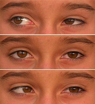
Duane syndrome is a congenital rare type of strabismus most commonly characterized by the inability of the eye to move outward. The syndrome was first described by ophthalmologists Jakob Stilling (1887) and Siegmund Türk (1896), and subsequently named after Alexander Duane, who discussed the disorder in more detail in 1905.

<span class="mw-page-title-main">Lateral rectus muscle</span> Muscle on lateral side of the eye

The lateral rectus muscle is a muscle on the lateral side of the eye in the orbit. It is one of six extraocular muscles that control the movements of the eye. The lateral rectus muscle is responsible for lateral movement of the eyeball, specifically abduction. Abduction describes the movement of the eye away from the midline, allowing the eyeball to move horizontally in the lateral direction, bringing the pupil away from the midline of the body.

<span class="mw-page-title-main">Medial rectus muscle</span> Extraocular muscle that rotates the eye medially

The medial rectus muscle is a muscle in the orbit near the eye. It is one of the extraocular muscles. It originates from the common tendinous ring, and inserts into the anteromedial surface of the eye. It is supplied by the inferior division of the oculomotor nerve (III). It rotates the eye medially (adduction).

<span class="mw-page-title-main">Extraocular muscles</span> Seven extrinsic muscles of the eye

The extraocular muscles, or extrinsic ocular muscles, are the seven extrinsic muscles of the eye in humans and other animals. Six of the extraocular muscles, the four recti muscles, and the superior and inferior oblique muscles, control movement of the eye. The other muscle, the levator palpebrae superioris, controls eyelid elevation. The actions of the six muscles responsible for eye movement depend on the position of the eye at the time of muscle contraction.

<span class="mw-page-title-main">Enucleation of the eye</span> Type of ocular surgery

Enucleation is the removal of the eye that leaves the eye muscles and remaining orbital contents intact. This type of ocular surgery is indicated for a number of ocular tumors, in eyes that have sustained severe trauma, and in eyes that are otherwise blind and painful.

<span class="mw-page-title-main">Joint replacement</span> Orthopedic surgery to replace a joint

Joint replacement is a procedure of orthopedic surgery known also as arthroplasty, in which an arthritic or dysfunctional joint surface is replaced with an orthopedic prosthesis. Joint replacement is considered as a treatment when severe joint pain or dysfunction is not alleviated by less-invasive therapies. Joint replacement surgery is often indicated from various joint diseases, including osteoarthritis and rheumatoid arthritis.

An ocularist specializes in the fabrication and fitting of ocular prostheses for people who have lost an eye or eyes due to trauma or illness. The fabrication process for a custom made eye typically includes taking an impression of the eye socket, shaping a plastic shell, painting the iris, and then fitting the ocular prostheses. Prefabricated ocular prostheses with different colored iris are also available. An ocularist may select the stock eye that is most closely matched to patient's iris color. However, due to better adaptation, comfort, and aesthetics, custom-made ocular prostheses are more accepted. In addition to creating the prosthetic eye, an ocularist shows the patient how to care for and handle the prosthesis.

<span class="mw-page-title-main">Phthisis bulbi</span> Shrunken, non-functional eye

Phthisis bulbi is a shrunken, non-functional eye. It may result from severe eye disease, inflammation or injury, or it may represent a complication of eye surgery. Treatment options include insertion of a prosthesis, which may be preceded by enucleation of the eye.

<span class="mw-page-title-main">Tenon's capsule</span> Membrane surrounding the eye forming a socket in which it moves

Tenon's capsule, also known as the Tenon capsule, fascial sheath of the eyeball or the fascia bulbi, is a thin membrane which envelops the eyeball from the optic nerve to the corneal limbus, separating it from the orbital fat and forming a socket in which it moves.

Oculoplastics, or oculoplastic surgery, includes a wide variety of surgical procedures that deal with the orbit, eyelids, tear ducts, and the face. It also deals with the reconstruction of the eye and associated structures.

<span class="mw-page-title-main">Eye neoplasm</span> Medical condition

An eye neoplasm is a tumor of the eye. A rare type of tumor, eye neoplasms can affect all parts of the eye, and can either be benign or malignant (cancerous), in which case it is known as eye cancer. Eye cancers can be primary or metastatic cancer. The two most common cancers that spread to the eye from another organ are breast cancer and lung cancer. Other less common sites of origin include the prostate, kidney, thyroid, skin, colon and blood or bone marrow.

<span class="mw-page-title-main">Anophthalmia</span> Medical condition

Anophthalmia is the medical term for the absence of one or both eyes. Both the globe and the ocular tissue are missing from the orbit. The absence of the eye will cause a small bony orbit, a constricted mucosal socket, short eyelids, reduced palpebral fissure and malar prominence. Genetic mutations, chromosomal abnormalities, and prenatal environment can all cause anophthalmia. Anophthalmia is an extremely rare disease and is mostly rooted in genetic abnormalities. It can also be associated with other syndromes.

<span class="mw-page-title-main">Orbital blowout fracture</span> Medical condition

An orbital blowout fracture is a traumatic deformity of the orbital floor or medial wall that typically results from the impact of a blunt object larger than the orbital aperture, or eye socket. Most commonly this results in a herniation of orbital contents through the orbital fractures. The proximity of maxillary and ethmoidal sinus increases the susceptibility of the floor and medial wall for the orbital blowout fracture in these anatomical sites. Most commonly, the inferior orbital wall, or the floor, is likely to collapse, because the bones of the roof and lateral walls are robust. Although the bone forming the medial wall is the thinnest, it is buttressed by the bone separating the ethmoidal air cells. The comparatively thin bone of the floor of the orbit and roof of the maxillary sinus has no support and so the inferior wall collapses mostly. Therefore, medial wall blowout fractures are the second-most common, and superior wall, or roof and lateral wall, blowout fractures are uncommon and rare, respectively. They are characterized by double vision, sunken ocular globes, and loss of sensation of the cheek and upper gums from infraorbital nerve injury.

<span class="mw-page-title-main">Wim Richter</span> South African chemist and researcher (1946–2019)

Paul Wilhelm Richter was a South African chemist and principal researcher involved in bioceramic research activities. He is most widely known for his development of the bioceramic hydroxyapatite orbital implant.

Minimally invasive strabismus surgery (MISS) is a technique in strabismus surgery that uses smaller incisions than the classical surgical approach to correct the condition, thus minimizing tissue disruption. The technique was introduced by Swiss ophthalmologist Daniel Mojon around 2007, after the Belgian ophthalmologist Marc Gobin described the idea in 1994 in a French-language textbook.

References

  1. 3rd Millennium BC Artificial Eyeball Discovered in Burnt City Archived 2012-04-11 at the Wayback Machine , December 10, 2006
  2. 1 2 London Times (February 20, 2007). "5,000-Year-Old Artificial Eye Found on Iran-Afghan Border". foxnews. Archived from the original on February 22, 2007. Retrieved December 14, 2012.
  3. Jerusalem Talmud Nedarim 41c
  4. 1 2 3 4 5 6 Frequently asked questions, American Society of Ocularists
  5. Lapointe, J; Durette, J-F; Harhira, A; Shaat, A; Boulos, PR; Kashyap, R (Sep 2010). "A 'living' prosthetic iris". Eye. 24 (11): 1716–23. doi: 10.1038/eye.2010.128 . PMID   20847748.
  6. 1 2 3 4 5 6 7 8 9 Shome, D; Honavar, SG; Raizada, K; Raizada, D (2010). "Implant and prosthesis movement after enucleation: a randomized controlled trial". Ophthalmology. 117 (8): 1638–44. doi:10.1016/j.ophtha.2009.12.035. PMID   20417565.
  7. 1 2 3 4 5 6 7 8 9 10 11 12 Colen, TP; Paridaens, DA; Lemij, HG; Mourits, MP; Van Den Bosch, WA (2000). "Comparison of artificial eye amplitudes with acrylic and hydroxyapatite spherical enucleation implants". Ophthalmology. 107 (10): 1889–94. doi:10.1016/S0161-6420(00)00348-1. PMID   11013194.
  8. 1 2 Chuah, CT; Chee, SP; Fong, KS; Por, YM; Choo, CT; Luu, C; Seah, LL (2004). "Integrated hydroxyapatite implant and non-integrated implants in enucleated Asian patients". Annals of the Academy of Medicine, Singapore. 33 (4): 477–83. PMID   15329760.
  9. 1 2 3 4 5 6 7 8 9 10 11 12 13 14 15 16 17 18 19 Custer, PL; Kennedy, RH; Woog, JJ; Kaltreider, SA; Meyer, DR (2003). "Orbital implants in enucleation surgery: a report by the American Academy of Ophthalmology". Ophthalmology. 110 (10): 2054–61. doi:10.1016/S0161-6420(03)00857-1. PMID   14522788.
  10. 1 2 3 4 5 6 Sadiq, SA; Mengher, LS; Lowry, J; Downes, R (2008). "Integrated orbital implants – a comparison of hydroxyapatite and porous polyethylene implants". Orbit (Amsterdam, Netherlands). 27 (1): 37–40. doi:10.1080/01676830701512585. PMID   18307145. S2CID   24577669.
  11. Hornblass, A; Biesman, BS; Eviatar, JA; Nunery, William R. (1995). "Current techniques of enucleation: a survey of 5,439 intraorbital implants and a review of the literature". Ophthalmic Plastic and Reconstructive Surgery. 11 (2): 77–86, discussion 87–88. doi:10.1097/00002341-199506000-00001. PMID   7654621. S2CID   6640588.
  12. 1 2 3 4 5 6 7 Duffy, M., Biesman, B. (2000). "Porous polyethylene expands orbitofacial options". Ophthalmology Times. 25 (7): 18.{{cite journal}}: CS1 maint: multiple names: authors list (link)
  13. Chen, YH; Cui, HG (2006). "High density porous polyethylene material (Medpor) as an unwrapped orbital implant". Journal of Zhejiang University Science B. 7 (8): 679–82. doi:10.1631/jzus.2006.B0679. PMC   1533749 . PMID   16845724.
  14. Su, GW; Yen, MT (2004). "Current trends in managing the anophthalmic socket after primary enucleation and evisceration". Ophthalmic Plastic and Reconstructive Surgery. 20 (4): 274–80. doi:10.1097/01.IOP.0000129528.16938.1E. PMID   15266140. S2CID   30449909.
  15. 1 2 3 Jordan, DR; Klapper, SR; Gilberg, SM; Dutton, JJ; Wong, A; Mawn, L (2010). "The bioceramic implant: evaluation of implant exposures in 419 implants". Ophthalmic Plastic and Reconstructive Surgery. 26 (2): 80–82. doi:10.1097/IOP.0b013e3181b80c30. PMID   20305504. S2CID   36175600.
  16. Marshak, H; Dresner, SC (2005). "Multipurpose conical orbital implant in evisceration". Ophthalmic Plastic and Reconstructive Surgery. 21 (5): 376–78. doi:10.1097/01.iop.0000173191.24824.40. PMID   16234704. S2CID   17738376.
  17. Byron C. Smith; Frank A. Nesi; Mark R. Levine; Richard D. Lisman (1998). Smith's Ophthalmic Plastic and Reconstructive Surgery. Mosby Incorporated. ISBN   978-0-8151-6356-5.
  18. Shields, CL; Shields, JA; De Potter, P (1992). "Hydroxyapatite orbital implant after enucleation. Experience with initial 100 consecutive cases". Archives of Ophthalmology. 110 (3): 333–38. doi:10.1001/archopht.1992.01080150031022. PMID   1311918.
  19. 1 2 Custer, PL; Trinkaus, KM; Fornoff, J (1999). "Comparative motility of hydroxyapatite and alloplastic enucleation implants". Ophthalmology. 106 (3): 513–16. doi:10.1016/S0161-6420(99)90109-4. PMID   10080207.
  20. Beard, C (1995). "Remarks on historical and newer approaches to orbital implants". Ophthalmic Plastic and Reconstructive Surgery. 11 (2): 89–90. doi:10.1097/00002341-199506000-00002. PMID   7654622. S2CID   36166165.
  21. Yadava U, Sachdeva P, Arora A (2004). "Myoconjunctival enucleation for enhanced implant movement: result of a randomised prospective study". Indian J Ophthalmol. 52 (3): 221–26. PMID   15510462.
  22. Jordan, DR; Chan, S; Mawn, L; Gilberg, S; Dean, T; Brownstein, S; Hill, VE (1999). "Complications associated with pegging hydroxyapatite orbital implants". Ophthalmology. 106 (3): 505–12. doi:10.1016/S0161-6420(99)90108-2. PMID   10080206.
  23. "American Society of Ocularists - Frequently Asked Questions". www.ocularist.org. Retrieved 2016-11-09.
  24. "American Society of Ocularists - Frequently Asked Questions". www.ocularist.org. Retrieved 2016-11-09.
  25. "Prosthetic Eye Care". ocularpro.com. Ocular Prosthetics, Inc. Retrieved 2016-11-09.
  26. van Orsouw, Michael (14 July 2021). "The 'Vaudois' King of Thailand". Swiss National Museum - Swiss history blog. Retrieved 15 July 2022. The driver involved in the accident was 21-year-old Bhumibol Adulyadej, the designated King of Thailand. The future monarch lost an eye in the accident in Vaud, and from then on had to live with a glass eye and some facial paralysis.
  27. Starkey, Joe (2006). Tales from the Pittsburgh Penguins . Sports Publishing LLC. p.  45. ISBN   978-1-58261-199-0 . Retrieved September 18, 2011.
  28. "Profile: Mokhtar Belmokhtar". BBC News. June 4, 2013.
  29. "Nice Fellow". Time. Time Warner. April 18, 1955. Archived from the original on November 4, 2012. Retrieved September 18, 2009.
  30. "Peter Falk". Bio. (UK). Archived from the original on June 10, 2009. Retrieved January 30, 2009.
  31. "Tex Avery Loses an Eye, 1933". Walter Lantz Archive. Cartoon Research. Retrieved March 16, 2018.
  32. Entry for "Ry Cooder", in The Rolling Stone Encyclopedia of Rock & Roll, Touchstone (revised, updated edition); November 8, 2001; ISBN   978-0743201209
  33. Ross, Deborah (April 30, 2010). "Deborah Ross: How exciting! I've never met proper racists before". London: independent.co.uk. Retrieved May 12, 2010.
  34. "Australian letters". Sun Books. 1: 1963. 1957. Retrieved September 18, 2011.
  35. "Pierre Carl Ouellet Profile". Slam! Sports. Canadian Online Explorer. Archived from the original on July 18, 2012. Retrieved August 6, 2008.{{cite web}}: CS1 maint: unfit URL (link)
  36. Commire, Anne (1994). Historic World Leaders: Europe (L–Z). Gale Research International, Limited. p. 769. ISBN   978-0-8103-8411-8 . Retrieved September 18, 2011.{{cite book}}: |journal= ignored (help)
  37. Roberts, John B.; Roberts, Elizabeth A. (2009). Freeing Tibet: 50 years of struggle, resilience, and hope. p. 160. ISBN   978-0-8144-0983-1 . Retrieved September 19, 2011.{{cite book}}: |journal= ignored (help)
  38. Kaufman, Burton Ira (2006). The Carter years. Facts on File. p. 485. ISBN   978-0-8160-5369-8 . Retrieved September 19, 2011.{{cite book}}: |journal= ignored (help)