In anatomy, the zygomatic arch, or cheek bone, is a part of the skull formed by the zygomatic process of the temporal bone and the temporal process of the zygomatic bone, the two being united by an oblique suture ; the tendon of the temporal muscle passes medial to the arch, to gain insertion into the coronoid process of the mandible (jawbone).

The palatopharyngeusmuscle is a small muscle in the roof of the mouth.

The superior pharyngeal constrictor muscle is a muscle in the pharynx. It is the highest located muscle of the three pharyngeal constrictors. The muscle is a quadrilateral muscle, thinner and paler than the inferior pharyngeal constrictor muscle and middle pharyngeal constrictor muscle.
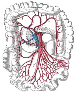
The right colic artery arises from about the middle of the concavity of the superior mesenteric artery, or from a stem common to it and the ileocolic.

The superficial palmar arch is formed predominantly by the ulnar artery, with a contribution from the superficial palmar branch of the radial artery. However, in some individuals the contribution from the radial artery might be absent, and instead anastomoses with either the princeps pollicis artery, the radialis indicis artery, or the median artery, the former two of which are branches from the radial artery.
The deep palmar arch is an arterial network found in the palm. It is usually formed mainly from the terminal part of the radial artery, with the ulnar artery contributing via its deep palmar branch, by an anastomosis. This is in contrast to the superficial palmar arch, which is formed predominantly by the ulnar artery.

The cardiac plexus is a plexus of nerves situated at the base of the heart that innervates the heart.

The superficial temporal vein is a vein of the side of the head. It begins on the side and vertex of the skull in a network of veins which communicates with the frontal vein and supraorbital vein, with the corresponding vein of the opposite side, and with the posterior auricular vein and occipital vein. It ultimately crosses the posterior root of the zygomatic arch, enters the parotid gland, and unites with the internal maxillary vein to form the posterior facial vein.
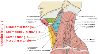
The anterior triangle is a region of the neck.

A palatal lift prosthesis is a prosthesis that addresses a condition referred to as palatopharyngeal incompetence. Palatopharyngeal incompetence broadly refers to a muscular inability to sufficiently close the port between the nasopharynx and oropharynx during speech and/or swallowing. An inability to adequately close the palatopharyngeal port during speech results in hypernasalance that, depending upon its severity, can render speakers difficult to understand or unintelligible. The potential for compromised intelligibility secondary to hypernasalance is underscored when consideration is given to the fact that only three English language phonemes – /m/, /n/, and /ng/ – are pronounced with an open palatopharyngeal port. Furthermore, an impaired ability to effect a closure of the palatopharyngeal port while swallowing can result in the nasopharyngeal regurgitation of liquid or solid boluses.

The medial arcuate ligament is a tendinous fascia that arches over the psoas major muscle as it passes through the diaphragm.

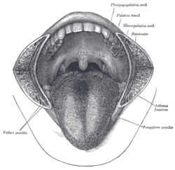
The palatoglossal arch on either side runs downward, lateral, and forward to the side of the base of the tongue, and is formed by the projection of the glossopalatine muscle with its covering mucous membrane. It is the anterior border of the isthmus of the fauces and marks the border between the mouth and the palatopharyngeal arch. The latter marks the beginning of the pharynx.

The temporal branches of the facial nerve crosses the zygomatic arch to the temporal region, supplying the auriculares anterior and superior, and joining with the zygomaticotemporal branch of the maxillary nerve, and with the auriculotemporal branch of the mandibular nerve.
The cervical branch of the facial nerve runs forward beneath the platysma, and forms a series of arches across the side of the neck over the suprahyoid region.
Special visceral efferent fibers (SVE) are the efferent nerve fibers that provide motor innervation to the muscles of the pharyngeal arches in humans, and the branchial arches in fish.
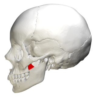
In human anatomy, the mandible's coronoid process is a thin, triangular eminence, which is flattened from side to side and varies in shape and size. Its anterior border is convex and is continuous below with the anterior border of the ramus. Its posterior border is concave and forms the anterior boundary of the mandibular notch. The lateral surface is smooth, and affords insertion to the temporalis and masseter muscles. Its medial surface gives insertion to the temporalis, and presents a ridge which begins near the apex of the process and runs downward and forward to the inner side of the last molar tooth.
The fauces, isthmus of fauces, or the oropharyngeal isthmus, is the opening at the back of the mouth into the throat. It is a narrow passage between the pharynx and the base of the tongue.

The tonsillar fossa is a space delineated by the triangular fold of the palatoglossal and palatopharyngeal arches within the lateral wall of the oral cavity..

The pharynx is the part of the throat behind the mouth and nasal cavity, and above the esophagus and larynx – the tubes going down to the stomach and the lungs. It is found in vertebrates and invertebrates, though its structure varies across species.
Passavant's ridge is a mucous elevation situated behind the floor of the naso-pharynx.