
Marfan syndrome (MFS) is a multi-systemic genetic disorder that affects the connective tissue. Those with the condition tend to be tall and thin, with long arms, legs, fingers, and toes. They also typically have exceptionally flexible joints and abnormally curved spines. The most serious complications involve the heart and aorta, with an increased risk of mitral valve prolapse and aortic aneurysm. The lungs, eyes, bones, and the covering of the spinal cord are also commonly affected. The severity of the symptoms is variable.
Patau syndrome is a syndrome caused by a chromosomal abnormality, in which some or all of the cells of the body contain extra genetic material from chromosome 13. The extra genetic material disrupts normal development, causing multiple and complex organ defects.

An eyelid is a thin fold of skin that covers and protects an eye. The levator palpebrae superioris muscle retracts the eyelid, exposing the cornea to the outside, giving vision. This can be either voluntarily or involuntarily. "Palpebral" means relating to the eyelids. Its key function is to regularly spread the tears and other secretions on the eye surface to keep it moist, since the cornea must be continuously moist. They keep the eyes from drying out when asleep. Moreover, the blink reflex protects the eye from foreign bodies. A set of specialized hairs known as lashes grow from the upper and lower eyelid margins to further protect the eye from dust and debris.

The lacrimal glands are paired exocrine glands, one for each eye, found in most terrestrial vertebrates and some marine mammals, that secrete the aqueous layer of the tear film. In humans, they are situated in the upper lateral region of each orbit, in the lacrimal fossa of the orbit formed by the frontal bone. Inflammation of the lacrimal glands is called dacryoadenitis. The lacrimal gland produces tears which are secreted by the lacrimal ducts, and flow over the ocular surface, and then into canals that connect to the lacrimal sac. From that sac, the tears drain through the lacrimal duct into the nose.

Fetal alcohol spectrum disorders (FASDs) are a group of conditions that can occur in a person who is exposed to alcohol during gestation, as a result of their mother drinking alcohol during pregnancy. The several forms of the condition are: fetal alcohol syndrome (FAS), partial fetal alcohol syndrome (pFAS), alcohol-related neurodevelopmental disorder (ARND), and neurobehavioral disorder associated with prenatal alcohol exposure (ND-PAE). Other terms used are fetal alcohol effects (FAE), partial fetal alcohol effects (PFAE), alcohol-related birth defects (ARBD), and static encephalopathy, but these terms have fallen out of favor and are no longer considered part of the spectrum.

Cri du chat syndrome is a rare genetic disorder due to a partial chromosome deletion on chromosome 5. Its name is a French term referring to the characteristic cat-like cry of affected children. It was first described by Jérôme Lejeune in 1963. The condition affects an estimated 1 in 50,000 live births across all ethnicities and is more common in females by a 4:3 ratio.

A coloboma is a hole in one of the structures of the eye, such as the iris, retina, choroid, or optic disc. The hole is present from birth and can be caused when a gap called the choroid fissure, which is present during early stages of prenatal development, fails to close up completely before a child is born. Ocular coloboma is relatively uncommon, affecting less than one in every 10,000 births.
Duane syndrome is a congenital rare type of strabismus most commonly characterized by the inability of the eye to move outward. The syndrome was first described by ophthalmologists Jakob Stilling (1887) and Siegmund Türk (1896), and subsequently named after Alexander Duane, who discussed the disorder in more detail in 1905.

The canthus is either corner of the eye where the upper and lower eyelids meet. More specifically, the inner and outer canthi are, respectively, the medial and lateral ends/angles of the palpebral fissure.

Sturge–Weber syndrome, sometimes referred to as encephalotrigeminal angiomatosis, is a rare congenital neurological and skin disorder. It is one of the phakomatoses and is often associated with port-wine stains of the face, glaucoma, seizures, intellectual disability, and ipsilateral leptomeningeal angioma. Sturge–Weber syndrome can be classified into three different types. Type 1 includes facial and leptomeningeal angiomas as well as the possibility of glaucoma or choroidal lesions. Normally, only one side of the brain is affected. This type is the most common. Type 2 involvement includes a facial angioma with a possibility of glaucoma developing. There is no evidence of brain involvement. Symptoms can show at any time beyond the initial diagnosis of the facial angioma. The symptoms can include glaucoma, cerebral blood flow abnormalities and headaches. More research is needed on this type of Sturge–Weber syndrome. Type 3 has leptomeningeal angioma involvement exclusively. The facial angioma is absent and glaucoma rarely occurs. This type is only diagnosed via brain scan.
Full trisomy 9 is a rare and fatal chromosomal disorder caused by having three copies (trisomy) of chromosome number 9. It can be a viable condition if trisomy affects only part of the cells of the body (mosaicism) or in cases of partial trisomy in which cells have a normal set of two entire chromosomes 9 plus part of a third copy, usually of the short arm of the chromosome.

The superior tarsal muscle is a smooth muscle adjoining the levator palpebrae superioris muscle that helps to raise the upper eyelid.
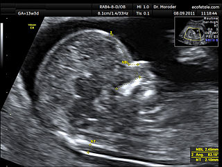
A nuchal scan or nuchal translucency (NT) scan/procedure is a sonographic prenatal screening scan (ultrasound) to detect chromosomal abnormalities in a fetus, though altered extracellular matrix composition and limited lymphatic drainage can also be detected.

Blepharophimosis is a congenital anomaly in which the eyelids are underdeveloped such that they cannot open as far as usual and permanently cover part of the eyes. Both the vertical and horizontal palpebral fissures are shortened; the eyes also appear spaced more widely apart as a result, known as telecanthus.

Not to be confused with Heterochromia, an optical condition which is commonly associated with cats

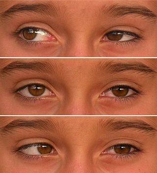
Telecanthus, or dystopia canthorum, refers to increased distance between the inner corners of the eyelids, while the inter-pupillary distance is normal. This is in contrast to hypertelorism, in which the distance between the whole eyes is increased. Telecanthus and hypertelorism are each associated with multiple congenital disorders.
Encephalocraniocutaneous lipomatosis (ECCL), is a rare condition primarily affecting the brain, eyes, and skin of the head and face. It is characterized by unilateral subcutaneous and intracranial lipomas, alopecia, unilateral porencephalic cysts, epibulbar choristoma and other ophthalmic abnormalities. This condition is described as sporadic because it occurs in people without a history of the disorder in their family.
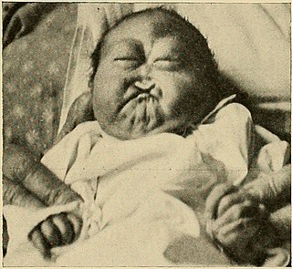
Ankyloblepharon is a medical condition, defined as the adhesion of the edges of the upper eyelid with the lower eyelid. Ankyloblepharon must be differentiated from blepharophimosis, in which palpebral aperture is reduced and there is telecanthus, but the eyelid margins are normal. Another condition similar to ankyloblepharon is symblepharon, in which the palpebral conjunctiva is attached to the bulbar conjunctiva. Recognition of ankyloblepharon necessitates systemic examination to detect associated abnormalities such as genitourinary and cardiac abnormalities and syndactyly.

Trisomy X, also known as triple X syndrome and characterized by the karyotype 47,XXX, is a chromosome disorder in which a female has an extra copy of the X chromosome. It is relatively common and occurs in 1 in 1,000 females, but is rarely diagnosed; fewer than 10% of those with the condition know they have it.
