Related Research Articles

Heart murmurs are unique heart sounds produced when blood flows across a heart valve or blood vessel. This occurs when turbulent blood flow creates a sound loud enough to hear with a stethoscope. Turbulent blood flow is not smooth. The sound differs from normal heart sounds by their characteristics. For example, heart murmurs may have a distinct pitch, duration and timing. The major way health care providers examine the heart on physical exam is heart auscultation; another clinical technique is palpation, which can detect by touch when such turbulence causes the vibrations called cardiac thrill. A murmur is a sign found during the cardiac exam. Murmurs are of various types and are important in the detection of cardiac and valvular pathologies.
Pulmonary heart disease, also known as cor pulmonale, is the enlargement and failure of the right ventricle of the heart as a response to increased vascular resistance or high blood pressure in the lungs.

Afterload is the pressure that the heart must work against to eject blood during systole. Afterload is proportional to the average arterial pressure. As aortic and pulmonary pressures increase, the afterload increases on the left and right ventricles respectively. Afterload changes to adapt to the continually changing demands on an animal's cardiovascular system. Afterload is proportional to mean systolic blood pressure and is measured in millimeters of mercury.

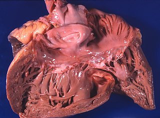
Mitral stenosis is a valvular heart disease characterized by the narrowing of the opening of the mitral valve of the heart. It is almost always caused by rheumatic valvular heart disease. Normally, the mitral valve is about 5 cm2 during diastole. Any decrease in area below 2 cm2 causes mitral stenosis. Early diagnosis of mitral stenosis in pregnancy is very important as the heart cannot tolerate increased cardiac output demand as in the case of exercise and pregnancy. Atrial fibrillation is a common complication of resulting left atrial enlargement, which can lead to systemic thromboembolic complications such as stroke.

Aortic regurgitation (AR), also known as aortic insufficiency (AI), is the leaking of the aortic valve of the heart that causes blood to flow in the reverse direction during ventricular diastole, from the aorta into the left ventricle. As a consequence, the cardiac muscle is forced to work harder than normal.

Mitral regurgitation(MR), also known as mitral insufficiency or mitral incompetence, is a form of valvular heart disease in which the mitral valve is insufficient and does not close properly when the heart pumps out blood. It is the abnormal leaking of blood backwards – regurgitation from the left ventricle, through the mitral valve, into the left atrium, when the left ventricle contracts. Mitral regurgitation is the most common form of valvular heart disease.
A transthoracic echocardiogram (TTE) is the most common type of echocardiogram, which is a still or moving image of the internal parts of the heart using ultrasound. In this case, the probe is placed on the chest or abdomen of the subject to get various views of the heart. It is used as a non-invasive assessment of the overall health of the heart, including a patient's heart valves and degree of heart muscle contraction. The images are displayed on a monitor for real-time viewing and then recorded.
Valvular heart disease is any cardiovascular disease process involving one or more of the four valves of the heart. These conditions occur largely as a consequence of aging, but may also be the result of congenital (inborn) abnormalities or specific disease or physiologic processes including rheumatic heart disease and pregnancy.
In medicine, the cardiac examination, also precordial exam, is performed as part of a physical examination, or when a patient presents with chest pain suggestive of a cardiovascular pathology. It would typically be modified depending on the indication and integrated with other examinations especially the respiratory examination.
The apex beat, also called the apical impulse, is the pulse felt at the point of maximum impulse (PMI), which is the point on the precordium farthest outwards (laterally) and downwards (inferiorly) from the sternum at which the cardiac impulse can be felt. The cardiac impulse is the vibration resulting from the heart rotating, moving forward, and striking against the chest wall during systole. The PMI is not the apex of the heart but is on the precordium not far from it. Another theory for the occurrence of the PMI is the early systolic contraction of the longitudinal fibers of the left ventricle located on the endocardial surface of this chamber. This period of the cardiac cycle is called isovolumic contraction. Because the contraction starts near the base of the left ventricle and spreads toward the apex most of the longitudinal fibers of the left ventricle have shortened before the apex. The rapidly increasing pressure developed by the shortening of these fibers causes the aortic valve to open and the apex to move outward causing the PMI. Anatomical dissection of the musculature of the apex reveals that muscle fibers are no longer longitudinal oriented but form a spiral mass of muscular tissues which may also have an effect on the ability of the apex to contract longitudinally. After the longitudinal fibers contract, the ejection of blood out of the left ventricle is accomplished by the torsional action of the circumferential muscle fibers of the left ventricle that are in the mid-portion of the ventricle and contract after the longitudinal fibers. During the longitudinal fiber contraction, the volume of the left ventricle has not changed keeping the apex in intimate contact with the chest wall allowing the ability to feel the apex move outward before the heart empties greater than 55% of its volume and the apex falling away from the chest wall.

Pulmonary valve stenosis (PVS) is a heart valve disorder. Blood going from the heart to the lungs goes through the pulmonary valve, whose purpose is to prevent blood from flowing back to the heart. In pulmonary valve stenosis this opening is too narrow, leading to a reduction of flow of blood to the lungs.

Right ventricular hypertrophy (RVH) is a condition defined by an abnormal enlargement of the cardiac muscle surrounding the right ventricle. The right ventricle is one of the four chambers of the heart. It is located towards the lower-end of the heart and it receives blood from the right atrium and pumps blood into the lungs.

Pulmonaryinsufficiency is a condition in which the pulmonary valve is incompetent and allows backflow from the pulmonary artery to the right ventricle of the heart during diastole. While a small amount of backflow may occur ordinarily, it is usually only shown on an echocardiogram and is harmless. More pronounced regurgitation that is noticed through a routine physical examination is a medical sign of disease and warrants further investigation. If it is secondary to pulmonary hypertension it is referred to as a Graham Steell murmur.
The following outline is provided as an overview of and topical guide to cardiology, the branch of medicine dealing with disorders of the human heart. The field includes medical diagnosis and treatment of congenital heart defects, coronary artery disease, heart failure, valvular heart disease and electrophysiology. Physicians who specialize in cardiology are called cardiologists.
Right atrial enlargement (RAE) is a form of cardiomegaly, or heart enlargement. It can broadly be classified as either right atrial hypertrophy (RAH), overgrowth, or dilation, like an expanding balloon. Common causes include pulmonary hypertension, which can be the primary defect leading to RAE, or pulmonary hypertension secondary to tricuspid stenosis; pulmonary stenosis or Tetralogy of Fallot i.e. congenital diseases; chronic lung disease, such as cor pulmonale. Other recognised causes are: right ventricular failure, tricuspid regurgitation, and atrial septal defect.
The cardiovascular examination is a portion of the physical examination that involves evaluation of the cardiovascular system. The exact contents of the examination will vary depending on the presenting complaint but a complete examination will involve the heart, lungs, belly and the blood vessels.

A ventricular outflow tract obstruction is a heart condition in which either the right or left ventricular outflow tract is blocked or obstructed. These obstructions represent a spectrum of disorders. Majority of these cases are congenital, but some are acquired throughout life.
Pressure overload refers to the pathological state of cardiac muscle in which it has to contract while experiencing an excessive afterload. Pressure overload may affect any of the four chambers of the heart, though the term is most commonly applied to one of the two ventricles. Chronic pressure overload leads to concentric hypertrophy of the cardiac muscle, which can in turn lead to heart failure, myocardial ischaemia or, in extreme cases, outflow obstruction.

Bernheim Syndrome is a presumed disorder whereby the right ventricle is severely compressed due to a shift in the ventricular septal wall of the heart leading to heart failure. It was first described by Hippolyte Bernheim in 1910. Today it is questioned whether or not Bernheim Syndrome is its own syndrome or a side effect of other cardiac conditions such as left ventricular heart failure whereby the left ventricle is substantially enlarged which encroaches on the space of the right ventricle.
References
- ↑ David Humes, H. (2001). Kelley's Essentials of Internal Medicine. ISBN 9780781719377.
- ↑ Hemanth, I. K.; Mattummal, Shafeeq (17 July 2017). Clinical Pearls in Cardiology. ISBN 9789351524366.
- ↑ "Precordial Impulses". Clinical Methods: The History, Physical, and Laboratory Examinations. Butterworths. 1990. ISBN 9780409900774.
- ↑ Clinical Examination: A Systematic Guide to Physical Diagnosis 5th Edition Nicholas Talley Simmon O' Connor
- ↑ "Precordial Impulses". Clinical Methods: The History, Physical, and Laboratory Examinations. Butterworths. 1990. ISBN 9780409900774.
- ↑ Sam, Amir H.; James T.H. Teo (2010). Rapid Medicine. Wiley-Blackwell. ISBN 978-1-4051-8323-9.