
An electron microscope is a microscope that uses a beam of electrons as a source of illumination. They use electron optics that are analogous to the glass lenses of an optical light microscope to control the electron beam, for instance focusing them to produce magnified images or electron diffraction patterns. As the wavelength of an electron can be up to 100,000 times smaller than that of visible light, electron microscopes have a much higher resolution of about 0.1 nm, which compares to about 200 nm for light microscopes. Electron microscope may refer to:

Microscopy is the technical field of using microscopes to view objects and areas of objects that cannot be seen with the naked eye. There are three well-known branches of microscopy: optical, electron, and scanning probe microscopy, along with the emerging field of X-ray microscopy.

A microscope is a laboratory instrument used to examine objects that are too small to be seen by the naked eye. Microscopy is the science of investigating small objects and structures using a microscope. Microscopic means being invisible to the eye unless aided by a microscope.

A scanning electron microscope (SEM) is a type of electron microscope that produces images of a sample by scanning the surface with a focused beam of electrons. The electrons interact with atoms in the sample, producing various signals that contain information about the surface topography and composition of the sample. The electron beam is scanned in a raster scan pattern, and the position of the beam is combined with the intensity of the detected signal to produce an image. In the most common SEM mode, secondary electrons emitted by atoms excited by the electron beam are detected using a secondary electron detector. The number of secondary electrons that can be detected, and thus the signal intensity, depends, among other things, on specimen topography. Some SEMs can achieve resolutions better than 1 nanometer.

The retina is the innermost, light-sensitive layer of tissue of the eye of most vertebrates and some molluscs. The optics of the eye create a focused two-dimensional image of the visual world on the retina, which then processes that image within the retina and sends nerve impulses along the optic nerve to the visual cortex to create visual perception. The retina serves a function which is in many ways analogous to that of the film or image sensor in a camera.

A saccade is a quick, simultaneous movement of both eyes between two or more phases of fixation in the same direction. In contrast, in smooth-pursuit movements, the eyes move smoothly instead of in jumps. The phenomenon can be associated with a shift in frequency of an emitted signal or a movement of a body part or device. Controlled cortically by the frontal eye fields (FEF), or subcortically by the superior colliculus, saccades serve as a mechanism for fixation, rapid eye movement, and the fast phase of optokinetic nystagmus. The word appears to have been coined in the 1880s by French ophthalmologist Émile Javal, who used a mirror on one side of a page to observe eye movement in silent reading, and found that it involves a succession of discontinuous individual movements.
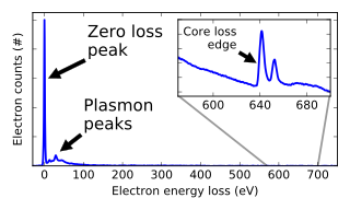
Electron energy loss spectroscopy (EELS) is a form of electron microscopy in which a material is exposed to a beam of electrons with a known, narrow range of kinetic energies. Some of the electrons will undergo inelastic scattering, which means that they lose energy and have their paths slightly and randomly deflected. The amount of energy loss can be measured via an electron spectrometer and interpreted in terms of what caused the energy loss. Inelastic interactions include phonon excitations, inter- and intra-band transitions, plasmon excitations, inner shell ionizations, and Cherenkov radiation. The inner-shell ionizations are particularly useful for detecting the elemental components of a material. For example, one might find that a larger-than-expected number of electrons comes through the material with 285 eV less energy than they had when they entered the material. This is approximately the amount of energy needed to remove an inner-shell electron from a carbon atom, which can be taken as evidence that there is a significant amount of carbon present in the sample. With some care, and looking at a wide range of energy losses, one can determine the types of atoms, and the numbers of atoms of each type, being struck by the beam. The scattering angle can also be measured, giving information about the dispersion relation of whatever material excitation caused the inelastic scattering.

The optical microscope, also referred to as a light microscope, is a type of microscope that commonly uses visible light and a system of lenses to generate magnified images of small objects. Optical microscopes are the oldest design of microscope and were possibly invented in their present compound form in the 17th century. Basic optical microscopes can be very simple, although many complex designs aim to improve resolution and sample contrast.

Ultrastructure is the architecture of cells and biomaterials that is visible at higher magnifications than found on a standard optical light microscope. This traditionally meant the resolution and magnification range of a conventional transmission electron microscope (TEM) when viewing biological specimens such as cells, tissue, or organs. Ultrastructure can also be viewed with scanning electron microscopy and super-resolution microscopy, although TEM is a standard histology technique for viewing ultrastructure. Such cellular structures as organelles, which allow the cell to function properly within its specified environment, can be examined at the ultrastructural level.

A scanning transmission electron microscope (STEM) is a type of transmission electron microscope (TEM). Pronunciation is [stɛm] or [ɛsti:i:ɛm]. As with a conventional transmission electron microscope (CTEM), images are formed by electrons passing through a sufficiently thin specimen. However, unlike CTEM, in STEM the electron beam is focused to a fine spot which is then scanned over the sample in a raster illumination system constructed so that the sample is illuminated at each point with the beam parallel to the optical axis. The rastering of the beam across the sample makes STEM suitable for analytical techniques such as Z-contrast annular dark-field imaging, and spectroscopic mapping by energy dispersive X-ray (EDX) spectroscopy, or electron energy loss spectroscopy (EELS). These signals can be obtained simultaneously, allowing direct correlation of images and spectroscopic data.

Asteroid hyalosis is a degenerative condition of the eye involving small white opacities in the vitreous humor. It is known to occur in humans, dogs, cats, horses, and chinchillas. Clinically, these opacities are quite refractile, giving the appearance of stars shining in the night sky—except that ocular asteroids are often quite mobile. Ocular asteroids must be distinguished from the more common typical vitreous floaters, which are usually fibrillar or cellular condensates. The cause of asteroid hyalosis is unknown, but it has been associated with diabetes mellitus, hypertension, hypercholesterolemia, and, in certain animals, tumors of the ciliary body. In dogs, asteroid hyalosis is considered to be an age-related change. The asteroid bodies are made up of hydroxylapatite, which in turn consists of calcium and phosphates or phospholipids. While asteroid hyalosis does not usually severely affect vision, the floating opacities can be quite annoying, and may interfere significantly with visualization and testing of the retina. While treatment of asteroid hyalosis is usually unnecessary, vitrectomy may occasionally be indicated, for both diagnostic and therapeutic purposes.

John Maxwell Cowley was an American Regents Professor at Arizona State University. The John M. Cowley Center for High-Resolution Electron Microscopy at Arizona State is named in his honor.
John Cowley was an extraordinarily productive scientist over more than five decades. He made pioneering contributions in the fields of electron microscopy, diffraction and crystallography, all of which brought him widespread recognition. He received the highest awards of the International Union of Crystallography, the Electron Microscopy Society of America and the American Crystallographic Society, and he was honored by election to Fellowship of the Australian Academy of Science, The Royal Society of London, and the American Physical Society. His monograph Diffraction Physics remains the standard reference in the field. His ideas, enthusiasm and basic understanding of electron optics and diffraction phenomena provided a valued source of leadership to many generations of students and co-workers, and he was universally admired by his peers and colleagues as a great and inspiring scientist.

The environmental scanning electron microscope (ESEM) is a scanning electron microscope (SEM) that allows for the option of collecting electron micrographs of specimens that are wet, uncoated, or both by allowing for a gaseous environment in the specimen chamber. Although there were earlier successes at viewing wet specimens in internal chambers in modified SEMs, the ESEM with its specialized electron detectors and its differential pumping systems, to allow for the transfer of the electron beam from the high vacuum in the gun area to the high pressure attainable in its specimen chamber, make it a complete and unique instrument designed for the purpose of imaging specimens in their natural state. The instrument was designed originally by Gerasimos Danilatos while working at the University of New South Wales.

Vision is the most important sense for birds, since good eyesight is essential for safe flight. Birds have a number of adaptations which give visual acuity superior to that of other vertebrate groups; a pigeon has been described as "two eyes with wings". Birds are theropod dinosaurs, and the avian eye resembles that of other sauropsids, with ciliary muscles that can change the shape of the lens rapidly and to a greater extent than in the mammals. Birds have the largest eyes relative to their size in the animal kingdom, and movement is consequently limited within the eye's bony socket. In addition to the two eyelids usually found in vertebrates, bird's eyes are protected by a third transparent movable membrane. The eye's internal anatomy is similar to that of other vertebrates, but has a structure, the pecten oculi, unique to birds.

Mammals normally have a pair of eyes. Although mammalian vision is not so excellent as bird vision, it is at least dichromatic for most of mammalian species, with certain families possessing a trichromatic color perception.
Scanning confocal electron microscopy (SCEM) is an electron microscopy technique analogous to scanning confocal optical microscopy (SCOM). In this technique, the studied sample is illuminated by a focussed electron beam, as in other scanning microscopy techniques, such as scanning transmission electron microscopy or scanning electron microscopy. However, in SCEM, the collection optics are arranged symmetrically to the illumination optics to gather only the electrons that pass the beam focus. This results in superior depth resolution of the imaging. The technique is relatively new and is being actively developed.

A diamond knife is a very sharp knife in which the edge is made from diamond, invented by Humberto Fernández-Morán in 1955. Diamond knives are used for medical and scientific applications where an extremely sharp and long-lasting edge is essential. The knives are very expensive to purchase, depending on the quality and size of the knife; in addition the knives must be professionally sharpened as the edge dulls.

Immunogold labeling or immunogold staining (IGS) is a staining technique used in electron microscopy. This staining technique is an equivalent of the indirect immunofluorescence technique for visible light. Colloidal gold particles are most often attached to secondary antibodies which are in turn attached to primary antibodies designed to bind a specific antigen or other cell component. Gold is used for its high electron density which increases electron scatter to give high contrast 'dark spots'.
Serial block-face scanning electron microscopy is a method to generate high resolution three-dimensional images from small samples. The technique was developed for brain tissue, but it is widely applicable for any biological samples. A serial block-face scanning electron microscope consists of an ultramicrotome mounted inside the vacuum chamber of a scanning electron microscope. Samples are prepared by methods similar to that in transmission electron microscopy (TEM), typically by fixing the sample with aldehyde, staining with heavy metals such as osmium and uranium then embedding in an epoxy resin. The surface of the block of resin-embedded sample is imaged by detection of back-scattered electrons. Following imaging the ultramicrotome is used to cut a thin section from the face of the block. After the section is cut, the sample block is raised back to the focal plane and imaged again. This sequence of sample imaging, section cutting and block raising can acquire many thousands of images in perfect alignment in an automated fashion. Practical serial block-face scanning electron microscopy was invented in 2004 by Winfried Denk at the Max-Planck-Institute in Heidelberg and is commercially available from Gatan Inc., Thermo Fisher Scientific (VolumeScope) and ConnectomX.

SEM-XRF is an established technical term for adding a X-ray generator to a Scanning Electron Microscope (SEM). Technological progress in the fields of small-spot low-power X-ray tubes and of polycapillary X-ray optics has enabled the development of compact micro-focus X-ray sources that can be attached to a SEM equipped for energy-dispersive X-ray spectroscopy.
Abumandour, M. M. A., Bassuoni, N. F., & Hanafy, B. G. (2021). Ultrastructural studies of the pecten oculi of the Garganey (Anas querquedula, Linnaeus 1758) and the Eurasian common moorhen (Gallinula chloropus chloropus, Linnaeus 1758). Microsc Res Tech, 84(9), 1967-1976. doi:https://doi.org/10.1002/jemt.23752
Abumandour, M. M. A., Morsy, K., & Hanafy, B. G. (2022). Biological features of the pecten oculi of the European wild quail (Coturnix coturnix): Adaptative habits to Northern Egyptian coast with novel vision to its SEM–EDX analysis. Microsc Res Tech, 85(12), 3817-3829. doi:10.1002/jemt.24236
Elghoul, M., Morsy, K., & Abumandour, M. M. A. (2022). Ultrastructural characterizations of the pecten oculi of the common ostrich (Struthio camelus): New insight to scanning electron microscope–energy dispersive X‐ray analysis. Microsc Res Tech, 85(5), 1654-1662.
Kandyle, R., El Basyouny, H. A., Morsy, K., Abourashed, N. M., Madkour, N., & Abumandour, M. M. A. (2022). Gross, ultrastructural, and histological characterizations of pecten oculi of the glossy ibis (Plegadis falcinellus): New insights into its scanning electron microscope–energy dispersive X‐ray analysis. Microscopy research and technique, 85(12), 3908-3920. doi:0.1002/jemt.24228
Gewily, D., Shalaby, W., Abumandour, M. M. A., Choudhary, O. P., & Kandyel, R. (2024). Pecten oculi of kestrel (Falco tinnunculus rupicolaeformes) and little owl (Athene noctua glaux): Scanning electron microscopy and histology with unique insights into SEM–EDX elemental analysis. Microscopy research and technique, 87(3), 546-564.
