Peripheral ulcerative keratitis

Last updated
Peripheral ulcerative keratitis

Peripheral Ulcerative Keratitis (PUK) is a group of destructive inflammatory diseases involving the peripheral cornea in human eyes. [1] The symptoms of PUK include pain, redness of the eyeball, photophobia, and decreased vision accompanied by distinctive signs of crescent-shaped damage of the cornea. [2] [3] The causes of this disease are broad, ranging from injuries, contamination of contact lenses, to association with other systemic conditions. [4] PUK is associated with different ocular and systemic diseases. [5] Mooren's ulcer is a common form of PUK. [5] The majority of PUK is mediated by local or systemic immunological processes, which can lead to inflammation and eventually tissue damage. Standard PUK diagnostic test involves reviewing the medical history and a completing physical examinations. [6] Two major treatments are the use of medications such as corticosteroids or other immunosuppressive agents and surgical resection of the conjunctiva. [7] The prognosis of PUK is unclear with one study providing potential complications. [8] PUK is a rare condition with an estimated incidence of 3 per million annually. [9] [10]

Contents

Signs and symptoms

The most easily identifiable sign is a visible lesion of the cornea presented usually in a crescent shape. [2] [3] [11] Common reasons for destruction are stromal degradation and epithelial defects on the inflammatory cells. [2] There would be a change in conformation of the peripheral cornea, depending on the severity of corneal thinning. [11] This process is usually accompanied by the possibility of concealing perforation. [12] The formation of an oval-shaped ulcer at the margin of the cornea is also a sign. [2]

Symptoms of PUK include pain, redness, tearing, increased sensitivity to bright light, impaired or blurred vision, and the feeling of foreign objects trapped in the eyes. [5] [13]

Association

There are several associations of PUK to ocular and systemic diseases. [5] [7] [14] Rheumatoid arthritis (RA), [9]   Wegner's granulomatosis (WG), and Polyarteritis Nodosa (PAN) are the most common systemic conditions. [5]

Causes

There are three major causes for PUK. One possible cause is injury due to any kind of scratches by sharp or hard objects on the surface of the cornea. [7] The scratched area forms an opening in the cornea, allowing microorganisms to access the cornea and lead to infection. [7] Contamination of contact lenses is another cause as fungi, bacteria and parasites, microscopic parasite acanthamoeba, in particular, could inhabit the surface of the carrying case of the contact lens. [4] When placing the contact lens to one's eyes, invisible microorganisms may contaminate the cornea resulting in PUK. [4] An extended period of wearing contact lenses could also cause damage on the cornea surface, allowing the entry of microorganisms to the cornea. [4] Other than contamination of contact lenses, contamination occurring in water could also cause PUK. Especially in places like the ocean, rivers, lakes and hot tubs, massive amounts of bacteria, fungi, and parasites exist. [16] When there is an injury on the cornea surface, contact with contaminated water could transfer unwanted microorganisms into the cornea resulting in PUK. [17] Virus and bacteria are sources of infection to the cornea. Herpes virus and bacteria that cause gonorrhea are some examples. [17]

Anatomy and pathogenesis

The corneal epithelium consists of five to six layers of cells with a total thickness of around 0.52mm. [1] The cornea thickens to 0.65mm towards the periphery of the cornea. [1] Stroma, which accounts for 90% of the corneal thickness, refers to the middle layer between epithelium and endothelium. [1] It is present in the peripheral cornea to act as a transitional zone between the sclera and cornea. [1] Limbal vasculature, deriving from capillaries that surround the peripheral cornea, supplies the stroma. [1] Various molecules normally diffuse from these capillaries at the periphery to the central cornea. [1] With limited diffusion, there is a higher concentration of IgM, factor C1 of the complement cascade, and Langerhans cells. [1] [7]  

Any kind of inflammatory stimulus present in the peripheral cornea results in recruitment of neutrophil and activation of both classical and alternative pathways of immune response, namely the humoral and cell-mediated autoimmune responses. [16] These responses will lead to the formation of antigen-specific antibodies to combat foreign antigens. [16] However, antigen-antibody complexes formed may deposit in the vascular endothelium and activate complements leading to severe local inflammation. [16] Under this circumstance, inflammatory cells, such as macrophages and neutrophils, enter the peripheral cornea. [16] These inflammatory cells release enzymes protease and collagenases, causing potential disruption of the corneal stroma. [16] The additional release of cytokines, for example, interleukin-1, from these cells further accelerates the process of stromal destruction. [7]

Mooren's ulcer and relevant classification

Mooren's ulcer is a common form of PUK. [5] [11] One classification of Mooren's ulcer, based on the clinical presentation, includes bilateral indolent mooren's ulcer, bilateral aggressive mooren's ulcer and unilateral mooren's ulcer. [5] Unilateral mooren's ulcer, meaning ulcer of one eye, mainly affects elderly above 60 years old. Rapid onset with redness and severe pain of the affected eye and either slow or extremely quick progression are some typical characteristics of unilateral mooren's ulcer. [5] Bilateral aggressive mooren's ulcer is prevalent in Indian between age 14 to 40. [5] The common presentation includes the appearance of lesions in one eye, followed by the development of lesions in another eye. [5] Finally, bilateral indolent mooren's ulcer is common in patients of at least 50-year-old. [5] It usually progresses slowly and causes little or no pain. [5]

Other classification methods also exist. The first one is classifying Mooren's ulcers based on clinical presentation and prognosis into two categories. [14] The first type is usually presented unilaterally, accompanied by symptoms ranging from mild to moderate. [14] Therefore, it has a more effective response to treatment. In contrast, type II appears in a bilateral manner, with severe symptoms and poor outcome of treatment. [14] The second classification is based on severity. [14] Grade I refers to corneal thinning, grade II describes impending corneal perforation, and grade III is corneal perforation with a diameter greater than 2mm. [14]

Diagnosis

There are many investigative modalities available for diagnosing PUK, including history review and physical examination. [6] [7] A thorough history of ocular infections, contact lens usage, other medication, or surgery is necessary to identify possible presence of associated diseases. [7] An ophthalmic examination helps identify whether it is due to local pathogenesis. [7] Physical examinations allow more understanding of the underlying systemic process. [7]

A standard testing procedure includes hematological investigations, immunological testing, followed by chest X-ray. Hematological investigations are blood tests estimating hemoglobin, platelet counts, total white blood cell counts, erythrocyte sedimentation rate and viscosity. [6] Other common body checks include urinalysis and liver and renal function tests. [6] The selection of immunological testing for various markers is based on numerous additional medical examinations and clinical history of the patient. [6]  Possible markers are antinuclear antibodies, anti-rheumatoid antibodies, and antibodies to cyclic citrullinated peptides. [6] Finally, a chest X-ray helps distinguish whether there are complications, such as pulmonary diseases, due to systemic conditions associated with PUK. [6]

One of the common causes of PUK is ocular infections by microorganisms such as bacteria, viruses, and fungi. [6] To detect the causative microorganism, doctors usually collect samples before the commencement of therapy and send them to laboratories. [6] Laboratory personnel then perform smear examination, inoculate the samples on culture media, and perform serological testing. [6] Serological testing is an antibody test providing information on PUK etiology. [6] The diagnosis of PUK due to systemic conditions requires a combination of serological and hematological testing, together with imaging techniques such as radiography and CT scanning. [6]

Treatments

Various PUK therapies are of different objectives, for example, inflammation control, halting of disease progression, stroma repairment, avoidance of secondary complications, and vision restoration. [18] A thorough understanding of PUK and different therapies is important. [7] Medical and surgical treatments are two major approaches to manage PUK. [7]

Medical therapy

As for medical therapy, there are several types of drugs available for PUK. Topical corticosteroids usually serve as therapy for milder unilateral cases of RA-associated PUK. [7] [9] [15] Systemic corticosteroids in the form of an oral dose are the acute management of more severe cases. [7] However, there are side effects with prolonged usage of oral corticosteroids. Immunosuppressive agents, such as azathioprine, cyclophosphamide, and methotrexate, have demonstrated efficacy in treating inflammatory eye diseases, including PUK. [7] [14] [9] The combined therapy of systemic corticosteroids up to 100 mg/day and immunosuppressive agents are used for severe cases of PUK. [7] Biological agents, such as anti-tissue necrosis factors (anti-TNF), is a well-established treatment of systemic inflammatory diseases, [7] Infliximab and Adalimumab are TNF blockers for treating RA-associated PUK. [9] However, the high cost and uncertainty of long-term side effects are the possible drawbacks. [7]

Surgical treatment

In terms of surgical treatment, conjunctival resection is a common procedure, which can temporarily remove local inflammatory mediators and collagenases and therefore slow down the disease progression. [7] Other surgical management includes corneal gluing, or keratoplasty procedures. [11] [14] [9] Corneal transplantation is a management option when there is severe corneal melting or perforation although one possible disadvantage is the risk of rejection. [14]

Surgical treatment helps maintain the integrity of the globe, but it is usually complementary because it alone cannot influence the underlying immunological process. [7] Therefore, medical and surgical treatments are commonly used in conjunction. [7]

Choice of treatment

The choice of treatment may be different depending on the nature of PUK, infectious or noninfectious. Selection of the right targeted antimicrobial therapy for infectious PUK is based on clinical judgement and culture results. [18]  For example, the appropriate treatment for bacterial infections is antibiotics, such as fluoroquinolones. [18] As for Mooren's ulcers, 56% of unilateral PUK and 50% of bilateral PUK in one eye showed recovery with intensive topical steroids. [18] Only 18% of patients with bilateral ulcers occurring simultaneously in both eyes show improvements with topical steroids alone; therefore a combination of immunosuppressive agents and systemic steroids should be given in early courses of management. [18] Corticosteroids are the first line of therapy, but side effects may arise from long-term usage. In addition, conjunctival resection can be performed to temporarily remove local inflammatory mediators, followed by the use of immunosuppressants. [18]

Prognosis

Currently, there are limited studies regarding the prognosis of PUK. However, one study has pointed out possible complications surrounding PUK include moderate to severe vision loss, corneal perforation and increased risk of recurrence. [8]

Epidemiology

PUK is a rare condition with an estimated incidence of 3 per million annually. [9] [10] Studies have reported that most patients with PUK are older than 60 years of age (32%). [10] Among them, men have a higher occurrence rate in men (60%). Most patients live in rural areas (66%) and are in the lower socioeconomic groups. [10] The age of those with PUK ranges from 5 to 89 years, with a mean age of 45.5 years. [10]

The mortality rate after PUK diagnosis in an investigation of 34 patients with and without immunosuppressive medication is 53% and 5%, respectively. [9] Another single-centre study involving 46 patients with RA reported a mortality rate of 15%. [9] Reports have also shown a possibility of PUK occurrence after any ocular surgery. [10] In a retrospective study of 771 eyes, 1.4% of participants reported developing late-onset PUK at an average of 3–6 months after surgery. [10]

Related Research Articles

<span class="mw-page-title-main">Keratitis</span> Medical condition

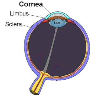
Keratitis is a condition in which the eye's cornea, the clear dome on the front surface of the eye, becomes inflamed. The condition is often marked by moderate to intense pain and usually involves any of the following symptoms: pain, impaired eyesight, photophobia, red eye and a 'gritty' sensation.

<span class="mw-page-title-main">Red eye (medicine)</span> Eye that appears red due to illness / injury. It is a common diseases

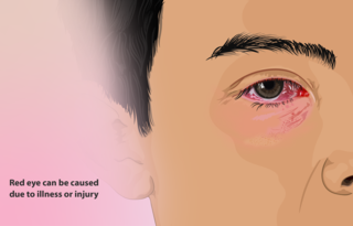
A red eye is an eye that appears red due to illness or injury. It is usually injection and prominence of the superficial blood vessels of the conjunctiva, which may be caused by disorders of these or adjacent structures. Conjunctivitis and subconjunctival hemorrhage are two of the less serious but more common causes.

<span class="mw-page-title-main">Recurrent corneal erosion</span> Medical condition

Recurrent corneal erosion is a disorder of the eyes characterized by the failure of the cornea's outermost layer of epithelial cells to attach to the underlying basement membrane. The condition is excruciatingly painful because the loss of these cells results in the exposure of sensitive corneal nerves. This condition can often leave patients with temporary blindness due to extreme light sensitivity (photophobia).

<span class="mw-page-title-main">Scleritis</span> Medical condition

Scleritis is a serious inflammatory disease that affects the white outer coating of the eye, known as the sclera. The disease is often contracted through association with other diseases of the body, such as granulomatosis with polyangiitis or rheumatoid arthritis. There are three types of scleritis: diffuse scleritis, nodular scleritis, and necrotizing scleritis. Scleritis may be the first symptom of onset of connective tissue disease.

<span class="mw-page-title-main">Phlyctenular keratoconjunctivitis</span> Medical condition

Phlyctenular keratoconjunctivitis is an inflammatory syndrome caused by a delayed hypersensitivity reaction to one or more antigens. The triggering antigen is usually a bacterial protein, but may also be a virus, fungus, or nematode.

<span class="mw-page-title-main">Corneal ulcers in animals</span> Veterinary inflammatory condition of the cornea

A corneal ulcer, or ulcerative keratitis, is an inflammatory condition of the cornea involving loss of its outer layer. It is very common in dogs and is sometimes seen in cats. In veterinary medicine, the term corneal ulcer is a generic name for any condition involving the loss of the outer layer of the cornea, and as such is used to describe conditions with both inflammatory and traumatic causes.

<span class="mw-page-title-main">Vernal keratoconjunctivitis</span> Medical condition

Vernal keratoconjunctivitis (VKC) is a recurrent, bilateral, and self-limiting inflammation of conjunctiva, having a periodic seasonal incidence.

<i>Acanthamoeba</i> keratitis Eye infection caused by a protist

Acanthamoeba keratitis (AK) is a rare disease in which amoebae of the genus Acanthamoeba invade the clear portion of the front (cornea) of the eye. It affects roughly 100 people in the United States each year. Acanthamoeba are protozoa found nearly ubiquitously in soil and water and can cause infections of the skin, eyes, and central nervous system.

Infectious bovine keratoconjunctivitis (IBK), also known as pinkeye, New Forest eye or blight, is a veterinary infection of cattle caused by Moraxella bovis, a Gram-negative, β-haemolytic, aerobic, rod-shaped bacterium. It is spread by direct contact or by flies serving as vectors. It is the most common ocular disease of cattle. IBK is similar to human pink eye and causes severe infection of the conjunctiva, edema, corneal opacity, and ulceration. This disease is highly contagious and occurs worldwide. Younger animals are more susceptible, but recovery with minimal damage is usual, if they are treated early.

<span class="mw-page-title-main">Corneal neovascularization</span> Medical condition

Corneal neovascularization (CNV) is the in-growth of new blood vessels from the pericorneal plexus into avascular corneal tissue as a result of oxygen deprivation. Maintaining avascularity of the corneal stroma is an important aspect of corneal pathophysiology as it is required for corneal transparency and optimal vision. A decrease in corneal transparency causes visual acuity deterioration. Corneal tissue is avascular in nature and the presence of vascularization, which can be deep or superficial, is always pathologically related.

<span class="mw-page-title-main">Corneal ulcer</span> Medical condition

Corneal ulcer is an inflammatory or, more seriously, infective condition of the cornea involving disruption of its epithelial layer with involvement of the corneal stroma. It is a common condition in humans particularly in the tropics and the agrarian societies. In developing countries, children afflicted by Vitamin A deficiency are at high risk for corneal ulcer and may become blind in both eyes, which may persist lifelong. In ophthalmology, a corneal ulcer usually refers to having an infectious cause while the term corneal abrasion refers more to physical abrasions.

<span class="mw-page-title-main">Chronic superficial keratitis</span>

Chronic superficial keratitis (CSK), also known as pannus or Uberreiter's disease, is an inflammatory condition of the cornea in dogs, particularly seen in the German Shepherd. Both eyes are usually affected. The corneas gradually become pigmented and infiltrated by blood vessels, and the dog may eventually become blind.

<span class="mw-page-title-main">Corneal dystrophies in dogs</span>

Corneal dystrophies are a group of diseases that affect the cornea in dogs.

<span class="mw-page-title-main">Herpes simplex keratitis</span> Medical condition

Herpetic simplex keratitis is a form of keratitis caused by recurrent herpes simplex virus (HSV) infection in the cornea.

Mooren's ulcer is a rare idiopathic ocular disorder that may lead to blindness due to progressive destruction of the peripheral cornea. Although the etiology of Mooren's ulcer is poorly understood, recent evidence suggests that the pathogenesis of this disease appears to be the result of an autoimmune process directed against molecules expressed in the corneal stroma.

Neurotrophic keratitis (NK) is a degenerative disease of the cornea caused by damage of the trigeminal nerve, which results in impairment of corneal sensitivity, spontaneous corneal epithelium breakdown, poor corneal healing and development of corneal ulceration, melting and perforation. This is because, in addition to the primary sensory role, the nerve also plays a role maintaining the integrity of the cornea by supplying it with trophic factors and regulating tissue metabolism.

<span class="mw-page-title-main">V. S. Sangwan</span> Indian ophthalmologist

Virender Singh Sangwan is an Indian ophthalmologist and the Dr. Paul Dubord Chair professor and director of the L. V. Prasad Eye Institute, Hyderabad. Known for his research on limbal stem cells, Sangwan is the founder secretary and an adviser of the Uveitis Society of India. The Council of Scientific and Industrial Research, the apex agency of the Government of India for scientific research, awarded him the Shanti Swarup Bhatnagar Prize for Science and Technology, one of the highest Indian science awards for his contributions to Medical Sciences in 2006.

<span class="mw-page-title-main">Corneal opacity</span> Medical condition

The human cornea is a transparent membrane which allows light to pass through it. The word corneal opacification literally means loss of normal transparency of cornea. The term corneal opacity is used particularly for the loss of transparency of cornea due to scarring. Transparency of the cornea is dependent on the uniform diameter and the regular spacing and arrangement of the collagen fibrils within the stroma. Alterations in the spacing of collagen fibrils in a variety of conditions including corneal edema, scars, and macular corneal dystrophy is clinically manifested as corneal opacity. The term corneal blindness is commonly used to describe blindness due to corneal opacity.

Exposure keratopathy is medical condition affecting the cornea of eyes. It can lead to corneal ulceration and permanent loss of vision due to corneal opacity.

Topical prednisolone is a type of glucocorticoid, mainly used in the ophthalmic pathway as eye drops in numerous eye conditions, such as corneal injuries caused by chemicals, burns and alien objects, inflammation of the eyes, mild to moderate non-infectious allergies, disorders of the eyelid, conjunctiva or sclera, ocular inflammation caused by operation and optic neuritis. Some side effects include glaucoma, blurring of vision, eye discomfort, impaired recovery of injured site, scarring of the optic nerve, clouding of lenses and urticaria. However, their prevalence is not known.

References

  1. 1 2 3 4 5 6 7 8 Devi, Saranya; Sethi, Anin; Gupta, Noopur; Sen, Seema; Vanathi, M. (2017), Tandon, Radhika; Galor, Anat; Sangwan, Virender Singh; Ray, Manotosh (eds.), "Anatomical Considerations", Peripheral Ulcerative Keratitis, Cham: Springer International Publishing, pp. 3–9, doi:10.1007/978-3-319-50404-9_1, ISBN   978-3-319-50402-5 , retrieved 2021-04-01
  2. 1 2 3 4 Ng, Ellen Yu-Keh (2020-05-08). "Peripheral Ulcerative Keratitis: Background, Pathophysiology, Epidemiology".{{cite journal}}: Cite journal requires |journal= (help)
  3. 1 2 Pelegrin, L.; Hernández-Rodríguez, J.; Torras, J.; Espinosa, G.; Adán, A.; Sainz, M.T. (June 2018). "AB0678 Peripheral ulcerative keratitis associated to autoimmune systemic diseases: visual prognosis and occurrence while systemic disease in remission". Vasculitis. BMJ Publishing Group Ltd and European League Against Rheumatism: 1482.3–1483. doi: 10.1136/annrheumdis-2018-eular.4334 .
  4. 1 2 3 4 Efron, Nathan (2019). Contact lens complications (Fourth ed.). Philadelphia, PA. ISBN   978-0-7020-7882-8. OCLC   1076544647.
  5. 1 2 3 4 5 6 7 8 9 10 11 12 13 14 15 16 17 Maharana, Prafulla K.; Pattebahadur, Rajesh; Sharma, Namrata (2017), Tandon, Radhika; Galor, Anat; Sangwan, Virender Singh; Ray, Manotosh (eds.), "Clinical Evaluation", Peripheral Ulcerative Keratitis, Cham: Springer International Publishing, pp. 17–25, doi:10.1007/978-3-319-50404-9_3, ISBN   978-3-319-50402-5 , retrieved 2021-04-01
  6. 1 2 3 4 5 6 7 8 9 10 11 12 Singh, Divya; Galor, Anat; Tandon, Radhika (2017), Tandon, Radhika; Galor, Anat; Sangwan, Virender Singh; Ray, Manotosh (eds.), "Investigative Modalities", Peripheral Ulcerative Keratitis, Cham: Springer International Publishing, pp. 27–34, doi:10.1007/978-3-319-50404-9_4, ISBN   978-3-319-50402-5 , retrieved 2021-04-01
  7. 1 2 3 4 5 6 7 8 9 10 11 12 13 14 15 16 17 18 19 20 21 22 Yanoff, Myron; Duker, Jay S (2018). Ophthalmology (Fifth ed.). Edinburgh: Elsevier Health Sciences. ISBN   978-0-323-52821-4. OCLC   1051774434.
  8. 1 2 Ogra, Siddharth; Sims, Joanne L.; McGhee, Charles N. J.; Niederer, Rachael L. (May 2020). "Ocular complications and mortality in peripheral ulcerative keratitis and necrotising scleritis: The role of systemic immunosuppression". Clinical & Experimental Ophthalmology. 48 (4): 434–441. doi:10.1111/ceo.13709. ISSN   1442-6404. PMID   31872475. S2CID   209462208.
  9. 1 2 3 4 5 6 7 8 9 Murray, Philip Ian; Rauz, Saaeha (2016). "The eye and inflammatory rheumatic diseases: The eye and rheumatoid arthritis, ankylosing spondylitis, psoriatic arthritis". Best Practice & Research. Clinical Rheumatology. 30 (5): 802–825. doi:10.1016/j.berh.2016.10.007. ISSN   1532-1770. PMID   27964790.
  10. 1 2 3 4 5 6 7 Gomes, Beatriz Fiuza; Santhiago, Marcony R. (March 2021). "Biology of peripheral ulcerative keratitis". Experimental Eye Research. 204: 108458. doi:10.1016/j.exer.2021.108458. PMID   33493475. S2CID   231710506.
  11. 1 2 3 4 5 Lohchab, Monica; Prakash, Gaurav; Arora, Tarun; Maharana, Prafulla; Jhanji, Vishal; Sharma, Namrata; Vajpayee, Rasik B. (January 2019). "Surgical management of peripheral corneal thinning disorders". Survey of Ophthalmology. 64 (1): 67–78. doi:10.1016/j.survophthal.2018.06.002. ISSN   1879-3304. PMID   29886126. S2CID   47007321.
  12. Açıkalın, Banu (2018). "Clinical findings, pathogenesis and treatment in non-infectious peripheral ulcerative keratitis". Beyoglu Eye Journal. 3 (2). doi: 10.14744/bej.2018.10820 .
  13. Antoun, Joëlle; Titah, Cherif; Cochereau, Isabelle (2016). "Ocular and orbital side-effects of checkpoint inhibitors: a review article". Current Opinion in Oncology. 28 (4): 288–294. doi:10.1097/CCO.0000000000000296. ISSN   1531-703X. PMID   27136135. S2CID   19600151.
  14. 1 2 3 4 5 6 7 8 9 10 11 Deshmukh, Rashmi; Stevenson, Louis J.; Vajpayee, Rasik B. (2020). "Techniques of noncircular corneal transplantation". Current Opinion in Ophthalmology. 31 (4): 293–301. doi:10.1097/ICU.0000000000000672. ISSN   1040-8738. PMID   32412959. S2CID   218659155.
  15. 1 2 Cao, Yan; Zhang, Wensong; Wu, Jie; Zhang, Hong; Zhou, Hongyan (2017-07-13). "Peripheral Ulcerative Keratitis Associated with Autoimmune Disease: Pathogenesis and Treatment". Journal of Ophthalmology. 2017: 1–12. doi: 10.1155/2017/7298026 . PMC   5530438 . PMID   28785483.
  16. 1 2 3 4 5 6 Ray, Manotosh; Chan, Hwei Wuen (2017), Tandon, Radhika; Galor, Anat; Sangwan, Virender Singh; Ray, Manotosh (eds.), "Etiopathogenesis", Peripheral Ulcerative Keratitis, Cham: Springer International Publishing, pp. 11–15, doi:10.1007/978-3-319-50404-9_2, ISBN   978-3-319-50402-5 , retrieved 2021-04-01
  17. 1 2 Yagci, Ayse (2012). "Update on peripheral ulcerative keratitis". Clinical Ophthalmology. 6: 747–754. doi:10.2147/OPTH.S24947. ISSN   1177-5483. PMC   3363308 . PMID   22654502.
  18. 1 2 3 4 5 6 Tandon, Radhika; Singh, Archita; Sangwan, Virender Singh (2017), Tandon, Radhika; Galor, Anat; Sangwan, Virender Singh; Ray, Manotosh (eds.), "General Principles of Medical Therapy", Peripheral Ulcerative Keratitis, Cham: Springer International Publishing, pp. 35–49, doi:10.1007/978-3-319-50404-9_5, ISBN   978-3-319-50402-5 , retrieved 2021-04-01