Phase resetting in neurons

Last updated
Neurons firing in synchrony in circadian pacemaker cells

Phase resetting in neurons is a behavior observed in different biological oscillators and plays a role in creating neural synchronization as well as different processes within the body. Phase resetting in neurons is when the dynamical behavior of an oscillation is shifted. This occurs when a stimulus perturbs the phase within an oscillatory cycle and a change in period occurs. The periods of these oscillations can vary depending on the biological system, with examples such as: (1) neural responses can change within a millisecond to quickly relay information; (2) In cardiac and respiratory changes that occur throughout the day, could be within seconds; (3) circadian rhythms may vary throughout a series of days; (4) rhythms such as hibernation may have periods that are measured in years. [1] [2] This activity pattern of neurons is a phenomenon seen in various neural circuits throughout the body and is seen in single neuron models and within clusters of neurons. Many of these models utilize phase response (resetting) curves where the oscillation of a neuron is perturbed and the effect the perturbation has on the phase cycle of a neuron is measured. [3] [4]

Contents

History

Leon Glass and Michael Mackey (1988) developed the theory behind limit cycle oscillators to observe the effects of perturbing oscillating neurons under the assumption the stimulus applied only affected the phase cycle and not the amplitude of response. [5]

Phase resetting plays a role in promoting neural synchrony in various pathways in the brain, from regulating circadian rhythms and heartbeat via cardiac pacemaker cells to playing significant roles in memory, pancreatic cells and neurodegenerative diseases such as epilepsy. [6] [7] Burst of activity in patterns of behaviors occur through coupled oscillators using pulsatile signals, better known as pulse-coupled oscillators. [8] [9]

Methodology of study

Phase response curve

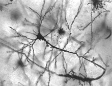
Shifts in phase (or behavior of neurons) caused due to a perturbation (an external stimulus) can be quantified within a Phase Response Curve (PRC) to predict synchrony in coupled and oscillating neurons. [8] [3] These effects can be computed, in the case of advances or delays to responses, to observe the changes in the oscillatory behavior of neurons, pending on when a stimulus was applied in the phase cycle of an oscillating neuron. The key to understanding this is in the behavioral patterns of neurons and the routes neural information travels. Neural circuits are able to communicate efficiently and effectively within milliseconds of experiencing a stimulus and lead to the spread of information throughout the neural network. [10] The study of neuron synchrony could provide information on the differences that occur in neural states such as normal and diseased states. Neurons that are involved significantly in diseases such as Alzheimer's disease or Parkinson's disease are shown to undergo phase resetting before launching into phase locking where clusters of neurons are able to begin firing rapidly to communicate information quickly. [8] [10]

The phase of ongoing oscillatory activity is reset. Phase resetting.png
The phase of ongoing oscillatory activity is reset.

A phase response curve can be calculated by noting changes to its period over time depending on where in the cycle the input is applied. The perturbation left by the stimulus moves the stable cycle within the oscillation followed by a return to the stable cycle limit. The curve tracks the amount of advancement or delay due to the input in the oscillating neuron. The PRC assumes certain patterns of behavior in firing pattern as well as the network of oscillating neurons to model the oscillations. Currently, only a few circuits exist which can be modeled using an assumed firing pattern. [5]

In order to model the behavior of firing neural circuits, the following is calculated to generate a PRC curve and its trajectory. The period is defined as the unperturbed period of an oscillator from the phase cycle defined as 0≤ ∅ ≤1 and the cycle that has undergone a perturbation is known as as shown in the following equation. [3] An advance in phase occurs when trajectory of the motion is displaced in the direction of the motion due a shortening of period whereas a phase delay occurs when the displacement occurs in the opposite direction of motion.

Types of phase response curves

If the perturbation to the oscillatory cycle is infinitesimally small, it is possible to derive a response function of the neural oscillator. This response function can be classified into different classes (Type 1 and Type 2) based upon its response. [8] [3] [11] [12]

  1. Type I Phase Response Curves are non-negative and strictly positive thus perturbations are only able to enhance a spike in phase, but never delay it. This occurs through a slight depolarization, such as postsynaptic potentials that increase the excitation of an axon. Type I PRCs are also shown to fire more slowly towards the onset of firing. Examples of Models that exhibit Type I PRCs in weakly coupled neural oscillators include the Connor and the Morris–Lecar model. [3] [11] [13]
  2. Type II Phase Response Curves can have negative and positive regions. Due to this characteristic, Type II PRCs are able to advance or delay changes in phase pending on the timing of perturbation that occurs. These curves can also have abrupt onset of firing and due to this, are unable to fire below their threshold. An example of a Type II PRC is seen within the Hodgkin–Huxley model. [3] [11]

Assumptions of phase response curves

Numerous research has suggested two primary assumptions that allow the use of PRCs to be used to predict the occurrence of synchrony within neural oscillation. These assumptions work to show synchrony within coupled neurons that are linked to other neurons. The first assumption claims that coupling between neurons must be weak and requires an infinitesimally small phase change in response to a perturbation. [8] [3] [14]

The second assumption assumes coupling between neurons to be pulsatile where the perturbation to calculate PRC should only include those inputs that are received within the circuit. This leads to a limitation of each phase being completed within a reset before another perturbation can be received. [8] [14]

The main difference between the two assumptions is for pulsatile the effects of any inputs must be known or measured prior. In weak coupling, only the magnitude of response due to a perturbation needs to be measured to calculate phase resetting. The weak coupling also induces the claim that many cycles must occur prior to convergence of oscillators to phase lock to lead to synchronization. [8] [14]

Conditions for validity of phase response curve

Much argument still exists in whether the assumptions behind phase resetting are valid for analysis of neural activity leading to synchronization and other neural properties. Event-related Potential (ERP) is a commonly used measure to the response of the brain to different events and can be measured via electroencephalography (EEG). EEGs can be used to measured electrical activity throughout the brain noninvasively. [14] The Phase Response Curve operates under the following criteria and must occur to prove that phase resetting is the cause of the behavior:

  1. An oscillation must already be occurring before it can reset in its phase. This implies that resetting in response to a stimulus can only occur if the oscillation pre-existed before the reset.
  2. If due to resetting of oscillation leads to the formation of ERP, the ERP must show similar characteristics.
  3. The neural sources responsible for the generation of the ERP must be the same as ongoing oscillation to be considered phase resetting. [14]

Arguments for and against phase resetting model

Arguments that claim the activity pattern observed in neurons is not phase resetting, but could instead be the response to evoked potentials (ERPs), include:

  1. If the ERP was generated due to phase resetting, measuring the phase concentration alone is not enough to prove that phase resetting is occurring. An example of this is measuring while filtering data as this may actually induce an artificial oscillation in response to perturbation. It has been suggested that this argument may be overcome if there is no increase in power of the phase reset from pre-stimulus to post-stimulus. [14]
  2. The amplitude and phase of ongoing oscillations at the time a stimulus is applied should influence the ERP once generated by current oscillations. This argument is overcome if the amplitude or phase of current oscillations is affected and creates an ERP, and cannot be assumed to be an independent event. [14]

Biological occurrences

Epilepsy

Epilepsy is traditionally viewed as a disease resulting from hypersynchronous neural activity. Research has shown that specific changes in the topology of neural networks and their increase in synaptic strength can move into hyper-excited states. Normal networks of neurons fire in synchronous patterns that lead to communication; if this behavior is excited further, it can lead to "bursting" and significantly increase this communication. This increase then leads to over-activation of neural networks and finally to seizures. Diseases such as epilepsy demonstrate how synchrony amongst neural networks must be highly regulated to prevent asynchronous activity. The study of neural regulation could help to outline methods to reduce symptoms of asynchronous activity such as that observed in epilepsy. [15] [16]

Neuron in the hippocampus of an epileptic patient Pyramidal hippocampal neuron 40x.jpg
Neuron in the hippocampus of an epileptic patient

Memory

Phase resetting is important in the formation of long-term memories. Due to synchronization within the gamma-frequency range has been shown to be followed by phase resetting of theta oscillations when phase-locked by a stimulus. This shows increased neural synchrony, due to connections within neural networks, during the formation of memories by reactivating certain networks continuously. [17] [18]

During memory tasks that required quick formation of memories, phase resetting within alpha activity increased the strength of the memories. [14] [19]

Hippocampus

Theta phase precession is a phenomenon observed in the hippocampus of rats and relates to the timing of neural spikes. [20] [21] When rats navigate around their environment, there are certain neurons in the hippocampus that fire (spike) when the animal is near a familiar landmark. Each neuron is tuned to a particular unique landmark, and for that reason, these neurons are called place cells.[ citation needed ]

Curiously, it turns out that when a place cell fires is determined by how far the animal is from the landmark. There is a background oscillation in the hippocampus in the theta band (4 – 8 Hz). As the animal approaches the landmark, the spiking of the place cell moves earlier in phase relative to the background theta oscillation, so that the phase offset essentially measures or represents the distance. This phase shifting relative to spatial distance is called phase precession. [22]

See also

Related Research Articles

<span class="mw-page-title-main">Synchronization</span> Coordination of events to operate a system in unison

Synchronization is the coordination of events to operate a system in unison. For example, the conductor of an orchestra keeps the orchestra synchronized or in time. Systems that operate with all parts in synchrony are said to be synchronous or in sync—and those that are not are asynchronous.

<span class="mw-page-title-main">Suprachiasmatic nucleus</span> Part of the brains hypothalamus

The suprachiasmatic nucleus or nuclei (SCN) is a small region of the brain in the hypothalamus, situated directly above the optic chiasm. It is the principle circadian pacemaker in mammals and is necessary for generating circadian rhythms. Reception of light inputs from photosensitive retinal ganglion cells allow the SCN to coordinate the subordinate cellular clocks of the body and entrain to the environment. The neuronal and hormonal activities it generates regulate many different body functions in an approximately 24-hour cycle.

A circadian clock, or circadian oscillator, is a biochemical oscillator that cycles with a stable phase and is synchronized with solar time.

A phase response curve (PRC) illustrates the transient change in the cycle period of an oscillation induced by a perturbation as a function of the phase at which it is received. PRCs are used in various fields; examples of biological oscillations are the heartbeat, circadian rhythms, and the regular, repetitive firing observed in some neurons in the absence of noise.

A gamma wave or gamma rhythm is a pattern of neural oscillation in humans with a frequency between 25 and 140 Hz, the 40 Hz point being of particular interest. Gamma rhythms are correlated with large scale brain network activity and cognitive phenomena such as working memory, attention, and perceptual grouping, and can be increased in amplitude via meditation or neurostimulation. Altered gamma activity has been observed in many mood and cognitive disorders such as Alzheimer's disease, epilepsy, and schizophrenia. Elevated gamma activity has also been observed in moments preceding death.

Brainwave entrainment, also referred to as brainwave synchronization or neural entrainment, refers to the observation that brainwaves will naturally synchronize to the rhythm of periodic external stimuli, such as flickering lights, speech, music, or tactile stimuli.

<span class="mw-page-title-main">Neural oscillation</span> Brainwaves, repetitive patterns of neural activity in the central nervous system

Neural oscillations, or brainwaves, are rhythmic or repetitive patterns of neural activity in the central nervous system. Neural tissue can generate oscillatory activity in many ways, driven either by mechanisms within individual neurons or by interactions between neurons. In individual neurons, oscillations can appear either as oscillations in membrane potential or as rhythmic patterns of action potentials, which then produce oscillatory activation of post-synaptic neurons. At the level of neural ensembles, synchronized activity of large numbers of neurons can give rise to macroscopic oscillations, which can be observed in an electroencephalogram. Oscillatory activity in groups of neurons generally arises from feedback connections between the neurons that result in the synchronization of their firing patterns. The interaction between neurons can give rise to oscillations at a different frequency than the firing frequency of individual neurons. A well-known example of macroscopic neural oscillations is alpha activity.

<span class="mw-page-title-main">Neural binding</span>

Neural binding is the neuroscientific aspect of what is commonly known as the binding problem: the interdisciplinary difficulty of creating a comprehensive and verifiable model for the unity of consciousness. "Binding" refers to the integration of highly diverse neural information in the forming of one's cohesive experience. The neural binding hypothesis states that neural signals are paired through synchronized oscillations of neuronal activity that combine and recombine to allow for a wide variety of responses to context-dependent stimuli. These dynamic neural networks are thought to account for the flexibility and nuanced response of the brain to various situations. The coupling of these networks is transient, on the order of milliseconds, and allows for rapid activity.

Theta waves generate the theta rhythm, a neural oscillation in the brain that underlies various aspects of cognition and behavior, including learning, memory, and spatial navigation in many animals. It can be recorded using various electrophysiological methods, such as electroencephalogram (EEG), recorded either from inside the brain or from electrodes attached to the scalp.

Neural coding is a neuroscience field concerned with characterising the hypothetical relationship between the stimulus and the individual or ensemble neuronal responses and the relationship among the electrical activity of the neurons in the ensemble. Based on the theory that sensory and other information is represented in the brain by networks of neurons, it is thought that neurons can encode both digital and analog information.

Repetition priming refers to improvements in a behavioural response when stimuli are repeatedly presented. The improvements can be measured in terms of accuracy or reaction time, and can occur when the repeated stimuli are either identical or similar to previous stimuli. These improvements have been shown to be cumulative, so as the number of repetitions increases the responses get continually faster up to a maximum of around seven repetitions. These improvements are also found when the repeated items are changed slightly in terms of orientation, size and position. The size of the effect is also modulated by the length of time the item is presented for and the length time between the first and subsequent presentations of the repeated items.

In the study of chronobiology, entrainment occurs when rhythmic physiological or behavioral events match their period to that of an environmental oscillation. It is ultimately the interaction between circadian rhythms and the environment. A central example is the entrainment of circadian rhythms to the daily light–dark cycle, which ultimately is determined by the Earth's rotation. Exposure to certain environmental stimuli will cue a phase shift, and abrupt change in the timing of the rhythm. Entrainment helps organisms maintain an adaptive phase relationship with the environment as well as prevent drifting of a free running rhythm. This stable phase relationship achieved is thought to be the main function of entrainment.

<span class="mw-page-title-main">Jürgen Aschoff</span>

Jürgen Walther Ludwig Aschoff was a German physician, biologist and behavioral physiologist. Together with Erwin Bünning and Colin Pittendrigh, he is considered to be a co-founder of the field of chronobiology.

Colin Stephenson Pittendrigh was a British-born biologist who spent most of his adult life in the United States. Pittendrigh is regarded as the "father of the biological clock," and founded the modern field of chronobiology alongside Jürgen Aschoff and Erwin Bünning. He is known for his careful descriptions of the properties of the circadian clock in Drosophila and other species, and providing the first formal models of how circadian rhythms entrain (synchronize) to local light-dark cycles.

Pigment dispersing factor (pdf) is a gene that encodes the protein PDF, which is part of a large family of neuropeptides. Its hormonal product, pigment dispersing hormone (PDH), was named for the diurnal pigment movement effect it has in crustacean retinal cells upon its initial discovery in the central nervous system of arthropods. The movement and aggregation of pigments in retina cells and extra-retinal cells is hypothesized to be under a split hormonal control mechanism. One hormonal set is responsible for concentrating chromatophoral pigment by responding to changes in the organism's exposure time to darkness. Another hormonal set is responsible for dispersion and responds to the light cycle. However, insect pdf genes do not function in such pigment migration since they lack the chromatophore.

<span class="mw-page-title-main">Theta model</span>

The theta model, or Ermentrout–Kopell canonical model, is a biological neuron model originally developed to model neurons in the animal Aplysia, and later used in various fields of computational neuroscience. The model is particularly well suited to describe neuron bursting, which are rapid oscillations in the membrane potential of a neuron interrupted by periods of relatively little oscillation. Bursts are often found in neurons responsible for controlling and maintaining steady rhythms. For example, breathing is controlled by a small network of bursting neurons in the brain stem. Of the three main classes of bursting neurons, the theta model describes parabolic bursting. Parabolic bursting is characterized by a series of bursts that are regulated by a slower external oscillation. This slow oscillation changes the frequency of the faster oscillation so that the frequency curve of the burst pattern resembles a parabola.

The neuroscience of rhythm refers to the various forms of rhythm generated by the central nervous system (CNS). Nerve cells, also known as neurons in the human brain are capable of firing in specific patterns which cause oscillations. The brain possesses many different types of oscillators with different periods. Oscillators are simultaneously outputting frequencies from .02 Hz to 600 Hz. It is now well known that a computer is capable of running thousands of processes with just one high-frequency clock. Humans have many different clocks as a result of evolution. Prior organisms had no need for a fast-responding oscillator. This multi-clock system permits quick response to constantly changing sensory input while still maintaining the autonomic processes that sustain life. This method modulates and controls a great deal of bodily functions.

<span class="mw-page-title-main">Phase precession</span> Neural mechanism

Phase precession is a neurophysiological process in which the time of firing of action potentials by individual neurons occurs progressively earlier in relation to the phase of the local field potential oscillation with each successive cycle. In place cells, a type of neuron found in the hippocampal region of the brain, phase precession is believed to play a major role in the neural coding of information. John O'Keefe, who later shared the 2014 Nobel Prize in Physiology or Medicine for his discovery that place cells help form a "map" of the body's position in space, co-discovered phase precession with Michael Recce in 1993.

Phase reduction is a method used to reduce a multi-dimensional dynamical equation describing a nonlinear limit cycle oscillator into a one-dimensional phase equation. Many phenomena in our world such as chemical reactions, electric circuits, mechanical vibrations, cardiac cells, and spiking neurons are examples of rhythmic phenomena, and can be considered as nonlinear limit cycle oscillators.

In the field of chronobiology, the dual circadian oscillator model refers to a model of entrainment initially proposed by Colin Pittendrigh and Serge Daan. The dual oscillator model suggests the presence of two coupled circadian oscillators: E (evening) and M (morning). The E oscillator is responsible for entraining the organism’s evening activity to dusk cues when the daylight fades, while the M oscillator is responsible for entraining the organism’s morning activity to dawn cues, when daylight increases. The E and M oscillators operate in an antiphase relationship. As the timing of the sun's position fluctuates over the course of the year, the oscillators' periods adjust accordingly. Other oscillators, including seasonal oscillators, have been found to work in conjunction with circadian oscillators in order to time different behaviors in organisms such as fruit flies.

References

  1. Krogh-Madsen, Trine, et al. "Phase Resetting Neural Oscillators: Topological Theory Versus the RealWorld." Phase response curves in neuroscience. Springer New York, 2012. 33-51.
  2. Czeisler, C. A.; RE Kronauer; JS Allan; JF Duffy; ME Jewett; EN Brown; JM Ronda (16 June 1989). "Bright light induction of strong (type 0) resetting of the human circadian pacemaker". Science. 244 (4910): 1328–1333. Bibcode:1989Sci...244.1328C. doi:10.1126/science.2734611. PMID   2734611.
  3. 1 2 3 4 5 6 7 Canavier, Carmen C. (2006). "Phase Response Curve". Scholarpedia. 1 (12): 1332. Bibcode:2006SchpJ...1.1332C. doi: 10.4249/scholarpedia.1332 .
  4. Achuthan, S.; Carmen Canavier (22 April 2009). "Phase-Resetting Curves Determine Synchronization, Phase Locking, and Clustering in Networks of Neural Oscillators". The Journal of Neuroscience. 29 (16): 5218–5233. doi:10.1523/jneurosci.0426-09.2009. PMC   2765798 . PMID   19386918.
  5. 1 2 Glass, L.; Michael C. Mackey (1988). From Clocks to Chaos: The Rhythms of Life . Princeton, NJ: Princeton University Press. ISBN   9780691084961.
  6. Kuramoto, Yoshiki (1984). Chemical oscillations, waves, and turbulence. Berlin, New York: Springer-Verlag. ISBN   978-0387133225.
  7. Winfree, Arthur T (1967). "Biological rhythms and the behavior of populations of coupled oscillators". Journal of Theoretical Biology. 16 (1): 15–42. Bibcode:1967JThBi..16...15W. doi:10.1016/0022-5193(67)90051-3. PMID   6035757.
  8. 1 2 3 4 5 6 7 Canavier, Carmen; S. Achuthan (2010). "Pulse coupled oscillators and the phase resetting curve". Mathematical Biosciences. 226 (2): 77–96. doi:10.1016/j.mbs.2010.05.001. PMC   3022482 . PMID   20460132.
  9. Rohling, Jos H. T; Vanderleest H; Michel S; Vansteensel M; Meijer J. (2011). "Phase Resetting of the Mammalian Circadian Clock Relies on a Rapid Shift of a Small Population of Pacemaker Neurons". PLOS ONE. 6 (9): 1–9. Bibcode:2011PLoSO...625437R. doi: 10.1371/journal.pone.0025437 . PMC   3178639 . PMID   21966529.
  10. 1 2 Varela, F; J. Lachaux; E. Rodriguez; J.Martinierie (2001). "The brainweb: Phase synchronization and large-scale integration". Nature Reviews Neuroscience. 2 (4): 229–239. doi:10.1038/35067550. PMID   11283746. S2CID   18651043.
  11. 1 2 3 Ermentout, B. (1996). "Type I membranes, phase resetting curves, and synchrony". Neural Computation. 5. 8 (5): 979–1001. doi:10.1162/neco.1996.8.5.979. PMID   8697231. S2CID   17168880.
  12. Hansel, D.; Mato, G.; Meunier, C. (March 1995). "Synchrony in excitatory neural networks". Neural Computation. 7 (2): 307–337. doi:10.1162/neco.1995.7.2.307. PMID   8974733. S2CID   15130785.
  13. Wang, S.G.; Musharoff M; Canavier C; Gasparini S (June 2013). "Hippocampal CA1 pyramidal neurons exhibit type 1 phase-response curves and type 1 excitability". Journal of Neurophysiology. 109 (11): 2757–2766. doi:10.1152/jn.00721.2012. PMC   3680797 . PMID   23468392.
  14. 1 2 3 4 5 6 7 8 Sauseng, P; Klimesch W; Gruber WR; Hanslmayr S; Freunberger R; Doppelmayr M (8 June 2007). "Are event-related potential components generated by phase resetting of brain oscillations? A critical discussion". Neuroscience. 146 (4): 1435–44. doi:10.1016/j.neuroscience.2007.03.014. PMID   17459593. S2CID   16148930.
  15. Neotoff, Theoden; Robert Clewley; Scott Arno; Tara Keck; John A. White (15 September 2004). "Epilepsy in Small-World Networks". The Journal of Neuroscience. 24 (37): 8075–8083. doi:10.1523/jneurosci.1509-04.2004. PMC   6729784 . PMID   15371508.
  16. Jahangiri, A; Durand D. (April 2011). "Phase resetting analysis of high potassium epileptiform activity in CA3 region of the rat hippocampus". International Journal of Neural Systems. 21 (2): 127–138. doi:10.1142/S0129065711002705. PMID   21442776.
  17. Axmacher, Nikolai; Florian Mormann; Guillen Fernández; Christian E. Elger; Juergen Fell (24 January 2006). "Memory formation by Neural Synchronization". Brain Research Reviews. 52 (1): 170–182. doi:10.1016/j.brainresrev.2006.01.007. PMID   16545463. S2CID   665254.
  18. Jutras, Michael; Elizabeth A. Buffalo (18 March 2010). "Synchronous Neural Activity and Memory formation". Neurobiology. 20 (2): 150–155. doi:10.1016/j.conb.2010.02.006. PMC   2862842 . PMID   20303255.
  19. Yu, Shan; Debin Huang; Wolf Singer; Danko Nikolic (9 April 2008). "A Small world of Neural Synchrony". Cerebral Cortex. 18 (12): 2891–2901. doi:10.1093/cercor/bhn047. PMC   2583154 . PMID   18400792.
  20. O'Keefe, John; Recce, Michael L. (1993). "Phase relationship between hippocampal place units and the EEG theta rhythm". Hippocampus. 3 (3): 317–330. doi:10.1002/hipo.450030307. PMID   8353611. S2CID   6539236.
  21. Skaggs, William E.; et al. (1996). "Theta phase precession in hippocampal". Hippocampus. 6 (2): 149–172. doi:10.1002/(sici)1098-1063(1996)6:2<149::aid-hipo6>3.0.co;2-k. PMID   8797016. S2CID   15813385.
  22. Jensen, Ole; Lisman, John E. (1996). "Hippocampal CA3 region predicts memory sequences: accounting for the phase precession of place cells". Learning & Memory. 3 (2–3): 279–287. doi: 10.1101/lm.3.2-3.279 . PMID   10456097.