Photon-counting computed tomography

Last updated

Photon-counting computed tomography (PCCT) is a form of X-ray computed tomography (CT) in which X-rays are detected using a photon-counting detector (PCD) which registers the interactions of individual photons. By keeping track of the deposited energy in each interaction, the detector pixels of a PCD each record an approximate energy spectrum, making it a spectral or energy-resolved CT technique. In contrast, more conventional CT scanners use energy-integrating detectors (EIDs), where the total energy (generally from a large number of photons as well as electronic noise) deposited in a pixel during a fixed period of time is registered. These EIDs thus register only photon intensity, comparable to black-and-white photography, whereas PCDs register also spectral information, similar to color photography.

Contents

The first clinically-approved PCCT system was cleared by the Food and Drug Administration (FDA) in September 2021. [1]

General advantages

Often EIDs are used as a baseline for comparison when evaluating PCD performance capabilities. With this lens, there are several potential advantages of using a PCD over using an EID in CT imaging. These include improved signal (and contrast) to noise ratio, reduced X-ray dose to the patient, improved spatial resolution and, through use of several energy bins, the ability to distinguish multiple contrast agents. [2] [3] Due to the large volumes and rates of data required (up to several hundred million photon interactions per mm2 and second [4] ) the use of PCDs in CT scanners has become feasible only with recent improvements in detector technology. As of January 2021 photon-counting CT is in use at five clinical sites. [5] [6] [7] [8] Some early research has found the dose reduction potential of photon-counting CT for breast imaging to be very promising. [9] On September 29, 2021 the FDA cleared the first photon-counting CT (developed by Siemens Healthineers) for clinical use. [1]

Detection characteristics

Discrete energy-dependent detection

When a photon interacts in a PCD, the amplitude of the resulting electrical pulse is roughly proportional to the photon energy. By comparing each pulse produced in a pixel with a suitable low-energy threshold, contributions from low-energy events (resulting from both photon interactions and electronic noise) can be filtered out. This effectively eliminates contributions from electronic noise at the expense of discarding photons with energy comparable to the noise level (which are of little use since they are indistinguishable from noise counts). In an EID, on the other hand, the contributions from individual photons are not known. Therefore, an energy threshold cannot be applied, making this technique susceptible to noise and other factors which can affect the linearity of the voltage to X-ray intensity relationship. [10]

The removal of electronic noise gives PCDs two advantages over EIDs. First, higher signal-to-noise and contrast-to-noise ratios are expected from using PCDs compared to EIDs. This can either be used to increase the image quality at the same X-ray exposure level, or to lower the patient X-ray dose whilst maintaining the same image quality. Second, it is difficult to manufacture energy-integrating detectors with smaller pixel size than approximately 1×1 mm2 without compromising dose efficiency. The reason for this is that reflective layers must be placed in the scintillator between the pixels to prevent cross-talk between pixels, and these cannot be made too thin. In addition, the measured signal is proportional to the pixel area whereas the electronic noise is fairly independent of pixel size, so that noise will dominate the measured signal if the pixels are made too small. These problems do not occur in a photon-counting detector with a low-energy threshold, which can therefore achieve higher detector resolution.

An animated representation of the practical principles of PCCT detection. The left image depicts the arrival of photons at the surface of the PCD while the right image shows a simplified version of the generated signal. Some key things to learn from this image include: the discrete nature of photon detection, the energy-dependent height of electrical pulses, the ability to theoretically eliminate the effects of electronic noise by using a high enough base threshold, and the ability to determine the energy of a photon using energy thresholds. Photon Energy-Dependent Detection.gif
An animated representation of the practical principles of PCCT detection. The left image depicts the arrival of photons at the surface of the PCD while the right image shows a simplified version of the generated signal. Some key things to learn from this image include: the discrete nature of photon detection, the energy-dependent height of electrical pulses, the ability to theoretically eliminate the effects of electronic noise by using a high enough base threshold, and the ability to determine the energy of a photon using energy thresholds.

Multi-energy, spectral detection

By introducing more energy thresholds above the low-energy threshold, a PCD can be divided into several discrete energy bins. Each registered photon is thus assigned to a specific bin depending on its energy, such that each pixel measures a histogram of the incident X-ray spectrum. This spectral information provides several advantages over the integrated deposited energy of an EID. [2] First, it makes it possible to quantitatively determine the material composition of each pixel in the reconstructed CT image, as opposed to the estimated average linear attenuation coefficient obtained in a conventional CT scan. It turns out such a material base decomposition, using at least two energy bins, can adequately account for all elements found in the body and increases the contrast between tissue types. [11] Further, the spectral information can be used to remove beam hardening artefacts. These arise because of the higher linear attenuation of most materials at lower energy which shifts the mean energy of the X-ray spectrum towards higher energies as the beam passes through the object. By comparing the ratios of counts in different energy bins with those of the attenuated beam, the amount of beam hardening can be accounted for (either explicitly or implicitly in the reconstruction) using a PCD. Finally, using more than two energy bins allows to discriminate between on the one hand dense bone and calcifications and on the other hand heavier elements (commonly iodine or gadolinium) used as contrast agents. This has the potential reduce the amount of X-ray dose from a contrast scan by removing the need for a reference scan before contrast injection. Although spectral CT is already clinically available in the form of dual-energy scanners, photon-counting CT offers a number of advantages. A PCD can implement more than two energy thresholds with a higher degree of separation than what is possible to achieve in dual-energy CT. This improvement in energy resolution translates to higher contrast-to-noise ratio in the image, in particular in contrast-enhanced and material-selective images. Also, it can be shown that at least three energies are necessary to simultaneously decompose both tissue and contrast medium. [12] More energy bins also allow for simultaneously differentiating between different contrast agents. [13]

Simplified illustration of pulse-pileup, one of the fundamental contributors to spectral distortion within PCDs. In this case, two photons that impact the detector at the same time or within a very small, indiscernible time window are recorded as a single high energy photon rather than as two lower energy photons. This creates an incorrect spectral reading. Pulse Pileup.png
Simplified illustration of pulse-pileup, one of the fundamental contributors to spectral distortion within PCDs. In this case, two photons that impact the detector at the same time or within a very small, indiscernible time window are recorded as a single high energy photon rather than as two lower energy photons. This creates an incorrect spectral reading.
A simplified illustration of charge-sharing, one of the fundamental contributors to spectral distortion within PCDs. An incident photon is identified as two individual photons of smaller energies rather than as a singular photon of the actual higher energy. Charge Sharing.png
A simplified illustration of charge-sharing, one of the fundamental contributors to spectral distortion within PCDs. An incident photon is identified as two individual photons of smaller energies rather than as a singular photon of the actual higher energy.

Detection challenges and spectral distortion

Despite encouraging research, there are several challenges which have until recently prevented incorporating PCDs in CT systems. Many challenges are related to demands on detector material and electronics resulting from large data volumes and count rates. As an example, each mm2 of a CT detector may receive several hundred million photon interactions per second during a scan. [4]

To avoid saturation in areas where little material is present between the X-ray source and the detector, the pulse resolving time must be small compared to the average time between photon interactions in a pixel. Even before saturation, the detector functionality starts to deteriorate because of pulse pileup (see figure to the left), where two (or more) photon interactions take place in the same pixel too close in time to be resolved as discrete events. Such quasi-coincident interactions lead to a loss of photon counts and distorts the pulse shape, skewing the recorded energy spectrum. [2] Due to these effects, the demands on the physical response time of the detector material as well as on the electronics responsible for pulse-shaping, binning and recording pixel data become very high. Using smaller image pixels decreases the per-pixel count rate and thus alleviates the demands on pulse resolving time at the expense of requiring more electronics.

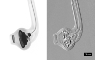
Partial energy deposition and single photons causing signals in multiple pixels poses another challenge in photon-counting CT. [2] Charge sharing, where an interaction takes place close to a pixel boundary, causing the released energy to be shared between neighboring pixels and thus be interpreted as several lower-energy photons, is one cause of such events (see figure to the right). Others include the emission of K-escape X-rays and Compton scattering, where the escaping or scattered photon results in a partial energy deposition in the primary pixel and may go on to cause further interactions in different pixels. The effects mentioned take place also in EIDs but cause additional problems in PCDs since they result in a distorted energy spectrum. In contrast to saturation and pileup effects, problems caused by partial energy deposition and multiply interacting photons is aggravated by smaller pixel size. Anti-coincidence logic, where simultaneous events in nearby pixels are added, can be used to somewhat counteract counting the same photon in different pixels.

Image reconstruction

Classical CT reconstruction

The fundamental challenge of tomographic reconstruction is the inverse problem of reconstructing the 3D, tomographic information of a volume using 2D projections from different angular positions. The same fundamental methods conventionally used for tomographic reconstruction can be used without alteration on data acquired from PCDs. [2] The fundamental geometric, physical, and mathematical approaches to typical reconstruction are unchanged by this new detection method. Arguably the most fundamental method of CT image reconstruction is filtered back projection. More in depth operations and the wide selection of iterative reconstruction methods remain entirely applicable. There is a great deal of literature on CT reconstruction for curious readers. [14]

Multi-energy reconstruction

Access to multiple energy bins opens up new possibilities when it comes to reconstructing a CT image from the acquired projections. The most basic possibility is to treat each of the N energy bins separately and use a conventional CT reconstruction method to reconstruct N different images. [15]

Material decomposition

As a next step to a multi-energy reconstruction, it is possible to determine the material components at a given voxel location by comparing and/or combining the intensities of the N images at this location. This is typically performed by writing each pixel as a linear combination of M base materials of known properties such as water, calcium, and a contrast agent such as iodine. This method is referred to as image-based material decomposition. Although intuitive, this pixel-wise approach relies heavily on the spectral fidelity (or spectral calibration) of the individual detector pixels across thresholds and does nothing to remove common image artifacts.

Another option is to perform the material base decomposition directly on the projection data, before the reconstruction. Using projection-based material decomposition, the material composition measured by a detector pixel for a given projection is expressed as a linear combination of M basis materials (e.g. soft tissue, bone and contrast agent). This is determined from the recorded energy histogram, for example through maximum likelihood estimation. [12] The reconstruction is then performed separately for each material basis, yielding M reconstructed basis images.

A third option would be to employ a single-step reconstruction, where the material basis decomposition is performed simultaneously with the image reconstruction. This approach, however, is not compatible with the reconstruction algorithms used in current clinical CT systems. Instead, novel iterative algorithms specific to photon-counting CT are required.

Research in the field of deep learning has also introduced possibilities of performing material decomposition using convolutional neural networks. [16]

Detector composition

Experimental PCDs for use in CT systems use semiconductor detectors based on either cadmium (zinc) telluride or silicon, neither of which need cryogenic cooling to operate. Cadmium telluride and cadmium zinc telluride detectors have the advantage of high attenuation and relatively high photoelectric-to-Compton ratio for X-ray energies used in CT imaging. This means the detectors can be made thinner and lose less spectral information due to Compton scattering. (Although they still lose spectral information due to K-escape electrons.) However, detectors made of Cadmium telluride (zinc) have longer collection times due to low charge carrier mobility, and thus suffer more from pileup effects. Further, it is currently difficult to produce such crystals without defects and impurities, which cause detector polarisation and incomplete charge collection. [17]

Silicon detectors, on the other hand, are more easily manufactured and less prone to pileup due to high charge carrier mobility. They do not suffer from K-escape X-rays but have a lower photoelectric-to-Compton ratio at X-ray energies used in CT imaging, which degrades the collected energy spectrum. Further, silicon attenuates X-rays less strongly and therefore silicon detectors have to be several centimetres thick to be useful in a CT system. [17]

Related Research Articles

<span class="mw-page-title-main">Positron emission tomography</span> Medical imaging technique

Positron emission tomography (PET) is a functional imaging technique that uses radioactive substances known as radiotracers to visualize and measure changes in metabolic processes, and in other physiological activities including blood flow, regional chemical composition, and absorption. Different tracers are used for various imaging purposes, depending on the target process within the body.

<span class="mw-page-title-main">X-ray</span> Form of short-wavelength electromagnetic radiation

X-ray is a high-energy electromagnetic radiation. In many languages, it is referred to as Röntgen radiation, after the German scientist Wilhelm Conrad Röntgen, who discovered it in 1895 and named it X-radiation to signify an unknown type of radiation.

<span class="mw-page-title-main">CT scan</span> Medical imaging procedure using X-rays to produce cross-sectional images

A computed tomography scan is a medical imaging technique used to obtain detailed internal images of the body. The personnel that perform CT scans are called radiographers or radiology technologists.

<span class="mw-page-title-main">Radiography</span> Imaging technique using ionizing and non-ionizing radiation

Radiography is an imaging technique using X-rays, gamma rays, or similar ionizing radiation and non-ionizing radiation to view the internal form of an object. Applications of radiography include medical and industrial radiography. Similar techniques are used in airport security,. To create an image in conventional radiography, a beam of X-rays is produced by an X-ray generator and it is projected towards the object. A certain amount of the X-rays or other radiation are absorbed by the object, dependent on the object's density and structural composition. The X-rays that pass through the object are captured behind the object by a detector. The generation of flat two-dimensional images by this technique is called projectional radiography. In computed tomography, an X-ray source and its associated detectors rotate around the subject, which itself moves through the conical X-ray beam produced. Any given point within the subject is crossed from many directions by many different beams at different times. Information regarding the attenuation of these beams is collated and subjected to computation to generate two-dimensional images on three planes which can be further processed to produce a three-dimensional image.

<span class="mw-page-title-main">Single-photon emission computed tomography</span> Nuclear medicine tomographic imaging technique

Single-photon emission computed tomography is a nuclear medicine tomographic imaging technique using gamma rays. It is very similar to conventional nuclear medicine planar imaging using a gamma camera, but is able to provide true 3D information. This information is typically presented as cross-sectional slices through the patient, but can be freely reformatted or manipulated as required.

<span class="mw-page-title-main">Tomography</span> Imaging by sections or sectioning using a penetrative wave

Tomography is imaging by sections or sectioning that uses any kind of penetrating wave. The method is used in radiology, archaeology, biology, atmospheric science, geophysics, oceanography, plasma physics, materials science, cosmochemistry, astrophysics, quantum information, and other areas of science. The word tomography is derived from Ancient Greek τόμος tomos, "slice, section" and γράφω graphō, "to write" or, in this context as well, "to describe." A device used in tomography is called a tomograph, while the image produced is a tomogram.

<span class="mw-page-title-main">Photodetector</span> Sensors of light or other electromagnetic energy

Photodetectors, also called photosensors, are sensors of light or other electromagnetic radiation. There are a wide variety of photodetectors which may be classified by mechanism of detection, such as photoelectric or photochemical effects, or by various performance metrics, such as spectral response. Semiconductor-based photodetectors typically use a p–n junction that converts photons into charge. The absorbed photons make electron–hole pairs in the depletion region. Photodiodes and photo transistors are a few examples of photo detectors. Solar cells convert some of the light energy absorbed into electrical energy.

<span class="mw-page-title-main">X-ray microtomography</span> X-ray 3D imaging method

In radiography, X-ray microtomography uses X-rays to create cross-sections of a physical object that can be used to recreate a virtual model without destroying the original object. It is similar to tomography and X-ray computed tomography. The prefix micro- is used to indicate that the pixel sizes of the cross-sections are in the micrometre range. These pixel sizes have also resulted in creation of its synonyms high-resolution X-ray tomography, micro-computed tomography, and similar terms. Sometimes the terms high-resolution computed tomography (HRCT) and micro-CT are differentiated, but in other cases the term high-resolution micro-CT is used. Virtually all tomography today is computed tomography.

Charge sharing is an effect of signal degradation through transfer of charges from one electronic domain to another.

<span class="mw-page-title-main">Medipix</span> Family of pixel detectors

Medipix is a family of photon counting and particle tracking pixel detectors developed by an international collaboration, hosted by CERN.

<span class="mw-page-title-main">Tomosynthesis</span>

Tomosynthesis, also digital tomosynthesis (DTS), is a method for performing high-resolution limited-angle tomography at radiation dose levels comparable with projectional radiography. It has been studied for a variety of clinical applications, including vascular imaging, dental imaging, orthopedic imaging, mammographic imaging, musculoskeletal imaging, and chest imaging.

<span class="mw-page-title-main">Automatic exposure control</span>

Automatic Exposure Control (AEC) is an X-ray exposure termination device. A medical radiographic exposure is always initiated by a human operator but an AEC detector system may be used to terminate the exposure when a predetermined amount of radiation has been received. The intention of AEC is to provide consistent x-ray image exposure, whether to film, a digital detector or a CT scanner. AEC systems may also automatically set exposure factors such as the X-ray tube current and voltage in a CT.

<span class="mw-page-title-main">Phase-contrast X-ray imaging</span> Imaging systems using changes in phase

Phase-contrast X-ray imaging or phase-sensitive X-ray imaging is a general term for different technical methods that use information concerning changes in the phase of an X-ray beam that passes through an object in order to create its images. Standard X-ray imaging techniques like radiography or computed tomography (CT) rely on a decrease of the X-ray beam's intensity (attenuation) when traversing the sample, which can be measured directly with the assistance of an X-ray detector. However, in phase contrast X-ray imaging, the beam's phase shift caused by the sample is not measured directly, but is transformed into variations in intensity, which then can be recorded by the detector.

<span class="mw-page-title-main">Photon counting</span> Counting photons using a single-photon detector

Photon counting is a technique in which individual photons are counted using a single-photon detector (SPD). A single-photon detector emits a pulse of signal for each detected photon. The counting efficiency is determined by the quantum efficiency and the system's electronic losses.

<span class="mw-page-title-main">Operation of computed tomography</span>

X-ray computed tomography operates by using an X-ray generator that rotates around the object; X-ray detectors are positioned on the opposite side of the circle from the X-ray source.

Jeffrey Harold Siewerdsen is an American physicist and biomedical engineer who is a Professor of Imaging Physics at The University of Texas MD Anderson Cancer Center as well as Biomedical Engineering, Computer Science, Radiology, and Neurosurgery at Johns Hopkins University.He is among the original inventors of cone-beam CT-guided radiotherapy as well as weight-bearing cone-beam CT for musculoskeletal radiology and orthopedic surgery. His work also includes the early development of flat-panel detectors on mobile C-arms for intraoperative cone-beam CT in image-guided surgery. He developed early models for the signal and noise performance of flat-panel detectors and later extended such analysis to dual-energy imaging and 3D imaging performance in cone-beam CT. He founded the ISTAR Lab in the Department of Biomedical Engineering, the Carnegie Center for Surgical Innovation at Johns Hopkins Hospital, and the Surgical Data Science Program at the Institute for Data Science in Oncology at The University of Texas MD Anderson Cancer Center.

<span class="mw-page-title-main">History of computed tomography</span> History of CT scanning technology

The history of X-ray computed tomography dates back to at least 1917 with the mathematical theory of the Radon transform In the early 1900s an Italian radiologist named Alessandro Vallebona invented tomography which used radiographic film to see a single slice of the body. It was not widely used until the 1930s, when Dr Bernard George Ziedses des Plantes developed a practical method for implementing the technique.

Spectral imaging is an umbrella term for energy-resolved X-ray imaging in medicine. The technique makes use of the energy dependence of X-ray attenuation to either increase the contrast-to-noise ratio, or to provide quantitative image data and reduce image artefacts by so-called material decomposition. Dual-energy imaging, i.e. imaging at two energy levels, is a special case of spectral imaging and is still the most widely used terminology, but the terms "spectral imaging" and "spectral CT" have been coined to acknowledge the fact that photon-counting detectors have the potential for measurements at a larger number of energy levels.

<span class="mw-page-title-main">MARS Bioimaging</span>

MARS Bioimaging Limited (MBI) is a medical imaging company focusing on spectral photon counting computed tomography for quantitative color imaging. The company was founded in Christchurch, New Zealand to commercialize the MARS imaging system for its applications in medicine.

Photon-counting mammography was introduced commercially in 2003 and was the first widely available application of photon-counting detector technology in medical x-ray imaging. Photon-counting mammography improves dose efficiency compared to conventional technologies, and enables spectral imaging.

References

  1. 1 2 "FDA Clears First Major Imaging Device Advancement for Computed Tomography in Nearly a Decade". Food and Drug Administration . 30 September 2021.
  2. 1 2 3 4 5 Taguchi K, Iwanczyk JS (October 2013). "Vision 20/20: Single photon counting x-ray detectors in medical imaging". Medical Physics. 40 (10): 100901. Bibcode:2013MedPh..40j0901T. doi:10.1118/1.4820371. PMC   3786515 . PMID   24089889.
  3. Shikhaliev PM, Xu T, Molloi S (February 2005). "Photon counting computed tomography: concept and initial results". Medical Physics. 32 (2): 427–36. Bibcode:2005MedPh..32..427S. doi:10.1118/1.1854779. PMID   15789589.
  4. 1 2 Persson M, Bujila R, Nowik P, Andersson H, Kull L, Andersson J, Bornefalk H, Danielsson M (July 2016). "Upper limits of the photon fluence rate on CT detectors: Case study on a commercial scanner". Medical Physics. 43 (7): 4398–4411. Bibcode:2016MedPh..43.4398P. doi:10.1118/1.4954008. PMID   27370155.
  5. "NIH uses photon-counting CT scanner in patients for the first time". National Institutes of Health (NIH). 2016-02-24. Retrieved 2017-11-22.
  6. "CT Clinical Innovation Center: four decades of CT innovation at Mayo Clinic". Mayo Clinic Alumni Association. 3 May 2017. Retrieved 2021-01-19.
  7. "Unique research photon-counting CT scanner to CMIV". liu.se. Retrieved 2021-01-19.
  8. "Schärfere Augen für die Computertomographie: Mit Photon-Counting Metastasen besser beurteilen". Radiologie Magazin (in German). 2021-01-11. Retrieved 2021-01-19.
  9. Kalender WA, Kolditz D, Steiding C, Ruth V, Lück F, Rößler AC, Wenkel E (March 2017). "Technical feasibility proof for high-resolution low-dose photon-counting CT of the breast". European Radiology. 27 (3): 1081–1086. doi:10.1007/s00330-016-4459-3. PMID   27306559. S2CID   7912239.
  10. Jenkins R, Gould RW, Gedcke D (1995). Quantitative x-ray spectrometry (2nd ed.). New York: Dekker. p. 90. ISBN   9780824795542. OCLC   31970216.
  11. Alvarez RE, Macovski A (1976). "Energy-selective reconstructions in X-ray computerized tomography". Physics in Medicine and Biology. 21 (5): 733–44. Bibcode:1976PMB....21..733A. doi:10.1088/0031-9155/21/5/002. PMID   967922. S2CID   250824716.
  12. 1 2 Roessl E, Proksa R (August 2007). "K-edge imaging in x-ray computed tomography using multi-bin photon counting detectors". Physics in Medicine and Biology. 52 (15): 4679–96. doi:10.1088/0031-9155/52/15/020. PMID   17634657. S2CID   5871406.
  13. Schlomka JP, Roessl E, Dorscheid R, Dill S, Martens G, Istel T, Bäumer C, Herrmann C, Steadman R, Zeitler G, Livne A, Proksa R (August 2008). "Experimental feasibility of multi-energy photon-counting K-edge imaging in pre-clinical computed tomography". Physics in Medicine and Biology. 53 (15): 4031–47. Bibcode:2008PMB....53.4031S. doi:10.1088/0031-9155/53/15/002. PMID   18612175. S2CID   25238021.
  14. Pan X, Sidky EY, Vannier M (2009-12-01). "Why do commercial CT scanners still employ traditional, filtered back-projection for image reconstruction?". Inverse Problems. 25 (12): 123009. doi:10.1088/0266-5611/25/12/123009. ISSN   0266-5611. PMC   2849113 . PMID   20376330.
  15. Schmidt TG (July 2009). "Optimal "image-based" weighting for energy-resolved CT". Medical Physics. 36 (7): 3018–27. Bibcode:2009MedPh..36.3018S. doi:10.1118/1.3148535. PMID   19673201. S2CID   17685742.
  16. Gong H, Tao S, Rajendran K, Zhou W, McCollough CH, Leng S (December 2020). "Deep-learning-based direct inversion for material decomposition". Medical Physics. 47 (12): 6294–6309. Bibcode:2020MedPh..47.6294G. doi:10.1002/mp.14523. ISSN   0094-2405. PMC   7796910 . PMID   33020942.
  17. 1 2 Persson M, Huber B, Karlsson S, Liu X, Chen H, Xu C, Yveborg M, Bornefalk H, Danielsson M (November 2014). "Energy-resolved CT imaging with a photon-counting silicon-strip detector". Physics in Medicine and Biology. 59 (22): 6709–27. Bibcode:2014PMB....59.6709P. doi:10.1088/0022-3727/59/22/6709. PMID   25327497.