Plasma cell leukemia

Last updated
Plasma cell leukemia
Plasma cell leukemia.jpg
A schematic showing peripheral blood with plasma cell leukemia. Many plasma cells are seen mixed with red cells.
Specialty Hematology and oncology

Plasma cell leukemia (PCL) is a plasma cell dyscrasia, i.e. a disease involving the malignant degeneration of a subtype of white blood cells called plasma cells. It is the terminal stage and most aggressive form of these dyscrasias, constituting 2% to 4% of all cases of plasma cell malignancies. PCL may present as primary plasma cell leukemia, i.e. in patients without prior history of a plasma cell dyscrasia or as secondary plasma cell dyscrasia, i.e. in patients previously diagnosed with a history of its predecessor dyscrasia, multiple myeloma. The two forms of PCL appear to be at least partially distinct from each other. In all cases, however, PCL is an extremely serious, life-threatening, and therapeutically challenging disease. [1] [2]

Contents

Signs and symptoms

Primary PCL

The clinical presentation of primary PCL (pPCL) indicates a far more aggressive disease than that of a typical multiple myeloma case with its clinical features being a combination of those found in multiple myeloma and acute leukemia. Like multiple myeloma patients, pPCL patients exhibit pathologically high levels of monoclonal plasma cells in their bone marrow plus a malignant plasma cell-secreted circulating monoclonal myeloma protein, either IgG, IgA, a light chain, or none in 28-56%, 4-7%, 23-44%, or 0-12% of cases, respectively. [1] [3] Similar to B cell leukemias, but unlike multiple myeloma, pPCL patients exhibit relative high frequencies of splenomegaly, lymphadenopathy, hepatomegaly, kidney failure, bone marrow failure (i.e. thrombocytopenia, anemia, and/or, rarely, leukopenia), central nervous system defects, and peripheral neuropathies due to the invasion of these tissues by plasma cells and/or the deposition of their circulating monoclonal immunoglobulin in them. Compared to multiple myeloma patients, pPCL patients also: exhibit 1) high rates of developing a hypercalcemic crisis, i.e. a potentially life-threatening episode of high ionic calcium (Ca2+) levels in the blood due to excess bone re-absorption and/or renal failure; b) higher levels of serum lactate dehydrogenase and Beta-2 microglobulin; and c) lower rates of bone but higher rates of soft tissue plasma cell tumors termed plasmacytomas. [1] [4]

Secondary PCL

Secondary PCL (sPCL) is diagnosed in 1-4% of patients known to have had multiple myeloma for a median time of ~21 months. It is the terminal phase of these patients' myeloma disease. sPCL patients typically are highly symptomatic due to extensive disease with malignant plasma cell infiltrations in, and failures of, not only the bone marrow but also other organs. They have failed or broken through one or more treatment regimens and therefore may also show some of the toxic effects of these treatments. [1] [5]

Cause

PCL is caused by the development of an excessively high number of genetic abnormalities in plasma cells or, more particularly, their precursor B cells and plasmablasts (see plasma cells). This genetic instability is due to a myriad of acquired abnormalities including gene mutations; single nucleotide polymorphisms; depletions and duplications of parts of a gene, larger portion of a chromosome, or even an entire arm of a chromosome; translocations, deletions, and duplications of entire chromosomes; and increases and decreases in the expression of intact genes due to, e.g. the methylation of gene promotors and various less direct effects. These genetic abnormalities effect the Wnt signaling pathway, regulation of the cell cycle, RNA metabolism, protein folding, and cadherin-related cell adherence to extracellular matrix. These effects in turn control plasma cell proliferation, survival, apoptosis, adhesion to bone marrow, genome stability, and secretion of monoclonal immunoglobulins. [6]

Secondary plasma cell leukemia (sPCL) results from the comparatively slow development of plasma cell/plasma cell precursor genetic abnormalities which initially create a clone of cells that cause the premalignant condition of monoclonal gammopathy of undetermined significance. In a very small percentage of these cases, the progressive development of further genetic abnormalities serially create a clone(s) of plasma cells that cause the more serious but still premalignant disorder of smoldering multiple myeloma, overt myeloma cancer, and ultimately sPCL. [7] [6] In contrast to sPCL, pPCL presents de novo with a broad range of genetic abnormalities. For example, advanced methods for examining the genome viz., whole-exome sequencing and gene expression profiling, have identified 166 non-silent gene variants per pPCL patient sample at the time of diagnosis. These abnormalities are similar but not identical to those detected in sPCL while the abnormalities detected in sPCL more closely resemble those detected in multiple myeloma than do those of pPCL: the genetic data support the clinical data in suggesting that sPCL and pPCL are distinct diseases with sPCL among the two PCLs being more closely related to multiple myeloma. [6] [8] Examination of plasma cell immunophenotype by measuring certain of their cell surface antigens, particularly Cluster of differentiation. CD markers on plasma cells from patients with pPCL differ from those taken form multiple myeloma or sPCL patients. For example: pPCL plasma cells more often express CD20 antigen, which is considered important in anchoring plasma cells to the bone marrow stroma, than do those on plasma cells taken from myeloma patients (50% vs. 17%); pPCL plasma cells often lack CD56 antigen which is present on the majority of plasma cells taken form multiple myeloma patients; and pPCL plasma cells more frequently express CD28 than do sPCL plasma cells. Thus, immunophenotyping supports that notion that multiple myeloma, sPCL, and pPCL show critically important fundamental differences that may explain their different clinical presentations, courses, responses to therapy, and prognoses. [6] [8] [9] [10]

Diagnosis

The International Myeloma Working Group has defined the diagnostic criteria for plasma cell leukemia as the presence in blood of >2x10⁹ plasma cells per liter or, alternatively, >20% of nucleated blood cells being plasma cells. More recently, the Group has suggested that values of 0.5x10⁹ or 5%, respectively, may be more appropriate from a therapeutic viewpoint and therefore should be studied as a definitive criterion for the disease. [1] A recent study supported this suggestion in finding that multiple myeloma patients with >5% circulating plasma cells had a prognosis much worse than that for multiple myeloma and similar to that for plasma cell leukemia. [2] Flow cytometry immunophenotyping of blood cells to detect clonal phenotypes of plasma cells seen in multiple myeloma (e.g. the CD138 +, CD38 +, CD19 , CD45 +/- phenotype) may be a more sensitive method to enumerate circulating clonal plasma cells and diagnose plasma cell leukemia. [3]

Treatments

Prior to the use of newly developed drugs and treatment regimens, median survival rates from the time of diagnosis for pPCL and sPCL were 8–11 months and 2–8 months, respectively, even when treated very aggressively with the VAD regimen of vincristine, doxorubicin, and dexamethasone or the VCMP regimen of vincristine, carmustine, melphalan, and prednisone alternating with vincristine, carmustine, doxorubicin, and prednisone. [1] [5] The treatment of PCL patients, particularly pPCL patients, with newer methods appears to have made modest improvements in survival rates. However, the rarity of these two leukemias has limited individual studies to case reports on a small number of patients or retrospective analyses of patient records. Randomized controlled trials on these patients have not been reported. One flaw of these methods is patient selection bias, i.e. patients selected for treatment with a new regimen may be less ill than average patients with the disease and therefore have an intrinsically less aggressive (i.e. longer overall survival time) disease. [4]

Primary plasma cell leukemia

Recent case report studies suggest that treatment regimens which include a proteasome inhibitor drug, particularly bortezomib, and/or autologous stem-cell transplantation have improved pPCL survival. For example, 28 patients treated with a bortezomib-based induction regimen followed by autologous stem-cell transplantation and then a maintenance regimen of lenaldomide (an immunosuppressant related to thalidomide), bortezomib, and dexamethasone (a corticosteroid) has a progression free survival rate of 66% at 3 years and an overall survival rate of 73% at 4 years. In one study, patients receiving intensive chemotherapy plus autologous stem-cell transplantation had a median survival of 34 months while those receiving chemotherapy alone had a median survival of 11 months. Two other studies that included bortezomib in their chemotherapy regimens likewise found that the addition of autologous stem-cell transplantation improved results. Current recommendations for treating pPCL often include induction with a three drug regimen such as borezomib-lenalidomide-dexamethasone followed by autologous stem-cell transplantion and consolidation/maintenance with of combination of immunomodulator agents (e.g. thalidomide, lenalidomide, or pomalidomide) plus a proteasome inhibitor (bortezomib, ixazomib, or carfilzomib. [4] [10] [11]

Secondary plasma cell leukemia

As the end stage of multiple myeloma that has failed or broken through one or more therapeutic regimens, sPCL continues to be highly refractory to various treatment regimens (<50%), very short response times of these regiments, and poor overall survival rates (median survival of 2–8 to months). [1] [5] [12] Patients with sPCL may have short-lived responses to treatment regimens (as communicated in case reports) that include bortezomid but there are no established therapeutic regimens that have clearly been shown to improve their overall or median survival. [4] [10]

See also

Related Research Articles

<span class="mw-page-title-main">Multiple myeloma</span> Cancer of plasma cells

Multiple myeloma (MM), also known as plasma cell myeloma and simply myeloma, is a cancer of plasma cells, a type of white blood cell that normally produces antibodies. Often, no symptoms are noticed initially. As it progresses, bone pain, anemia, kidney dysfunction, and infections may occur. Complications may include hypercalcemia and amyloidosis.

<span class="mw-page-title-main">Hematopoietic stem cell transplantation</span> Medical procedure to replace blood or immune stem cells

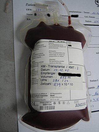
Hematopoietic stem-cell transplantation (HSCT) is the transplantation of multipotent hematopoietic stem cells, usually derived from bone marrow, peripheral blood, or umbilical cord blood in order to replicate inside of a patient and to produce additional normal blood cells. It may be autologous, allogeneic or syngeneic.

<span class="mw-page-title-main">POEMS syndrome</span> Paraneoplastic syndrome

POEMS syndrome is a rare paraneoplastic syndrome caused by a clone of aberrant plasma cells. The name POEMS is an acronym for some of the disease's major signs and symptoms, as is PEP.

<span class="mw-page-title-main">Lenalidomide</span> Pair of enantiomers

Lenalidomide, sold under the brand name Revlimid among others, is a medication used to treat multiple myeloma, smoldering myeloma, and myelodysplastic syndromes (MDS). For multiple myeloma, it is a first line treatment, and is given with dexamethasone. It is taken by mouth.

<span class="mw-page-title-main">Bortezomib</span> Chemical compound

Bortezomib, sold under the brand name Velcade among others, is an anti-cancer medication used to treat multiple myeloma and mantle cell lymphoma. This includes multiple myeloma in those who have and have not previously received treatment. It is generally used together with other medications. It is given by injection.

<span class="mw-page-title-main">Monoclonal gammopathy of undetermined significance</span> Medical condition

Monoclonal gammopathy of undetermined significance (MGUS) is a plasma cell dyscrasia in which plasma cells or other types of antibody-producing cells secrete a myeloma protein, i.e. an abnormal antibody, into the blood; this abnormal protein is usually found during standard laboratory blood or urine tests. MGUS resembles multiple myeloma and similar diseases, but the levels of antibodies are lower, the number of plasma cells in the bone marrow is lower, and it rarely has symptoms or major problems. However, since MGUS can lead to multiple myeloma, which develops at the rate of about 1.5% a year, or other symptomatic conditions, yearly monitoring is recommended.

Waldenström macroglobulinemia is a type of cancer affecting two types of B cells: lymphoplasmacytoid cells and plasma cells. Both cell types are white blood cells. It is characterized by having high levels of a circulating antibody, immunoglobulin M (IgM), which is made and secreted by the cells involved in the disease. Waldenström macroglobulinemia is an "indolent lymphoma" and a type of lymphoproliferative disease which shares clinical characteristics with the indolent non-Hodgkin lymphomas. It is commonly classified as a form of plasma cell dyscrasia, similar to other plasma cell dyscrasias that, for example, lead to multiple myeloma. Waldenström macroglobulinemia is commonly preceded by two clinically asymptomatic but progressively more pre-malignant phases, IgM monoclonal gammopathy of undetermined significance and smoldering Waldenström macroglobulinemia. The Waldenström macroglobulinemia spectrum of dysplasias differs from other spectrums of plasma cell dyscrasias in that it involves not only aberrant plasma cells but also aberrant lymphoplasmacytoid cells and that it involves IgM while other plasma dyscrasias involve other antibody isoforms.

<span class="mw-page-title-main">Follicular lymphoma</span> Medical condition

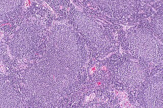
Follicular lymphoma (FL) is a cancer that involves certain types of white blood cells known as lymphocytes. The cancer originates from the uncontrolled division of specific types of B-cells known as centrocytes and centroblasts. These cells normally occupy the follicles in the germinal centers of lymphoid tissues such as lymph nodes. The cancerous cells in FL typically form follicular or follicle-like structures in the tissues they invade. These structures are usually the dominant histological feature of this cancer.

<span class="mw-page-title-main">Plasmacytoma</span> Growth of a plasma cell tumour within soft tissue or the axial skeleton

Plasmacytoma is a plasma cell dyscrasia in which a plasma cell tumour grows within soft tissue or within the axial skeleton.

Chronic neutrophilic leukemia (CNL) is a rare myeloproliferative neoplasm that features a persistent neutrophilia in peripheral blood, myeloid hyperplasia in bone marrow, hepatosplenomegaly, and the absence of the Philadelphia chromosome or a BCR/ABL fusion gene.

<span class="mw-page-title-main">Myeloma protein</span> Abnormal immunoglobulin fragment

A myeloma protein is an abnormal antibody (immunoglobulin) or a fragment thereof, such as an immunoglobulin light chain, that is produced in excess by an abnormal monoclonal proliferation of plasma cells, typically in multiple myeloma or Monoclonal gammopathy of undetermined significance. Other terms for such a protein are monoclonal protein, M protein, M component, M spike, spike protein, or paraprotein. This proliferation of the myeloma protein has several deleterious effects on the body, including impaired immune function, abnormally high blood viscosity, and kidney damage.

<span class="mw-page-title-main">Acute megakaryoblastic leukemia</span> Medical condition

Acute megakaryoblastic leukemia (AMKL) is life-threatening leukemia in which malignant megakaryoblasts proliferate abnormally and injure various tissues. Megakaryoblasts are the most immature precursor cells in a platelet-forming lineage; they mature to promegakaryocytes and, ultimately, megakaryocytes which cells shed membrane-enclosed particles, i.e. platelets, into the circulation. Platelets are critical for the normal clotting of blood. While malignant megakaryoblasts usually are the predominant proliferating and tissue-damaging cells, their similarly malignant descendants, promegakaryocytes and megakaryocytes, are variable contributors to the malignancy.

In hematology, plasma cell dyscrasias are a spectrum of progressively more severe monoclonal gammopathies in which a clone or multiple clones of pre-malignant or malignant plasma cells over-produce and secrete into the blood stream a myeloma protein, i.e. an abnormal monoclonal antibody or portion thereof. The exception to this rule is the disorder termed non-secretory multiple myeloma; this disorder is a form of plasma cell dyscrasia in which no myeloma protein is detected in serum or urine of individuals who have clear evidence of an increase in clonal bone marrow plasma cells and/or evidence of clonal plasma cell-mediated tissue injury. Here, a clone of plasma cells refers to group of plasma cells that are abnormal in that they have an identical genetic identity and therefore are descendants of a single genetically distinct ancestor cell.

Amyloid light-chain (AL) amyloidosis, also known as primary amyloidosis, is the most common form of systemic amyloidosis. The disease is caused when a person's antibody-producing cells do not function properly and produce abnormal protein fibers made of components of antibodies called light chains. These light chains come together to form amyloid deposits which can cause serious damage to different organs. An abnormal light chain in urine is known as Bence Jones protein.

<span class="mw-page-title-main">Blastic plasmacytoid dendritic cell neoplasm</span> Medical condition

Blastic plasmacytoid dendritic cell neoplasm (BPDCN) is a rare hematologic malignancy. It was initially regarded as a form of lymphocyte-derived cutaneous lymphoma and alternatively named CD4+CD56+ hematodermic tumor, blastic NK cell lymphoma, and agranular CD4+ NK cell leukemia. Later, however, the disease was determined to be a malignancy of plasmacytoid dendritic cells rather than lymphocytes and therefore termed blastic plasmacytoid dendritic cell neoplasm. In 2016, the World Health Organization designated BPDCN to be in its own separate category within the myeloid class of neoplasms. It is estimated that BPDCN constitutes 0.44% of all hematological malignancies.

Free light chains (FLCs) are immunoglobulin light chains that are found in the serum (blood) in an unbound (free) state. In recent decades, measuring the amount of free light chains (FLCs) in the blood has become a practical clinical test. FLC tests can be used to diagnose and monitor diseases like multiple myeloma and amyloidosis.

<span class="mw-page-title-main">Light chain deposition disease</span> Medical condition

Light chain deposition disease (LCDD) is a rare blood cell disease which is characterized by deposition of fragments of infection-fighting immunoglobulins, called light chains (LCs), in the body. LCs are normally cleared by the kidneys, but in LCDD, these light chain deposits damage organs and cause disease. The kidneys are almost always affected and this often leads to kidney failure. About half of people with light chain deposition disease also have a plasma cell dyscrasia, a spectrum of diseases that includes multiple myeloma, Waldenström's macroglobulinemia, and the monoclonal gammopathy of undetermined significance premalignant stages of these two diseases. Unlike in AL amyloidosis, in which light chains are laid down in characteristic amyloid deposits, in LCDD, light chains are deposited in non-amyloid granules.

Smouldering myeloma is a disease classified as intermediate in a spectrum of step-wise progressive diseases termed plasma cell dyscrasias. In this spectrum of diseases, a clone of plasma cells secreting monoclonal paraprotein causes the relatively benign disease of monoclonal gammopathy of undetermined significance. This clone proliferates and may slowly evolve into more aggressive sub-clones that cause smouldering multiple myeloma. Further and more rapid evolution causes the overtly malignant stage of multiple myeloma and can subsequently lead to the extremely malignant stage of secondary plasma cell leukemia. Thus, some patients with smouldering myeloma progress to multiple myeloma and plasma cell leukemia. Smouldering myeloma, however, is not a malignant disease. It is characterised as a pre-malignant disease that lacks symptoms but is associated with bone marrow biopsy showing the presence of an abnormal number of clonal myeloma cells, blood and/or urine containing a myeloma protein, and a significant risk of developing into a malignant disease.

<span class="mw-page-title-main">Serum B-cell maturation antigen</span> Cleaved form of B-cell maturation antigen

Serum B-cell maturation antigen (sBCMA) is the cleaved form of B-cell maturation antigen (BCMA), found at low levels in the serum of normal patients and generally elevated in patients with multiple myeloma (MM). Changes in sBCMA levels have been found to correlate with a MM patient’s clinical status in response to treatment.

Monoclonal immunoglobulin deposition disease, or MIDD, is a disease characterised by the deposition of monoclonal immunoglobulins on the basement membrane of the kidney. Monoclonal immunoglobulins are produced by monoclonal plasma cells, which are found in a variety of plasma cell dyscrasias. The deposition of monoclonal immunoglobulins on the basement membrane of the kidney causes renal impairment. As well as the kidney, MIDD may also affect the liver, heart, peripheral nerves, lung and skin.

References

  1. 1 2 3 4 5 6 7 Fernández de Larrea C, Kyle RA, Durie BG, Ludwig H, Usmani S, Vesole DH, Hajek R, San Miguel JF, Sezer O, Sonneveld P, Kumar SK, Mahindra A, Comenzo R, Palumbo A, Mazumber A, Anderson KC, Richardson PG, Badros AZ, Caers J, Cavo M, LeLeu X, Dimopoulos MA, Chim CS, Schots R, Noeul A, Fantl D, Mellqvist UH, Landgren O, Chanan-Khan A, Moreau P, Fonseca R, Merlini G, Lahuerta JJ, Bladé J, Orlowski RZ, Shah JJ (2013). "Plasma cell leukemia: consensus statement on diagnostic requirements, response criteria and treatment recommendations by the International Myeloma Working Group". Leukemia. 27 (4): 780–91. doi:10.1038/leu.2012.336. PMC   4112539 . PMID   23288300.
  2. 1 2 Granell M, Calvo X, Garcia-Guiñón A, Escoda L, Abella E, Martínez CM, Teixidó M, Gimenez MT, Senín A, Sanz P, Campoy D, Vicent A, Arenillas L, Rosiñol L, Sierra J, Bladé J, de Larrea CF (2017). "Prognostic impact of circulating plasma cells in patients with multiple myeloma: implications for plasma cell leukemia definition". Haematologica. 102 (6): 1099–1104. doi:10.3324/haematol.2016.158303. PMC   5451342 . PMID   28255016.
  3. 1 2 Gonsalves, Wilson I. (2017-04-10). "Primary Plasma Cell Leukemia: A Practical Approach to Diagnosis and Clinical Management". American Journal of Hematology / Oncology. 13 (3).
  4. 1 2 3 4 Joseph NS, Gentili S, Kaufman JL, Lonial S, Nooka AK (2017). "High-risk Multiple Myeloma: Definition and Management". Clinical Lymphoma, Myeloma & Leukemia. 17S: S80–S87. doi:10.1016/j.clml.2017.02.018. PMID   28760306. S2CID   22803869.
  5. 1 2 3 Rotaru I, Găman G, Dumitrescu D, Foarfă C (2012). "Secondary plasma cell leukemia". Romanian Journal of Morphology and Embryology = Revue Roumaine de Morphologie et Embryologie. 53 (4): 1073–6. PMID   23303035.
  6. 1 2 3 4 Cifola I, Lionetti M, Pinatel E, Todoerti K, Mangano E, Pietrelli A, Fabris S, Mosca L, Simeon V, Petrucci MT, Morabito F, Offidani M, Di Raimondo F, Falcone A, Caravita T, Battaglia C, De Bellis G, Palumbo A, Musto P, Neri A (2015). "Whole-exome sequencing of primary plasma cell leukemia discloses heterogeneous mutational patterns". Oncotarget. 6 (19): 17543–58. doi:10.18632/oncotarget.4028. PMC   4627327 . PMID   26046463.
  7. Dutta AK, Hewett DR, Fink JL, Grady JP, Zannettino AC (2017). "Cutting edge genomics reveal new insights into tumour development, disease progression and therapeutic impacts in multiple myeloma". British Journal of Haematology. 178 (2): 196–208. doi: 10.1111/bjh.14649 . PMID   28466550.
  8. 1 2 Barbieri M, Manzoni M, Fabris S, Ciceri G, Todoerti K, Simeon V, Musto P, Cortelezzi A, Baldini L, Neri A, Lionetti M (2016). "Compendium of FAM46C gene mutations in plasma cell dyscrasias". British Journal of Haematology. 174 (4): 642–5. doi: 10.1111/bjh.13793 . PMID   26456599. S2CID   3292996.
  9. Robillard N, Jego G, Pellat-Deceunynck C, Pineau D, Puthier D, Mellerin MP, Barillé S, Rapp MJ, Harousseau JL, Amiot M, Bataille R (1998). "CD28, a marker associated with tumoral expansion in multiple myeloma". Clinical Cancer Research. 4 (6): 1521–6. PMID   9626472.
  10. 1 2 3 Jelinek T, Kryukov F, Rihova L, Hajek R (2015). "Plasma cell leukemia: from biology to treatment". European Journal of Haematology. 95 (1): 16–26. doi: 10.1111/ejh.12533 . PMID   25778450.
  11. Gonsalves WI, Rajkumar SV, Go RS, Dispenzieri A, Gupta V, Singh PP, Buadi FK, Lacy MQ, Kapoor P, Dingli D, Lust JA, Zeldenrust SR, Hayman SR, Kyle RA, Gertz MA, Kumar SK (2014). "Trends in survival of patients with primary plasma cell leukemia: a population-based analysis". Blood. 124 (6): 907–12. doi:10.1182/blood-2014-03-565051. PMC   4126330 . PMID   24957143.
  12. Bommannan K, Sachdeva MU, Malhotra P, Kumar N, Sharma P, Naseem S, Ahluwalia J, Das R, Varma N, Prakash G, Khadwal A, Srinivasan R, Varma S (2016). "Plasma cell leukemia in North India: retrospective analysis of a distinct clinicohematological entity from a tertiary care center and review of literature". Blood Research. 51 (1): 23–30. doi:10.5045/br.2016.51.1.23. PMC   4828524 . PMID   27104188.
Bibliography