Plastic bronchitis

Last updated
Plastic bronchitis
Other namesFibrinous bronchitis, bronchitis pseudomembranosa, Hoffmann bronchitis
Specialty Respirology

Plastic bronchitis (PB) is a disorder in which branching casts of the airways are expectorated. PB is not a single disease with a defined mechanism that explains the cast formation in all conditions. Examples of diseases associated with expectoration of casts, and which sometimes are labeled PB include tuberculosis, atypical mycobacterial disease, allergic bronchopulmonary aspergillosis, and asthma.

Contents

When casts are very large with many branches, an abnormal communication or leakage of lymphatic fluid into the airway is often the cause. This entity is termed lymphatic plastic bronchitis (LPB). LPB is a lymphatic flow disorder characterized by the recurrent formation of branching, rubbery bronchial casts composed primarily of proteinaceous and sometimes chylous material and lymphocytes. Lymphatic fluids deposited into the airspaces become gelatinous as they cool, forming large string cheese-like casts of the airways, which can obstruct airflow. Attempts to expectorate casts can be quite frightening, leading to fears of asphyxiation.

Signs and symptoms

The clinical presentation of plastic bronchitis beyond expectoration of casts includes a productive cough, dyspnea, fever and wheezing. Focal wheezing is a characteristic, if not specific, physical examination finding. If the casts completely obstruct the airway, breath sounds will be decreased and dullness will be present with percussion. With partial obstruction, a “fan sound” or “flag flapping” sound can be heard during auscultation. Bronchial casts can sometimes fill the airways of almost an entire lung, and present as an acute, life-threatening emergency.[ citation needed ]

Pathology

The majority of PB cases are associated with an underlying disease. Several systemic illnesses have been associated with plastic bronchitis: [1]

The most common form of plastic bronchitis follows cardiac surgery for congenital heart disease, especially the Fontan procedure. Systemic blood flow is diverted to pulmonary flow, elevating pressures in the pulmonary venous system, and promoting leaks of proteinaceous and lipid-rich fluids from the lymphatics into the bronchial tree. [2]

Diagnosis

Plastic bronchitis cast Plastic Bronchitis specimen.jpg
Plastic bronchitis cast

The diagnosis of plastic bronchitis is confirmed by recovery of casts that have been coughed up or visualized during a bronchoscopy. There is no specific cytologic, pathologic or laboratory test that is diagnostic for casts due to lymphatic PB.[ citation needed ]

Imaging

Simple chest X-rays may reveal collapse due to airway obstruction. The contralateral lung may be hyperinflated. Casts can be visualized within the major airways using computerized axial tomography scans. [3]

Heavy T2-weighted MRI, and, as appropriate, intranodal lymphangiogram and/or dynamic contrast-enhanced MR lymphangiography may be useful for identifying pathological lymphatic tissue or lymphatic flow.[ citation needed ]

Management

Acute therapy for PB is often focused on removal or facilitated expectoration of the casts. This is followed by short and long term efforts to identify and remediate the underlying condition resulting in the excessive airway leakage or inflammation that is causing the casts to form.[ citation needed ]

PB can present as a life threatening emergency when the casts obstruct the major airways resulting in acute respiratory distress. [4] [5] [6] Intervention by a skilled physician experienced with foreign body removal from the lungs is essential. Evaluation by means of bronchoscopy can be difficult and time consuming and is best performed under general anesthesia.

Casts can be removed mechanically by bronchoscopy or physical therapy. High-frequency chest wall oscillation can also be used to vibrate the chest wall at a high frequency to try to loosen and thin the casts. [7] [8] Inhaled therapy using bronchodilators, corticosteroids or mucolytics can be used to try to disrupt the cast formation. Guaifenesin syrup or tablets can assist in loosening existing casts for expectoration.

Recently[ when? ], heavy T2-weighted MRI has revealed that occult lymphatic anomalies that represent developmental remnants or subclinical GLA are present in adults who present with expectoration of large multiantennary, branching casts. [9] Intranodal lymphangiogram [10] and dynamic contrast-enhanced MR lymphangiography [11] [12] have been used to more precisely image the leaks, and in the small number of patients who have been treated to date, embolization of the thoracic duct has been highly successful in controlling cast formation. [13]

Cannulation of the thoracic duct followed by embolization should be considered in those patients who are shown to have leakage of lymphatic fluid into the airway. [11]

Medications

Therapeutic interventions with medium-chain triglyceride-enriched low-fat diets, intratracheal heparin, inhaled tissue plasminogen activator, and steroids have also been reported and have met with variable success. [14] [15] [16] [17]

Expectorants such as guaifenesin increase thinner secretions and lubricate the airways, allowing loosening and possible self-expulsion of casts.

Inhaled mucolytics: Potassium iodide and acetylcysteine inhaled therapy are often used to help the patient cough up the casts by breaking down the thick mucus formations.

Inhaled and oral steroids: If PB is associated with asthma or an infection, inhaled and oral steroids have been shown to be effective. [18] [19]

Prognosis

Patients with plastic bronchitis that is being caused due to a co-morbid condition generally have a good prognosis once the underlying disease is treated.

Epidemiology

To date, about 420 cases have been reported in the medical literature. [1] Given its unusual nature, the true prevalence of PB is unknown, and it is likely that many patients are undiagnosed. PB does affect patients of all age groups and genders.

Related Research Articles

<span class="mw-page-title-main">Cough</span> Sudden expulsion of air from the lungs as a reflex to clear irritants

A cough is a sudden expulsion of air through the large breathing passages that can help clear them of fluids, irritants, foreign particles and microbes. As a protective reflex, coughing can be repetitive with the cough reflex following three phases: an inhalation, a forced exhalation against a closed glottis, and a violent release of air from the lungs following opening of the glottis, usually accompanied by a distinctive sound.

<span class="mw-page-title-main">Hemoptysis</span> Medical symptom consisting of bloody mucus from coughing

Hemoptysis is the coughing up of blood or blood-stained mucus from the bronchi, larynx, trachea, or lungs. In other words, it is the airway bleeding. This can occur with lung cancer, infections such as tuberculosis, bronchitis, or pneumonia, and certain cardiovascular conditions. Hemoptysis is considered massive at 300 mL. In such cases, there are always severe injuries. The primary danger comes from choking, rather than blood loss.

<span class="mw-page-title-main">Bronchiectasis</span> Disease of the lungs

Bronchiectasis is a disease in which there is permanent enlargement of parts of the airways of the lung. Symptoms typically include a chronic cough with mucus production. Other symptoms include shortness of breath, coughing up blood, and chest pain. Wheezing and nail clubbing may also occur. Those with the disease often get frequent lung infections.

<span class="mw-page-title-main">Pulmonology</span> Study of respiratory diseases

Pulmonology, pneumology or pneumonology is a medical specialty that deals with diseases involving the respiratory tract. It is also known as respirology, respiratory medicine, or chest medicine in some countries and areas.

<span class="mw-page-title-main">Fontan procedure</span>

The Fontan procedure or Fontan–Kreutzer procedure is a palliative surgical procedure used in children with univentricular hearts. It involves diverting the venous blood from the inferior vena cava (IVC) and superior vena cava (SVC) to the pulmonary arteries without passing through the morphologic right ventricle; i.e., the systemic and pulmonary circulations are placed in series with the functional single ventricle. The procedure was initially performed in 1968 by Francis Fontan and Eugene Baudet from Bordeaux, France, published in 1971, simultaneously described in 1971 by Guillermo Kreutzer from Buenos Aires, Argentina, and finally published in 1973.

<span class="mw-page-title-main">Lymphangioleiomyomatosis</span> Medical condition

Lymphangioleiomyomatosis (LAM) is a rare, progressive and systemic disease that typically results in cystic lung destruction. It predominantly affects women, especially during childbearing years. The term sporadic LAM is used for patients with LAM not associated with tuberous sclerosis complex (TSC), while TSC-LAM refers to LAM that is associated with TSC.

<span class="mw-page-title-main">Bronchoconstriction</span> Constriction of the terminal airways in the lungs

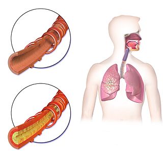
Bronchoconstriction is the constriction of the airways in the lungs due to the tightening of surrounding smooth muscle, with consequent coughing, wheezing, and shortness of breath.

<span class="mw-page-title-main">Chest physiotherapy</span>

Chest physiotherapy (CPT) are treatments generally performed by physical therapists and respiratory therapists, whereby breathing is improved by the indirect removal of mucus from the breathing passages of a patient. Other terms include respiratory or cardio-thoracic physiotherapy.

Reactive airway disease (RAD) is an informal label that physicians apply to patients with symptoms similar to those of asthma. An exact definition of the condition does not exist. Individuals who are typically labeled as having RAD generally have a history of wheezing, coughing, dyspnea, and production of sputum that may or may not be caused by asthma. Symptoms may also include, but are not limited to, coughing, shortness of breath, excess mucus in the bronchial tube, swollen mucous membrane in the bronchial tube, and/or hypersensitive bronchial tubes. Physicians most commonly label patients with RAD when they are hesitant about formally diagnosing a patient with asthma, which is most prevalent in the pediatric setting. While some physicians may use RAD and asthma synonymously, there is controversy over this usage.

<span class="mw-page-title-main">Aspergillosis</span> Medical condition

Aspergillosis is a fungal infection of usually the lungs, caused by the genus Aspergillus, a common mould that is breathed in frequently from the air around, but does not usually affect most people. It generally occurs in people with lung diseases such as asthma, cystic fibrosis or tuberculosis, or those who have had a stem cell or organ transplant, and those who cannot fight infection because of medications they take such as steroids and some cancer treatments. Rarely, it can affect skin.

<span class="mw-page-title-main">Bronchopulmonary segment</span>

A bronchopulmonary segment is a portion of lung supplied by a specific segmental bronchus and its vessels. These arteries branch from the pulmonary and bronchial arteries, and run together through the center of the segment. Veins and lymphatic vessels drain along the edges of the segment. The segments are separated from each other by layers of connective tissue that forms them into discrete anatomical and functional units. This separation means that a bronchopulmonary segment can be surgically removed without affecting the function of the others.

Bronchorrhea is the production of more than 100 mL per day of watery sputum. Chronic bronchitis is a common cause, but it may also be caused by asthma, pulmonary contusion, bronchiectasis, tuberculosis, cancer, scorpion stings, severe hypothermia and poisoning by organophosphates and other poisons. Massive bronchorrhea may occur in either bronchioloalveolar carcinoma, or in metastatic cancer that is growing in a bronchioloalveolar pattern. It commonly occurs in the setting of chest wall trauma, in which setting it can cause lobar atelectasis.

<span class="mw-page-title-main">Bronchitis</span> Inflammation of the large airways in the lungs

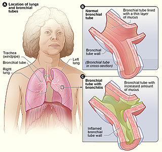
Bronchitis is inflammation of the bronchi in the lungs that causes coughing. Bronchitis usually begins as an infection in your nose, ears, throat, or sinuses. The infection then makes its way down to the bronchi. Symptoms include coughing up sputum, wheezing, shortness of breath, and chest pain. Bronchitis can be acute or chronic.

Williams–Campbell syndrome (WCS) is a disease of the airways where cartilage in the bronchi is defective. It is a form of congenital cystic bronchiectasis. This leads to collapse of the airways and bronchiectasis. It acts as one of the differential to allergic bronchopulmonary aspergillosis. WCS is a deficiency of the bronchial cartilage distally.

Pulmonary hygiene, formerly referred to as pulmonary toilet, is a set of methods used to clear mucus and secretions from the airways. The word pulmonary refers to the lungs. The word toilet, related to the French toilette, refers to body care and hygiene; this root is used in words such as toiletry that also relate to cleansing.

<span class="mw-page-title-main">Eosinophilic bronchitis</span> Medical condition

Eosinophilic bronchitis (EB) is a type of airway inflammation due to excessive mast cell recruitment and activation in the superficial airways as opposed to the smooth muscles of the airways as seen in asthma. It often results in a chronic cough. Lung function tests are usually normal. Inhaled corticosteroids are often an effective treatment.

<span class="mw-page-title-main">Chronic obstructive pulmonary disease</span> Lung disease involving long-term poor airflow

Chronic obstructive pulmonary disease (COPD) is a type of progressive lung disease characterized by long-term respiratory symptoms and airflow limitation. The main symptoms include shortness of breath and a cough, which may or may not produce mucus. COPD progressively worsens, with everyday activities such as walking or dressing becoming difficult. While COPD is incurable, it is preventable and treatable.

<span class="mw-page-title-main">Chronic pulmonary aspergillosis</span> Fungal infection

Chronic pulmonary aspergillosis is a long-term fungal infection caused by members of the genus Aspergillus—most commonly Aspergillusfumigatus. The term describes several disease presentations with considerable overlap, ranging from an aspergilloma—a clump of Aspergillus mold in the lungs—through to a subacute, invasive form known as chronic necrotizing pulmonary aspergillosis which affects people whose immune system is weakened. Many people affected by chronic pulmonary aspergillosis have an underlying lung disease, most commonly tuberculosis, allergic bronchopulmonary aspergillosis, asthma, or lung cancer.

<span class="mw-page-title-main">Lung cavity</span> Medical condition

A lung cavity or pulmonary cavity is an abnormal, thick-walled, air-filled space within the lung. Cavities in the lung can be caused by infections, cancer, autoimmune conditions, trauma, congenital defects, or pulmonary embolism. The most common cause of a single lung cavity is lung cancer. Bacterial, mycobacterial, and fungal infections are common causes of lung cavities. Globally, tuberculosis is likely the most common infectious cause of lung cavities. Less commonly, parasitic infections can cause cavities. Viral infections almost never cause cavities. The terms cavity and cyst are frequently used interchangeably; however, a cavity is thick walled, while a cyst is thin walled. The distinction is important because cystic lesions are unlikely to be cancer, while cavitary lesions are often caused by cancer.

Bronchial artery embolization is a treatment for hemoptysis, abbreviated as BAE. It is a kind of catheter intervention to control hemoptysis by embolizing the bronchial artery, which is a bleeding source. Embolic agents are particulate embolic material such as gelatin sponge or polyvinyl alcohol (PVA), and liquid embolic material such as NBCA, or metallic coils.

References

  1. 1 2 Eberlein, MH; Drummond, MB; Haponik, EF (2008). "Plastic bronchitis: a management challenge". Am J Med Sci. 335 (2): 163–169. doi:10.1097/maj.0b013e318068b60e. PMID   18277130.
  2. Dori, Y; Keller, MS; Rome, JJ; Gillespie, MJ; Glatz, AC; Dodds, K; Goldberg, DJ; Goldfarb, S; Rychik, J; Itkin, M (2016). "Percutaneous lymphatic embolization of abnormal pulmonary lymphatic flow as treatment of plastic bronchitis in patients with congenital heart disease". Circulation. 133 (12): 1160–70. doi:10.1161/circulationaha.115.019710. PMID   26864093. S2CID   26817256.
  3. "Plastic Bronchitis - Treatment, Symptoms and Causes".
  4. Itabashi, S; Kano, M; Endo, K; Sasaki, H (1999). "A resuscitated case from asphyxia by large bronchial cast". Tohoku J Exp Med. 187 (2): 183–8. doi: 10.1620/tjem.187.183 . PMID   10228989.
  5. Park, JY; Elshami, AA; Kang, DS; Jung, TH (1996). "Plastic bronchitis". Eur Respir J. 9 (3): 612–4. doi: 10.1183/09031936.96.09030612 . PMID   8730027.
  6. Noizet, O; Leclerc, F; Leteurtre, S; Brichet, A; Pouessel, G; Dorkenoo, A; Fourier, C; Cremer, R (2003). "Plastic bronchitis mimicking foreign body aspiration that needs a specific diagnostic procedure". Intensive Care Med. 29 (2): 329–31. doi:10.1007/s00134-002-1610-1. PMID   12594596. S2CID   32534712.
  7. Gross, D; Zidulka, A; O'Brien, C; Wright, D; Fraser, R; Rosenthal, L; King, M (1985). "Peripheral mucociliary clearance with high-frequency chest wall compression". J Appl Physiol. 58 (4): 1157–63. doi:10.1152/jappl.1985.58.4.1157. PMID   3988672.
  8. Koga, T; Kawazu, T; Iwashita, K; Yahata, R (2004). "Pulmonary hyperinflation and respiratory distress following solvent aspiration in a patient with asthma: expectoration of bronchial casts and clinical improvement with high-frequency chest wall oscillation". Respir Care. 49 (11): 1335–8. PMID   15507168.
  9. El Mouhadi, S; Taillé, C; Cazes, A; Arrivé, L (2015). "Plastic bronchitis related to idiopathic thoracic lymphangiectasia. Noncontrast magnetic resonance lymphography". Am J Respir Crit Care Med. 192 (5): 632–3. doi:10.1164/rccm.201503-0631im. PMID   26561678.
  10. Nadolski, GJ; Itkin, M (2012). "Feasibility of ultrasound-guided intranodal lymphangiogram for thoracic duct embolization". J Vasc Interv Radiol. 23 (5): 613–6. doi:10.1016/j.jvir.2012.01.078. PMID   22440590.
  11. 1 2 Dori, Y; Keller, MS; Rychik, J; Itkin, M (2014). "Successful treatment of plastic bronchitis by selective lymphatic embolization in a Fontan patient". Pediatrics. 134 (2): e590-5. doi:10.1542/peds.2013-3723. PMID   25002668. S2CID   2257097.
  12. Dori, Y; Zviman, MM; Itkin, M (2014). "Dynamic contrast-enhanced MR lymphangiography: feasibility study in swine". Radiology. 273 (2): 410–6. doi:10.1148/radiol.14132616. PMID   24988434.
  13. Itkin, M; McCormack, FX; Dori, Y (2016). "Diagnosis and treatment of lymphatic plastic bronchitis in adults using advanced lymphatic imaging and percutaneous embolization". Ann Am Thorac Soc. 13 (10): 1689–96. doi:10.1513/AnnalsATS.201604-292OC. PMID   27599255.
  14. Avitabile, CM; Goldberg, DJ; Dodds, K; Dori, Y; Ravishankar, C; Rychik, J (2014). "A multifaceted approach to the management of plastic bronchitis after cavopulmonary palliation". Ann Thorac Surg. 98 (2): 634–40. doi:10.1016/j.athoracsur.2014.04.015. PMC   4425256 . PMID   24841545.
  15. Houin, PR; Veress, LA; Rancourt, RC; Hendry-Hofer, TB; Loader, JE; Rioux, JS; Garlick, RB; White, CW (2015). "Intratracheal heparin improves plastic bronchitis due to sulfur mustard analog". Pediatr Pulmonol. 50 (2): 118–26. doi:10.1002/ppul.23043. PMC   4182164 . PMID   24692161.
  16. Parikh, K; Witte, MH; Samson, R; Teodori, M; Carpenter, JB; Lowe, MC; Morgan, W; Hardin, C; Brown, M; Naughton, Y; Sinha, S; Barber, BJ (2012). "Successful treatment of plastic bronchitis with low fat diet and subsequent thoracic duct ligation in child with fontan physiology". Lymphology. 45 (2): 47–52. PMID   23057148.
  17. Turgut, T; In, E; Özercan, IH; Kaplan, M (2014). "A case of plastic bronchitis". Arch Iran Med. 17 (8): 589–90. PMID   25065285.
  18. Onoue, Y; Adachi, Y; Ichida, F; Miyawaki, T (2003). "Effective use of corticosteroid in a child with life-threatening plastic bronchitis after Fontan operation". Pediatr Int. 45 (1): 107–9. doi:10.1046/j.1442-200x.2003.01659.x. PMID   12654082. S2CID   39667914.
  19. Wang, G; Wang, YJ; Luo, FM; Wang, L; Jiang, LL; Wang, L; Mao, B (2006). "Effective use of corticosteroids in treatment of plastic bronchitis with hemoptysis in Chinese adults". Acta Pharmacologica Sinica. 27 (9): 1206–12. doi: 10.1111/j.1745-7254.2006.00418.x . PMID   16923342.