Related Research Articles
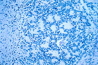
Neuroblastoma (NB) is a type of cancer that forms in certain types of nerve tissue. It most frequently starts from one of the adrenal glands but can also develop in the neck, chest, abdomen, or spine. Symptoms may include bone pain, a lump in the abdomen, neck, or chest, or a painless bluish lump under the skin.

Desmoplastic small-round-cell tumor (DSRCT) is an aggressive and rare cancer that primarily occurs as masses in the abdomen. Other areas affected may include the lymph nodes, the lining of the abdomen, diaphragm, spleen, liver, chest wall, skull, spinal cord, large intestine, small intestine, bladder, brain, lungs, testicles, ovaries, and the pelvis. Reported sites of metastatic spread include the liver, lungs, lymph nodes, brain, skull, and bones. It is characterized by the EWS-WT1 fusion protein.
Anaplasia is a condition of cells with poor cellular differentiation, losing the morphological characteristics of mature cells and their orientation with respect to each other and to endothelial cells. The term also refers to a group of morphological changes in a cell that point to a possible malignant transformation.

In pathology, grading is a measure of the cell appearance in tumors and other neoplasms. Some pathology grading systems apply only to malignant neoplasms (cancer); others apply also to benign neoplasms. The neoplastic grading is a measure of cell anaplasia in the sampled tumor and is based on the resemblance of the tumor to the tissue of origin. Grading in cancer is distinguished from staging, which is a measure of the extent to which the cancer has spread.

Ganglioneuroma is a rare and benign tumor of the autonomic nerve fibers arising from neural crest sympathogonia, which are completely undifferentiated cells of the sympathetic nervous system. However, ganglioneuromas themselves are fully differentiated neuronal tumors that do not contain immature elements.

Paired-like homeobox 2b (PHOX2B), also known as neuroblastoma Phox (NBPhox), is a protein that in humans is encoded by the PHOX2B gene located on chromosome 4.
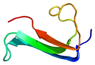
Midkine, also known as neurite growth-promoting factor 2 (NEGF2), is a protein that in humans is encoded by the MDK gene.
Ectomesenchymoma is a rare, fast-growing tumor of the nervous system or soft tissue that occurs mainly in children, although cases have been reported in patients up to age 60. Ectomesenchymomas may form in the head and neck, abdomen, perineum, scrotum, or limbs. Also called malignant ectomesenchymoma.

Achaete-scute homolog 1 is a protein that in humans is encoded by the ASCL1 gene. Because it was discovered subsequent to studies on its homolog in Drosophila, the Achaete-scute complex, it was originally named MASH-1 for mammalian achaete scute homolog-1.

Ganglioneuroblastoma is a variant of neuroblastoma that is surrounded by ganglion cells.
Large cell lung carcinoma with rhabdoid phenotype (LCLC-RP) is a rare histological form of lung cancer, currently classified as a variant of large cell lung carcinoma (LCLC). In order for a LCLC to be subclassified as the rhabdoid phenotype variant, at least 10% of the malignant tumor cells must contain distinctive structures composed of tangled intermediate filaments that displace the cell nucleus outward toward the cell membrane. The whorled eosinophilic inclusions in LCLC-RP cells give it a microscopic resemblance to malignant cells found in rhabdomyosarcoma (RMS), a rare neoplasm arising from transformed skeletal muscle. Despite their microscopic similarities, LCLC-RP is not associated with rhabdomyosarcoma.

Giant-cell carcinoma of the lung (GCCL) is a rare histological form of large-cell lung carcinoma, a subtype of undifferentiated lung cancer, traditionally classified within the non-small-cell lung carcinomas (NSCLC).

Crizotinib, sold under the brand name Xalkori among others, is an anti-cancer medication acting as an ALK and ROS1 inhibitor, approved for treatment of some non-small cell lung carcinoma (NSCLC) in the US and some other countries, and undergoing clinical trials testing its safety and efficacy in anaplastic large cell lymphoma, neuroblastoma, and other advanced solid tumors in both adults and children.

Pleomorphism is a term used in histology and cytopathology to describe variability in the size, shape and staining of cells and/or their nuclei. Several key determinants of cell and nuclear size, like ploidy and the regulation of cellular metabolism, are commonly disrupted in tumors. Therefore, cellular and nuclear pleomorphism is one of the earliest hallmarks of cancer progression and a feature characteristic of malignant neoplasms and dysplasia. Certain benign cell types may also exhibit pleomorphism, e.g. neuroendocrine cells, Arias-Stella reaction.
A sialoblastoma is a low-grade salivary gland neoplasm that recapitulates primitive salivary gland anlage. It has previously been referred to as congenital basal cell adenoma, embryoma, or basaloid adenocarcinoma. It is an extremely rare tumor, with less than 100 cases reported worldwide.
Rhabdomyoma is a benign mesenchymal tumor of skeletal muscle, separated into two major categories based on site: Cardiac and extracardiac. They are further separated by histology: fetal, juvenile (intermediate), and adult types. Genital types are recognized, but are often part of either the fetal or juvenile types. The fetal type is thought to recapitulate immature skeletal muscle at about week six to ten of gestational development.
Smooth muscle tumor of uncertain malignant potential, abbreviated STUMP, is an uncommon tumor of the uterine smooth muscle that may behave like a benign tumor or a cancerous tumor.
Ectomesenchymal chondromyxoid tumor (ECT) is a benign intraoral tumor with presumed origin from undifferentiated (ecto)mesenchymal cells. There are some who think it is a myoepithelial tumor type.
Askin's tumor is a rare, primitive neuroectodermal tumor which arises from the soft tissues of the chest wall, particularly of the paravertebral region. It was first described by Askin et al in 1979. Askin's tumor is now recognized as part of the Ewing's sarcoma family of tumors. This neoplasm tended to recur locally, but did not seem to disseminate as widely as some of the other small cell tumors of childhood such as rhabdomyosarcoma or neuroblastoma.

The histopathology of colorectal cancer of the adenocarcinoma type involves analysis of tissue taken from a biopsy or surgery. A pathology report contains a description of the microscopical characteristics of the tumor tissue, including both tumor cells and how the tumor invades into healthy tissues and finally if the tumor appears to be completely removed. The most common form of colon cancer is adenocarcinoma, constituting between 95% to 98% of all cases of colorectal cancer. Other, rarer types include lymphoma, adenosquamous and squamous cell carcinoma. Some subtypes have been found to be more aggressive.
References
- 1 2 3 Cozzutto C, Carbone A (1988). Pleomorphic (Anaplastic) neuroblastoma. Arch Pathol Lab Med 112:621-625.
- ↑ Cowan J, Dayal Y, Schaitzberg S, Tischler AS (1997). Cytogenetic and immunohistological analysis of an adult anaplastic neuroblastoma. Am J Surg Pathol 21(8):957-963.
- ↑ Navarro S, Noguera R, Pellin A, Mejia C, Ruiz A, Llombart-Bosch A (2000). Pleomorphic anaplastic neuroblastoma. Med Pediat Oncol 35:449-502.
- 1 2 Fletcher CDM (2007). Diagnostic Histopatholoy of tumors. Churchill Livingstone, p. 1774. Rosai (2004).
- 1 2 Rosai and Ackerman's Surgical Pathology. Philadelphia, Mosby, p.1128.
- ↑ Chatten J (1989). Anaplastic neuroblastomas. Letter to the editor. Arch Pathol Lab Med 113:9-10.
- ↑ Abramowsky CR, Katzenstein HM, Alvarado CS, Shehata BM (2009). Anaplastic large cell neuroblastoma. Pediat Devel Pathol 12(1):1-5.
- ↑ Joshi VV, Cantor AB, Altshuler G, Larkin EW et al. (1992). Age-linked prognostic categorization based on a new histologic grading system of neuroblastomas. A clinicopathologic study of 211 cases from the pediatric oncology group. Cancer 69:2197-2211.
- ↑ Dehner LP (1989). Anaplasia in solid malignant tumors of childhood. Arch Pathol Lab Med 113:11-12.
- ↑ Shimada H, Ambros IM, Dehner LP, Joshi VV, Roald B (1999). Terminology and morphologic criteria of neuroblastic tumors. Recommendations by the International Neuroblastoma Pathology Committee. Cancer 86(2):349-363.
- ↑ Tornòczky T, Kàlmàn E, Kajtàr PG, Nyàri T, Pearson AD, Tweddle DA, Board J, Shimada H (2004) Large cell neuroblastoma. A distinctive phenotype of neuroblastoma with aggressive clinical behavior. Cancer 100(2):390-397.
- Shimada H, Ambros IM, Dehner LP, Hata J, Joshi VV, Roald B (1999). Terminology and morphologic criteria of neuroblastic tumors. Recommendations by the International Neuroblastoma Pathology Committee. Cancer 86(2) 349–363.