Primary tumors of the heart

Last updated
Primary tumors of the heart
Papillary fibroelastoma.jpg
Micrograph of a papillary fibroelastoma, a benign tumor of heart valves. H&E stain.
Specialty Oncology, cardiac surgery

Primary tumors of the heart are extremely rare tumors that arise from the normal tissues that make up the heart. The incidence of primary cardiac tumors has been found to be approximately 0.02%. [1] This is in contrast to secondary tumors of the heart, which are typically either metastatic from another part of the body, or infiltrate the heart via direct extension from the surrounding tissues. Metastatic tumors to the heart are about 20 times more common than primary cardiac tumors.

Contents

Types

Micrograph of an atrial myxoma, the most common primary tumor of the heart. Atrial myxoma intermed mag.jpg
Micrograph of an atrial myxoma, the most common primary tumor of the heart.

Benign

The most common primary tumor of the heart is the myxoma. In surgical series, the myxoma makes up as much as 77% of all primary tumors of the heart. [2] Less common tumors of the heart include: [3]

Malignant

About 20 percent of primary tumors of the heart are malignant in nature. [2] Malignant tumors of the heart include rhabdomyosarcomas, angiosarcomas, myxosarcomas, fibrosarcomas, leiomyosarcomas, reticulum cell sarcomas, desmoplastic small round cell tumor, and liposarcomas. Angiosarcoma is the most common primary cardiac malignancy. [4] It tends to occur in the right atrium and involve the pericardium. Cardiac sarcomas may occur at any age but are more commonly seen in individuals in their 20s to 40s. They occur equally in males and females. Rhabdomyosarcoma is the most common primary cardiac malignancy in children and is more likely than other primary cardiac sarcomas to involve the valves.

Valvular

A subset of the primary tumors of the heart are tumors that are found on the valves of the heart. Tumors that affect the valves of the heart are found in an equal distribution among the four heart valves. [2] The vast majority of these are papillary fibroelastomas. Primary tumors of the valves of the heart are more likely to occur in males. While most primary tumors of the valves of the heart are not malignant, they are more likely to have symptoms related to the valve, including neurologic symptoms and (in a few cases) sudden cardiac death.

Clinical manifestations

In general, tumors might manifest in one of three ways:

Systemic symptoms include constitutional symptoms(fever, arthralgias, weight loss, weariness) and paraneoplastic disorders (PCTs).

Cardiovascular symptoms: mass effect compromising myocardial function or blood flow, causing arrhythmias, obstruction of heart valves leading to regurgitation, or pericardial effusion with or without tamponade. Primary symptoms include breathlessness, chest pain, presyncope, and syncope.

Thromboembolic symptoms: lung and systemic thromboembolic condition resulting from the tumor. [5]

Symptoms caused by primary cardiac tumors are usually dependent on size and location. Cardiac tumors can cause a variety of symptoms. The mass size can obstruct blood flow or interfere with cardiac valve function and produce symptoms of heart failure. A tumor that invades the heart walls may cause arrhythmias, heart block, or pericardial effusion with or without tamponade. Tumors that invade adjacent lung tissue may cause pulmonary symptoms that mimic bronchogenic carcinoma. [6] The tumor may break apart and can embolize. Embolization can be systemic or pulmonic. Tumors with the highest anatomic risk for embolization are located in the left atrium or aortic valve. Some tumors produce no symptoms and are incidentally found. [7]

Systemic symptoms [8]

Primary cardiac tumors may secrete factors including interleukin-6 and endothelin. This may contribute to systemic inflammatory or autoimmune manifestations. Systemic symptoms including fever, fatigue, arthralgia, anemia, elevated white blood cell count, thrombocytosis, thrombocytopenia, hypergammaglobulinemia, and elevated erythrocyte sedimentation rate. Myxomas have been found to produce vascular endothelial growth factor, which contributes to the development of new blood vessels and the early stages of tumor growth. [9]

Diagnostic evaluation

The initial diagnostic evaluation is to determine whether or not a cardiac tumor is present, the location of the tumor, and if the tumor is benign or malignant.

Initial imaging is often performed by echocardiography. [10] Benign lesions have a distinct appearance on an echocardiogram. An echocardiogram can identify the presence of a mass and its mobility. The echocardiogram can also identify a mass causing obstruction of circulation. The echocardiogram can provide accurate information as to whether the tumor is malignant or benign.

Cardiac magnetic resonance imaging can identify the chemical microenvironment within the tumor, thus providing more information as to the type of tumor present.

Cardiac computed tomography is useful when MRI is unavailable or contraindicated.

Coronary angiography is used to identify the blood supply to cardiac tumors.

Treatment and prognosis

The vast majority of the tumors of the heart have a benign course and are not directly fatal. However, even the benign tumors of the heart can be lethal due to either direct extension into the electrical conduction system of the heart (causing complete heart block or a fatal dysrhythmia), or due to emboli from the tumor mass that may have lethal sequelae. Prompt resection is indicated for myxomas due to the risk of embolization or cardiovascular complications. Close observation has also been suggested for cardiac tumors that are small (<1 cm) and non-mobile. [11]

The malignant tumors of the heart have a worse prognosis. Cardiac sarcomas generally lead to death within 2 years of diagnosis, due to rapid infiltration of the myocardium of the heart and obstruction of the normal flow of blood within the heart. [12] [13]

See also

Related Research Articles

<span class="mw-page-title-main">Kidney cancer</span> Medical condition

Kidney cancer, also known as renal cancer, is a group of cancers that starts in the kidney. Symptoms may include blood in the urine, lump in the abdomen, or back pain. Fever, weight loss, and tiredness may also occur. Complications can include spread to the lungs or brain.

<span class="mw-page-title-main">Malignancy</span> Tendency of a medical condition to become progressively worse

Malignancy is the tendency of a medical condition to become progressively worse; the term is most familiar as a characterization of cancer.

<span class="mw-page-title-main">Mitral regurgitation</span> Form of valvular heart disease

Mitral regurgitation(MR), also known as mitral insufficiency or mitral incompetence, is a form of valvular heart disease in which the mitral valve is insufficient and does not close properly when the heart pumps out blood. It is the abnormal leaking of blood backwards – regurgitation from the left ventricle, through the mitral valve, into the left atrium, when the left ventricle contracts. Mitral regurgitation is the most common form of valvular heart disease.

<span class="mw-page-title-main">Myxoma</span> Myxoid tumor of primitive connective tissue

A myxoma is a myxoid tumor of primitive connective tissue. It is most commonly found in the heart but can also occur in other locations.

<span class="mw-page-title-main">Rhabdomyoma</span> Medical condition

A rhabdomyoma is a benign tumor of striated muscle. Rhabdomyomas may be either cardiac or extracardiac. Extracardiac forms of rhabdomyoma are sub-classified into three distinct types: adult type, fetal type, and genital type.

Libman–Sacks endocarditis is a form of non-bacterial endocarditis that is seen in association with systemic lupus erythematosus, antiphospholipid syndrome, and malignancies. It is one of the most common cardiac manifestations of lupus.

Spinal tumors are neoplasms located in either the vertebral column or the spinal cord. There are three main types of spinal tumors classified based on their location: extradural and intradural. Extradural tumors are located outside the dura mater lining and are most commonly metastatic. Intradural tumors are located inside the dura mater lining and are further subdivided into intramedullary and extramedullary tumors. Intradural-intramedullary tumors are located within the dura and spinal cord parenchyma, while intradural-extramedullary tumors are located within the dura but outside the spinal cord parenchyma. The most common presenting symptom of spinal tumors is nocturnal back pain. Other common symptoms include muscle weakness, sensory loss, and difficulty walking. Loss of bowel and bladder control may occur during the later stages of the disease.

<span class="mw-page-title-main">Angiosarcoma</span> Cancer of the lining of the blood or lymphatic vessels

Angiosarcoma is a rare and aggressive cancer that starts in the endothelial cells that line the walls of blood vessels or lymphatic vessels. Since they are made from vascular lining, they can appear anywhere and at any age, but older people are more commonly affected, and the skin is the most affected area, with approximately 60% of cases being cutaneous. Specifically, the scalp makes up ~50% of angiosarcoma cases, but this is still <0.1% of all head and neck tumors. Since angiosarcoma is an umbrella term for many types of tumor that vary greatly in origin and location, many symptoms may occur, from completely asymptomatic to non-specific symptoms like skin lesions, ulceration, shortness of breath and abdominal pain. Multiple-organ involvement at time of diagnosis is common and makes it difficult to ascertain origin and how to treat it.

<span class="mw-page-title-main">Superior vena cava syndrome</span> Group of symptoms caused by obstruction of the superior vena cava

Superior vena cava syndrome (SVCS), is a group of symptoms caused by obstruction of the superior vena cava ("SVC"), a short, wide vessel carrying circulating blood into the heart. The majority of cases are caused by malignant tumors within the mediastinum, most commonly lung cancer and non-Hodgkin's lymphoma, directly compressing or invading the SVC wall. Non-malignant causes are increasing in prevalence due to expanding use of intravascular devices, which can result in thrombosis. Other non-malignant causes include benign mediastinal tumors, aortic aneurysm, infections, and fibrosing mediastinitis.

<span class="mw-page-title-main">Carcinoid</span> A slow-growing type of neuroendocrine tumor

A carcinoid is a slow-growing type of neuroendocrine tumor originating in the cells of the neuroendocrine system. In some cases, metastasis may occur. Carcinoid tumors of the midgut are associated with carcinoid syndrome.

The International Classification of Diseases for Oncology (ICD-O) is a domain-specific extension of the International Statistical Classification of Diseases and Related Health Problems for tumor diseases. This classification is widely used by cancer registries.

<span class="mw-page-title-main">Hemangioendothelioma</span> Medical condition

Hemangioendotheliomas are a family of vascular neoplasms of intermediate malignancy.

Carcinoid syndrome is a paraneoplastic syndrome comprising the signs and symptoms that occur secondary to neuroendocrine tumors. The syndrome is caused by neuroendocrine tumors most often found in the gut releasing biologically active substances into the blood causing symptoms such as flushing and diarrhea, and less frequently, heart failure, vomiting and bronchoconstriction.

<span class="mw-page-title-main">Pericardial effusion</span> Medical condition

A pericardial effusion is an abnormal accumulation of fluid in the pericardial cavity. The pericardium is a two-part membrane surrounding the heart: the outer fibrous connective membrane and an inner two-layered serous membrane. The two layers of the serous membrane enclose the pericardial cavity between them. This pericardial space contains a small amount of pericardial fluid, normally 15-50 mL in volume. The pericardium, specifically the pericardial fluid provides lubrication, maintains the anatomic position of the heart in the chest, and also serves as a barrier to protect the heart from infection and inflammation in adjacent tissues and organs.

Heart cancer is an extremely rare form of cancer that is divided into primary tumors of the heart and secondary tumors of the heart.

<span class="mw-page-title-main">Cardiac myxoma</span> Medical condition

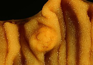
A myxoma is a rare benign tumor of the heart. Myxomata are the most common primary cardiac tumor in adults, and are most commonly found within the left atrium near the valve of the fossa ovalis. Myxomata may also develop in the other heart chambers. The tumor is derived from multipotent mesenchymal cells. Cardiac myxoma can affect adults between 30 and 60 years of age.

<span class="mw-page-title-main">Transitional cell carcinoma</span> Medical condition

Transitional cell carcinoma, also called urothelial carcinoma, is a type of cancer that typically occurs in the urinary system. It is the most common type of bladder cancer and cancer of the ureter, urethra, and urachus. Symptoms of urothelial carcinoma in the bladder include hematuria. Diagnosis includes urine analysis and imaging of the urinary tract (cystoscopy). Transitional cell carcinomas arise from the transitional epithelium, a tissue lining the inner surface of these hollow organs. When the term "urothelial" is used, it specifically refers to a carcinoma of the urothelium, meaning a transitional cell carcinomas of the urinary system.

<span class="mw-page-title-main">Papillary fibroelastoma</span> Medical condition

A papillary fibroelastoma is a primary tumor of the heart that typically involves one of the valves of the heart. Papillary fibroelastomas, while considered uncommon, make up about 10 percent of all primary tumors of the heart. They are the third most common type of primary tumor of the heart, behind cardiac myxomas and cardiac lipomas.

Cardiac fibroma, also known as cardiac fibromatosis, cardiac fibrous hamartoma, fibroelastic hamartoma of heart and fibroma of heart is the second highest type of primary cardiac tumor seen in infants and children. This benign tumor made by connective tissue and fibroblast is largely observed in the ventricles of the heart. The left ventricle is the most common location of cardiac fibroma and accounts for approximately 57% of cardiac fibroma cases followed by the right ventricle with 27.5% of cases. Symptoms of the disease depend on the size of the tumor, its location relative to the conduction system, and whether it obstructs blood flow. Two-thirds of children with this tumor are asymptomatic, showing no signs and symptoms. Therefore the cause of cardiac fibroma is unexplained but has been associated with Gorlin Syndrome. Echocardiography is the primarily diagnostic method used to detect if an individual has cardiac fibroma. Resection of the tumor is recommended however heart transplantation is done if surgery is not possible. Overall prognosis of resection is favorable and the chance of recurrence is scarcely reported.

Hyperthermic intrathoracic chemotherapy (HITOC) is part of a surgical strategy employed in the treatment of various pleural malignancies. The pleura in this situation could be considered to include the surface linings of the chest wall, lungs, mediastinum, and diaphragm. HITOC is the chest counterpart of HIPEC. Traditionally used in the treatment of malignant mesothelioma, a primary malignancy of the pleura, this modality has recently been evaluated in the treatment of secondary pleural malignancies.

References

  1. Reynen, Klaus (January 1996). "Frequency of primary tumors of the heart". The American Journal of Cardiology. 77 (1): 107. doi:10.1016/s0002-9149(97)89149-7. ISSN   0002-9149. PMID   8540447.
  2. 1 2 3 Molina JE, Edwards JE, Ward HB (1990). "Primary cardiac tumors: experience at the University of Minnesota". Thorac Cardiovasc Surg. 38 (Suppl 2): 183–91. doi:10.1055/s-2007-1014064. PMID   2237900. S2CID   34899353.
  3. Elbardissi, Andrew W.; Dearani, Joseph A.; Daly, Richard C.; Mullany, Charles J.; Orszulak, Thomas A.; Puga, Francisco J.; Schaff, Hartzell V. (January 2009). "Embolic potential of cardiac tumors and outcome after resection: a case-control study". Stroke. 40 (1): 156–162. doi: 10.1161/STROKEAHA.108.525709 . ISSN   1524-4628. PMID   18948602. S2CID   1548077.
  4. Araoz, P. A.; Eklund, H. E.; Welch, T. J.; Breen, J. F. (November 1999). "CT and MR imaging of primary cardiac malignancies". Radiographics. 19 (6): 1421–1434. doi:10.1148/radiographics.19.6.g99no031421. ISSN   0271-5333. PMID   10555666.
  5. Tyebally, Sara; Chen, Daniel; Bhattacharyya, Sanjeev; Mughrabi, Abdallah; Hussain, Zeeshan; Manisty, Charlotte; Westwood, Mark; Ghosh, Arjun K.; Guha, Avirup (June 2020). "Cardiac Tumors: JACC CardioOncology State-of-the-Art Review". JACC: CardioOncology. 2 (2): 293–311. doi:10.1016/j.jaccao.2020.05.009. ISSN   2666-0873. PMC   8352246 . PMID   34396236.
  6. Sheu, Chau-Chyun; Lin, Sheng-Fung; Chiu, Chaw-Chi; Lee, Chee-Siong; Chai, Chee-Yin; Liu, Yi-Chang; Huang, Ming-Shyan (2007-04-01). "Left Atrial Sarcoma Mimicking Obstructive Pulmonary Disease". Journal of Clinical Oncology. 25 (10): 1277–1279. doi: 10.1200/JCO.2006.09.9077 . ISSN   0732-183X. PMID   17401018.
  7. Vander Salm, Thomas J. (April 2000). "Unusual Primary Tumors Of The Heart". Seminars in Thoracic and Cardiovascular Surgery. 12 (2): 89–100. doi:10.1053/ct.2000.5080. ISSN   1043-0679. PMID   10807431.
  8. Paraskevaidis, Ioannis A.; Michalakeas, Christos A.; Papadopoulos, Constantinos H.; Anastasiou-Nana, Maria (2011). "Cardiac Tumors". ISRN Oncology. 2011: 208929. doi: 10.5402/2011/208929 . ISSN   2090-5661. PMC   3195386 . PMID   22091416.
  9. Sakamoto, Hironosuke; Sakamaki, Tetsuo; Kanda, Tsugiyasu; Tsuchiya, Yoko; Sato, Mahito; Sato, Hiroko; Oyama, Yuko; Sawada, Yoshie; Tamura, Jun-ichi; Nagai, Ryozo; Kurabayashi, Masahiko (May 2004). "Vascular endothelial growth factor is an autocrine growth factor for cardiac myxoma cells". Circulation Journal. 68 (5): 488–493. doi: 10.1253/circj.68.488 . ISSN   1346-9843. PMID   15118294.
  10. Vander Salm, T. J. (April 2000). "Unusual primary tumors of the heart". Seminars in Thoracic and Cardiovascular Surgery. 12 (2): 89–100. doi:10.1053/ct.2000.5080. ISSN   1043-0679. PMID   10807431.
  11. Gowda, Ramesh M.; Khan, Ijaz A.; Nair, Chandra K.; Mehta, Nirav J.; Vasavada, Balendu C.; Sacchi, Terrence J. (September 2003). "Cardiac papillary fibroelastoma: a comprehensive analysis of 725 cases". American Heart Journal. 146 (3): 404–410. doi:10.1016/S0002-8703(03)00249-7. ISSN   1097-6744. PMID   12947356.
  12. Burke AP, Cowan D, Virmani R (1992). "Primary sarcomas of the heart". Cancer. 69 (2): 387–95. doi: 10.1002/1097-0142(19920115)69:2<387::AID-CNCR2820690219>3.0.CO;2-N . PMID   1728367. S2CID   10893409.
  13. Burke AP, Virmani R (1991). "Osteosarcomas of the heart". Am J Surg Pathol. 15 (3): 289–95. doi:10.1097/00000478-199103000-00009. PMID   1705103. S2CID   43134869.