Pupilometer

Last updated

Pupillometer, also spelled pupilometer, is a medical device intended to measure by reflected light the size of the pupil of the eye. [1] In addition to measuring pupil size, current automated pupillometers may also be able to characterize pupillary light reflex. Some instruments for measuring pupillary distance (PD) are often, but incorrectly, referred to as pupilometers. [2]

Contents

Manual pupillometry

A manual pupillometer measures pupil size via a comparison chart method. There are several types of manual pupillometers. The most common type is the Haab scale, or Haab's pupillometer, which is a series of graduated filled circles on a slide ruler. [2]

Automated Pupillometry

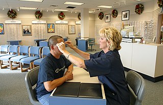
An automated pupillometer is a portable, handheld device that provides a reliable and objective measurement of pupillary size, symmetry, and reactivity through measurement of the pupillary light reflex (PLR). PLR is historically assessed by a nurse or a clinician using a manual flash lamp (sPLR, “s” stands for standard). sPLR is opposed to quantitative PLR (qPLR) that is provided by an automated pupillometer. qPLR corresponds to the percentage of pupillary constriction to a calibrated light stimulus. [3] Independent of examiner, an automated pupillometer offers reproducible and precise measurements by eliminating variability and subjectivity, expressing pupil reactivity numerically so that both pupil size and reactivity can be trended for changes, just like other vital signs. An automated pupillometer also provides a reliable and effective way to quantitatively classify and trend the pupil light response. [4] [5] [6] [7]

The pupillary light reflex is the constriction of the pupils when exposed to bright light, protecting the retina from excessive light exposure. It involves the automatic constriction and dilation of the pupils in response to changes in light intensity or accommodation.

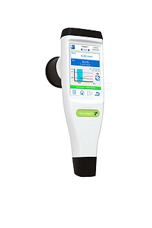
With an automated pupillometer and an algorithm analyzing the pupil continuously for 5 seconds, the Quantitative Pupillometry Index (QPi) can measure pupillary reactivity and provides a numerical value. It provides objective data and can detect subtle changes that might not be apparent to the naked eye. Its quantitative nature provides objective and more reliable assessment. Moreover, the index of Neurolight pupillometer is color-coded for a quick clinical interpretation. It displays through a qualitative scale a quantitative interval for each color associated with its number. [8]


Automated pupillometry removes subjectivity from the pupillary evaluation[5], providing a more accurate trend of pupil data, and allowing earlier detection of changes for more timely patient treatment. Pupil data can be uploaded to the patient record, eliminating the possibility of data entry errors. The pupil size and reactivity are daily measurements and part of the protocol for critically injured or ill patients. They are essential in the clinical monitoring and neurological assessment of the patient. Abnormalities in pupillary responses can be indicative of underlying neurological disorders, such as traumatic brain injury, stroke, cardiac arrest [9] or certain neurodegenerative diseases.

Neurological assessment with NeuroLight pupillometer (IDMED, IDMED Corp.) Pupillometer neurolight.jpg
Neurological assessment with NeuroLight pupillometer (IDMED, IDMED Corp.)
NPi-300 automated infrared pupillometer (NeurOptics, Inc.) NeurOptics' NPi-300 Automated Pupillometer.png
NPi-300 automated infrared pupillometer (NeurOptics, Inc.)

Another automated pupillometer named NeurOptics' Neurological Pupil index (NPi) can offer a consolidated parametric approach to mitigate subjectivity. [10] The NeuroLight and NPi pupillometer are both device for measuring pupils but differ significantly in terms of ergonomics and functionality. The main distinction lies in the NPi’s use of a transparent eyecup that contains an electronic component for patient identification and results recording, making it unique to each patient. This consumable allows ambient light to pass through, which can lead to data reproducibility issues. On the other hand, NeuroLight features a touchscreen display and utilizes a reusable opaque eyecup that isolates from ambient light. The NPi and automated pupillometry such as NeuroLight (QPi) have also recently been included in the updated 2020 American Heart Association (AHA) Guidelines for Cardiopulmonary Resuscitation (CPR) and Emergency Cardiovascular Care (ECC) as an objective measurement supporting brain injury prognosis in patients following cardiac arrest. [11] Studies published in peer-reviewed journals continue to demonstrate the effectiveness of NeurOptics' NPi in helping clinicians improve patient outcomes. [12] [13] [14] [15] [16] [17] [18] [19] [20] [21] [22] [23] [24]

The most effective way to use an automated pupillometer is to establish the earliest possible baseline measurement when the patient is admitted into the critical care unit or emergency department, and then trend for changes over time.

NeurOptics, Inc. Pupillometer Results Screens NeurOptics' NPi-300 Screens.png
NeurOptics, Inc. Pupillometer Results Screens

The use of automated pupillometry in critical care is a natural progression in technology for routine examination. [25] The pupillometer does not modify the clinical interest of the routine assessment; it removes the margin of error by giving measurements instead of evaluations. [26] Taking a measurement with a pupillometer is very easy and healthcare professionals can start the measurement without the need of calibration. To avoid artifacts in the measurement, it is recommended to use a pupillometer with an opaque eyecup to ambient light. If the eyecup is translucent the ambient light can have a negative impact on the measurements and on their reproducibility. The NeuroLight pupillometer can overcome these constraints thanks to its opaque eyecup. [27] [28]

Pupil response

Many automated pupilometers can also function as a type of pupil response monitor by measuring pupil dilation in response to a visual stimulus.

In ophthalmology, a pupillary response to light is differentiated from a pupillary response to focus (i.e. pupils may constrict on near focus, as with the Argyll Robertson pupil) in the diagnosis of tertiary syphilis. Although a pupillometer can be used, the diagnosis is often made with a penlight & near-point card

The extent of dilation of the pupil in the eye could be an indicator of interest and attention. [29] Methods of reliable measurement of cognitive load, such as the dilation or constriction of the pupils, are used in marketing research to assess the attractiveness of TV commercials. Dilation of the pupils reflects an increase in mental processes, whether it be attentiveness, or psychomotor responsiveness. [30] The pupil response has also been found to reflect long-term memory processes both at encoding, predicting the success of memory formation, [31] and at retrieval reflecting the operation of different recognition outcomes. [32] In summary, pupillary response refers to the changes in pupil size that occur in response to light, emotional stimuli, or cognitive processes. In addition, monitoring can provide valuable insights into the functioning of the automatic nervous system and aid in the diagnosis and management of neurological disorders.

Pupillary Distance Measurement

In the context of dispensing eyeglasses, some instruments for measuring PD are colloquially referred to as a pupillometer even though "interpupillometer" is the appropriate term for this instrument. [2] There are many ways to measure PD ranging from a simple ruler (or "PD stick") traditionally used by eye care professionals (ECP) to the so-called pupillometers to state of the art digital systems that may offer better accuracy and precision while also allowing for various other measurements (e.g., vertex distance, pantoscopic tilt, wrap, etc.) to be taken. [33] Measurement accuracy is more of a concern for progressive lenses where small deviations can severely impact visual performance.

The PD measuring instruments referred to as a pupillometers are optical devices that rest on the nose bridge similar to eyeglass frames and work by sighting the corneal reflection produced by an internally-mounted coaxial light source (e.g. Essilor Corneal Reflection Pupillometer [34] ). These instruments are most commonly used for fitting glasses (i.e., center the lenses on the visual axes). However, they may also be used to verify a PD measurements taken with a PD stick. Since these instruments do not measure any actual pupil parameters (e.g., size, symmetry, reflex, etc.), they do not fall under the medical device definition of a pupillometer. [1]

In addition to having PD measured in a retail setting, a variety of web and mobile (Android and iOS) apps are now widely available. Web apps are used by a variety of online sellers of eyeglasses where an object of known size, such as a credit card, is needed to assist (size reference) the measurement process. [35] [36] Some mobile apps have eliminated the need for a reference object to make accurate PD measurements by leveraging depth imaging and advanced algorithms now available on some mobile platforms. [37]

See also

Related Research Articles

Brain death is the permanent, irreversible, and complete loss of brain function which may include cessation of involuntary activity necessary to sustain life. It differs from persistent vegetative state, in which the person is alive and some autonomic functions remain. It is also distinct from comas as long as some brain and bodily activity and function remain, and it is also not the same as the condition locked-in syndrome. A differential diagnosis can medically distinguish these differing conditions.

<span class="mw-page-title-main">Mydriasis</span> Excessive dilation of the pupil

Mydriasis is the dilation of the pupil, usually having a non-physiological cause, or sometimes a physiological pupillary response. Non-physiological causes of mydriasis include disease, trauma, or the use of certain types of drug. It may also be of unknown cause.

Photophobia is a medical symptom of abnormal intolerance to visual perception of light. As a medical symptom, photophobia is not a morbid fear or phobia, but an experience of discomfort or pain to the eyes due to light exposure or by presence of actual physical sensitivity of the eyes, though the term is sometimes additionally applied to abnormal or irrational fear of light, such as heliophobia. The term photophobia comes from the Greek φῶς (phōs), meaning "light", and φόβος (phóbos), meaning "fear".

<span class="mw-page-title-main">Optician</span> Profession that makes or fits eyeglasses

An optician is an individual who fits eyeglasses or contact lenses by filling a refractive prescription from an optometrist or ophthalmologist. They are able to translate and adapt ophthalmic prescriptions, dispense products, and work with accessories. There are several specialties within the field.

<span class="mw-page-title-main">Pupillary light reflex</span> Eye reflex which alters the pupils size in response to light intensity

The pupillary light reflex (PLR) or photopupillary reflex is a reflex that controls the diameter of the pupil, in response to the intensity (luminance) of light that falls on the retinal ganglion cells of the retina in the back of the eye, thereby assisting in adaptation of vision to various levels of lightness/darkness. A greater intensity of light causes the pupil to constrict, whereas a lower intensity of light causes the pupil to dilate. Thus, the pupillary light reflex regulates the intensity of light entering the eye. Light shone into one eye will cause both pupils to constrict.

<span class="mw-page-title-main">Argyll Robertson pupil</span> Medical condition

Argyll Robertson pupils are bilateral small pupils that reduce in size on a near object, but do not constrict when exposed to bright light. They are a highly specific sign of neurosyphilis; however, Argyll Robertson pupils may also be a sign of diabetic neuropathy. In general, pupils that accommodate but do not react are said to show light-near dissociation (i.e., it is the absence of a miotic reaction to light, both direct and consensual, with the preservation of a miotic reaction to near stimulus.

<span class="mw-page-title-main">Pretectal area</span> Structure in the midbrain which mediates responses to ambient light

In neuroanatomy, the pretectal area, or pretectum, is a midbrain structure composed of seven nuclei and comprises part of the subcortical visual system. Through reciprocal bilateral projections from the retina, it is involved primarily in mediating behavioral responses to acute changes in ambient light such as the pupillary light reflex, the optokinetic reflex, and temporary changes to the circadian rhythm. In addition to the pretectum's role in the visual system, the anterior pretectal nucleus has been found to mediate somatosensory and nociceptive information.

Intrinsically photosensitive retinal ganglion cells (ipRGCs), also called photosensitive retinal ganglion cells (pRGC), or melanopsin-containing retinal ganglion cells (mRGCs), are a type of neuron in the retina of the mammalian eye. The presence of ipRGCs was first suspected in 1927 when rodless, coneless mice still responded to a light stimulus through pupil constriction, This implied that rods and cones are not the only light-sensitive neurons in the retina. Yet research on these cells did not advance until the 1980s. Recent research has shown that these retinal ganglion cells, unlike other retinal ganglion cells, are intrinsically photosensitive due to the presence of melanopsin, a light-sensitive protein. Therefore, they constitute a third class of photoreceptors, in addition to rod and cone cells.

<span class="mw-page-title-main">Adie syndrome</span> Neurological disorder

Adie syndrome, also known as Holmes-Adie syndrome, is a neurological disorder characterized by a tonically dilated pupil that reacts slowly to light but shows a more definite response to accommodation. It is frequently seen in females with absent knee or ankle jerks and impaired sweating.

<span class="mw-page-title-main">Laryngospasm</span> Involuntary contraction of the vocal folds restricting inhalation

Laryngospasm is an uncontrolled or involuntary muscular contraction (spasm) of the vocal folds. It may be triggered when the vocal cords or the area of the trachea below the vocal folds detects the entry of water, mucus, blood, or other substance. It may be associated with stridor or retractions.

<span class="mw-page-title-main">Relative afferent pupillary defect</span> Medical condition

A relative afferent pupillary defect (RAPD), also known as a Marcus Gunn pupil, is a medical sign observed during the swinging-flashlight test whereupon the patient's pupils dilate when a bright light is swung from the unaffected eye to the affected eye. The affected eye still senses the light and produces pupillary sphincter constriction to some degree, albeit reduced.

Sudomotor function refers to the autonomic nervous system control of sweat gland activity in response to various environmental and individual factors. Sweat production is a vital thermoregulatory mechanism used by the body to prevent heat-related illness as the evaporation of sweat is the body’s most effective method of heat reduction and the only cooling method available when the air temperature rises above skin temperature. In addition, sweat plays key roles in grip, microbial defense, and wound healing.

<span class="mw-page-title-main">Neurointensive care</span> Branch of medicine that deals with life-threatening diseases of the nervous system

Neurocritical care is a medical field that treats life-threatening diseases of the nervous system and identifies, prevents, and treats secondary brain injury.

<span class="mw-page-title-main">Pupillary response</span> Physiological response that varies the size of the pupil

Pupillary response is a physiological response that varies the size of the pupil, via the optic and oculomotor cranial nerve.

<span class="mw-page-title-main">Midline shift</span> Sideways displacement of the brain

Midline shift is a shift of the brain past its center line. The sign may be evident on neuroimaging such as CT scanning. The sign is considered ominous because it is commonly associated with a distortion of the brain stem that can cause serious dysfunction evidenced by abnormal posturing and failure of the pupils to constrict in response to light. Midline shift is often associated with high intracranial pressure (ICP), which can be deadly. In fact, midline shift is a measure of ICP; presence of the former is an indication of the latter. Presence of midline shift is an indication for neurosurgeons to take measures to monitor and control ICP. Immediate surgery may be indicated when there is a midline shift of over 5 mm. The sign can be caused by conditions including traumatic brain injury, stroke, hematoma, or birth deformity that leads to a raised intracranial pressure.

<span class="mw-page-title-main">Task-invoked pupillary response</span>

Task-invoked pupillary response is a pupillary response caused by a cognitive load imposed on a human and as a result of the decrease in parasympathetic activity in the peripheral nervous system. It is found to result in a linear increase in pupil dilation as the demand a task places on the working memory increases. Beatty evaluated task-invoked pupillary response in different tasks for short-term memory, language processing, reasoning, perception, sustained attention and selective attention and found that it fulfills Kahneman's three criteria for indicating processing load. That is, it can reflect differences in processing load within a task, between different tasks and between individuals. It is used as an indicator of cognitive load levels in psychophysiology research.

The FOUR Score is a clinical grading scale designed for use by medical professionals in the assessment of patients with impaired level of consciousness. It was developed by Dr. Eelco F.M. Wijdicks and colleagues in Neurocritical care at the Mayo Clinic in Rochester, Minnesota. "FOUR" in this context is an acronym for "Full Outline of UnResponsiveness".

Pupillometry, the measurement of pupil size and reactivity, is a key part of the clinical neurological exam for patients with a wide variety of neurological injuries. It is also used in psychology.

Clinicians routinely check the pupils of critically injured and ill patients to monitor neurological status. However, manual pupil measurements have been shown to be subjective, inaccurate, and not repeatable or consistent. Automated assessment of the pupillary light reflex has emerged as an objective means of measuring pupillary reactivity across a range of neurological diseases, including stroke, traumatic brain injury and edema, tumoral herniation syndromes, and sports or war injuries. Automated pupillometers are used to assess an array of objective pupillary variables including size, constriction velocity, latency, and dilation velocity, which are normalized and standardized to compute an indexed score such as the Neurological Pupil index (NPi).

Stephan A. Mayer is an American neurologist and critical care physician who currently serves as Director of Neurocritical Care and Emergency Neurology Services for the Westchester Medical Center Health System. Mayer is most noted for his research in subarachnoid and intracerebral hemorrhage, acute ischemic stroke, cardiac arrest, coma, status epilepticus, brain multimodality monitoring, therapeutic temperature modulation, and outcomes after severe brain injury. He has gained media attention for popularizing the concept that physicians have historically underestimated the brain’s resilience and capacity for recovery. He has authored over 400 original research publications, 200 chapters and review articles, and 370 abstracts.

References

  1. 1 2 21 CFR Section 886.1700 Pupillometer, Food and Drug Administration, Retrieved 20 February 2023.
  2. 1 2 3 thefreedictionary.com, Definition of "pupilometer", Millodot: Dictionary of Optometry and Visual Science, 7th edition. © 2009 Butterworth-Heinemann. Retrieved 20 February 2023.
  3. Oddo, M.; Sandroni, C.; Citerio, G.; Miroz, J. P.; Horn, J.; Rundgren, M.; Cariou, A.; Payen, J. F.; Storm, C.; Stammet, P.; Taccone, F. S. (2018). "Quantitative versus standard pupillary light reflex for early prognostication in comatose cardiac arrest patients: An international prospective multicenter double-blinded study". Intensive Care Medicine. 44 (12): 2102–2111. doi:10.1007/s00134-018-5448-6. PMC   6280828 . PMID   30478620.
  4. Olson D, Fishel M. The use of automated pupillometry in critical care. Critical Care Nursing Clinics North America. 2015;28(2016):101-107.
  5. Meeker M, Du R, Bacchetti P, et al. Pupil examination: validity and clinical utility of an automated pupillometer. J Neurosci Nurs. 2005;37:34–40.
  6. Chen J, Gombart Z, Rogers S, Gardiner S, Cecil S, Bullock R. Pupillary reactivity as an early indicator of increased intracranial pressure: the introduction of the neurological pupil index. Surg Neurol Int. 2011;2:82.
  7. Du R, Meeker M, Bacchetti P, Larson M, Holland M, Manley G. Evaluation of the portable infrared pupillometer. Neurosurgery. 2005 57:198–203.
  8. Larson, M. D.; Singh, V. (2016). "Portable infrared pupillometry in critical care". Critical Care. 20 (1): 161. doi: 10.1186/s13054-016-1349-7 . PMC   4916536 . PMID   27329287.
  9. Suys, T.; Bouzat, P.; Marques-Vidal, P.; Sala, N.; Payen, J. F.; Rossetti, A. O.; Oddo, M. (2014). "Automated quantitative pupillometry for the prognostication of coma after cardiac arrest". Neurocritical Care. 21 (2): 300–308. doi:10.1007/s12028-014-9981-z. PMID   24760270. S2CID   19461539.
  10. Olson, D.; Stutzman, S; Saju, C; Wilson, M; Zhao, W; Aiyagari, V (2016). "Interrater Reliability of Pupillary Assessments". Neurocritical Care. 24 (2): 251–7. doi:10.1007/s12028-015-0182-1. PMID   26381281. S2CID   6853532.
  11. Panchal, Ashish R.; Bartos, Jason A.; Cabañas, José G.; Donnino, Michael W.; Drennan, Ian R.; Hirsch, Karen G.; Kudenchuk, Peter J.; Kurz, Michael C.; Lavonas, Eric J.; Morley, Peter T.; O’Neil, Brian J. (2020-10-20). "Part 3: Adult Basic and Advanced Life Support: 2020 American Heart Association Guidelines for Cardiopulmonary Resuscitation and Emergency Cardiovascular Care". Circulation. 142 (16_suppl_2): S366–S468. doi: 10.1161/CIR.0000000000000916 . ISSN   0009-7322. PMID   33081529.
  12. Al-Obaidi, Sameer (October 2019). "Impact of Increased Intracranial Pressure on Pupillometry: A Replication Study". Critical Care Explorations. 1 (10): e0054. doi:10.1097/CCE.0000000000000054. PMC   7063890 . PMID   32166235.
  13. Al-Obaidi, Sameer (October 2019). "Investigating the Association Between Eye Colour and the Neurological Pupil Index" (PDF). Australian Critical Care.
  14. Lussier, Bethany (December 2019). "Distributions and Reference Ranges for Automated Pupillometer Values in Neurocritical Care Patients". Journal of Neuroscience Nursing. 51 (6): 335–340. doi:10.1097/JNN.0000000000000478. PMID   31688284. S2CID   207896754.
  15. Miroz, John-Paul (February 2020). "Neurological Pupil index for Early Prognostication After Venoarterial Extracorporeal Membrane Oxygenation" (PDF). Chest. 157 (5): 1167–1174. doi:10.1016/j.chest.2019.11.037. PMID   31870911. S2CID   209461340.
  16. Kim, Tae Jung (February 2020). "Neurological Pupil Index as an Indicator of Neurological Worsening in Large Hemispheric Strokes" (PDF). Journal of Neurocritical Care. 33 (2): 575–581. doi:10.1007/s12028-020-00936-0. PMID   32096118. S2CID   211266302.
  17. Ahmadieh, Tarek (2021). "Automated Pupillometry as a Triage and Assessment Tool in Patients with Traumatic Brain Injury" (PDF). World Neurosurgery. 145: e163–e169. doi:10.1016/j.wneu.2020.09.152. PMID   33011358. S2CID   222145396.
  18. Godau, Jana (November 2020). "Quantitative Infrared Pupillometry in Nonconvulsive Status Epilepticus" (PDF). Journal of Neurocritical Care. 35 (1): 113–120. doi:10.1007/s12028-020-01149-1. PMID   33215395. S2CID   227066130.
  19. Khadijah, Mazhar (December 2020). "Supratentorial intracerebral hemorrhage volume and other CT variables predict the neurological pupil index" (PDF). Clinical Neurology and Neurosurgery.
  20. Nichols, Aaron (2020). "Objective Measurement of Sustained Pupillary Constriction: A Pilot Study Using an App-Based Pupilometer". Vision Development and Rehabilitation. 6: 57 via COVD.
  21. Achamallah, Natalie; Fried, Jeffrey; Love, Rebecca; Matusov, Yuri; Sharma, Rohit (April 2021). "Pupillary Light Reflex Is Not Abolished by Epinephrine and Atropine Given During Advanced Cardiac Life Support in Patients Who Achieve Return of Spontaneous Circulation". Journal of Intensive Care Medicine. 36 (4): 459–465. doi:10.1177/0885066620906802. ISSN   0885-0666. PMID   32066312. S2CID   211158534.
  22. Lussier, Bethany L.; Stutzman, Sonja E.; Atem, Folefac; Venkatachalam, Aardhra M.; Perera, Anjali C.; Barnes, Arianna; Aiyagari, Venkatesh; Olson, DaiWai M. (December 2019). "Distributions and Reference Ranges for Automated Pupillometer Values in Neurocritical Care Patients". Journal of Neuroscience Nursing. 51 (6): 335–340. doi:10.1097/JNN.0000000000000478. ISSN   1945-2810. PMID   31688284. S2CID   207896754.
  23. Khadijah, Mazhar (2021-01-01). "Supratentorial intracerebral hemorrhage volume and other CT variables predict the neurological pupil index". Clinical Neurology and Neurosurgery. 200: 106410. doi:10.1016/j.clineuro.2020.106410. ISSN   0303-8467. PMID   33341651. S2CID   227279539.
  24. Cortes, Michaela X.; Siaron, Kathrina B.; Nadim, Hend T.; Ahmed, Khalid M.; Romito, Jia W. (June 2021). "Neurological Pupil Index as an Indicator of Irreversible Cerebral Edema: A Case Series". Journal of Neuroscience Nursing. 53 (3): 145–148. doi:10.1097/JNN.0000000000000584. ISSN   1945-2810. PMID   33782353. S2CID   232419340.
  25. Couret, D.; Boumaza, D.; Grisotto, C.; Triglia, T.; Pellegrini, L.; Ocquidant, P.; Bruder, N. J.; Velly, L. J. (2016). "Reliability of standard pupillometry practice in neurocritical care: An observational, double-blinded study". Critical Care. 20: 99. doi: 10.1186/s13054-016-1239-z . PMC   4828754 . PMID   27072310.
  26. Larson, M. D.; Singh, V. (2016). "Portable infrared pupillometry in critical care". Critical Care. 20 (1): 161. doi: 10.1186/s13054-016-1349-7 . PMC   4916536 . PMID   27329287.
  27. Couret, David; Simeone, Pierre; Freppel, Sebastien; Velly, Lionel (2019). "The effect of ambient-light conditions on quantitative pupillometry: A history of rubber cup". Neurocritical Care. 30 (2): 492–493. doi:10.1007/s12028-018-0664-z. PMID   30604030. S2CID   58598812.
  28. Ong, C.; Hutch, M.; Smirnakis, S. (2019). "The Effect of Ambient Light Conditions on Quantitative Pupillometry". Neurocritical Care. 30 (2): 316–321. doi:10.1007/s12028-018-0607-8. PMID   30218349. S2CID   52275495.
  29. Hess, Eckhard H.; Polt, James M. (5 August 1960). "Pupil Size as Related to Interest Value of Visual Stimuli". Science. 132 (3423): 349–50. Bibcode:1960Sci...132..349H. doi:10.1126/science.132.3423.349. PMID   14401489. S2CID   12857616.
  30. "Dr. John Andreassi, editor of International Journal of Psychphysiology, with guest editor Eric Granholm "Pupillometric Measures of Cognitive and Emotional Processes"" (PDF). Retrieved 2007-07-16.
  31. Kafkas, Alexandros; Montaldi, Daniela (2011). "Recognition Memory Strength is Predicted by Pupillary Responses at Encoding While Fixation Patterns Distinguish Recollection from Familiarity". Quarterly Journal of Experimental Psychology. 64 (10): 1971–1989. doi:10.1080/17470218.2011.588335. PMID   21838656. S2CID   28231193.
  32. Kafkas, Alexandros; Montaldi, Daniela (2012). "Familiarity and recollection produce distinct eye movement, pupil and medial temporal lobe responses when memory strength is matched". Neuropsychologia. 50 (13): 3080–3093. doi:10.1016/j.neuropsychologia.2012.08.001. PMID   22902538. S2CID   8517388.
  33. In German: Moderne Videozentriersysteme und Pupilometer im Vergleich, Teil 1, PD Dr. Wolfgang Wesemann, DOZ 6-2009 Archived 2015-09-24 at the Wayback Machine
  34. Digital C.R.P., Essilor Instruments, Retrieved 21 February 2023.
  35. The Pupil Meter
  36. "The Pupil Meter". Archived from the original on 2015-02-01. Retrieved 2013-06-10.
  37. PD+, on the App Store, Retrieved February 21, 2023.