Calculation

where:
- RI is the Reid Index
- wall is the thickness of the airway wall between the epithelium and the cartilage's perichondrium
- gland is the thickness of the mucus-producing gland at the location of inspection.
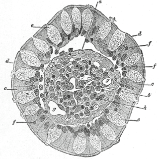
The Reid Index is a mathematical relationship that exists in a human bronchus section observed under the microscope. It is defined as ratio between the thickness of the submucosal mucus secreting glands and the thickness between the epithelium and cartilage that covers the bronchi. The Reid index is not of diagnostic use in vivo since it requires a dissection of the airway tube, but it has value in post mortem evaluations and for research. [1]
The Reid Index was developed in the late 1950s from the work of Dr. Lynne McArthur Reid, M.D. who first described the relationship between hypertrophic bronchial mucous glands and the resultant narrowing of the airways seen in chronic bronchitis. [2] In 1967, Dr. Reid became the first woman to achieve the rank of professor of experimental pathology in England and later became the first dean of the Cardiothoracic Institute at London University. [3]
where:
A normal Reid Index should be smaller than 0.4, the thickness of the wall is always more than double the thickness of the glands it contains. Chronic smoking causes submucosal gland hypertrophy and hyperplasia, leading to a Reid Index of >0.5 indicating chronic bronchitis. [4]

The cervix or cervix uteri is the lower part of the uterus in the human female reproductive system. The cervix is usually 2 to 3 cm long and roughly cylindrical in shape, which changes during pregnancy. The narrow, central cervical canal runs along its entire length, connecting the uterine cavity and the lumen of the vagina. The opening into the uterus is called the internal os, and the opening into the vagina is called the external os. The lower part of the cervix, known as the vaginal portion of the cervix, bulges into the top of the vagina. The cervix has been documented anatomically since at least the time of Hippocrates, over 2,000 years ago.

The lungs are the primary organs of the respiratory system in humans and many other animals including a few fish and some snails. In mammals and most other vertebrates, two lungs are located near the backbone on either side of the heart. Their function in the respiratory system is to extract oxygen from the atmosphere and transfer it into the bloodstream, and to release carbon dioxide from the bloodstream into the atmosphere, in a process of gas exchange. Respiration is driven by different muscular systems in different species. Mammals, reptiles and birds use their different muscles to support and foster breathing. In early tetrapods, air was driven into the lungs by the pharyngeal muscles via buccal pumping, a mechanism still seen in amphibians. In humans, the main muscle of respiration that drives breathing is the diaphragm. The lungs also provide airflow that makes vocal sounds including human speech possible.

The trachea, colloquially called the windpipe, is a cartilaginous tube that connects the larynx to the bronchi of the lungs, allowing the passage of air, and so is present in almost all air-breathing animals with lungs. The trachea extends from the larynx and branches into the two primary bronchi. At the top of the trachea the cricoid cartilage attaches it to the larynx. The trachea is formed by a number of horseshoe-shaped rings, joined together vertically by ligaments over their substance and by the trachealis muscle at their ends. The epiglottis closes the opening to the larynx during swallowing.

A pulmonary alveolus is a hollow cup-shaped cavity found in the lung parenchyma where gas exchange takes place. Lung alveoli are found in the acini at the beginning of the respiratory zone. They are located sparsely in the respiratory bronchioles, line the walls of the alveolar ducts, and are more numerous in the blind-ended alveolar sacs. The acini are the basic units of respiration, with gas exchange taking place in all the alveoli present. The alveolar membrane is the gas exchange surface, surrounded by a network of capillaries. Across the membrane oxygen is diffused into the capillaries and carbon dioxide released from the capillaries into the alveoli to be breathed out.

A cough is a sudden expulsion of air through the large breathing passages that can help clear them of fluids, irritants, foreign particles and microbes. As a protective reflex, coughing can be repetitive with the cough reflex following three phases: an inhalation, a forced exhalation against a closed glottis, and a violent release of air from the lungs following opening of the glottis, usually accompanied by a distinctive sound.

Sputum is mucus that is coughed up from the lower airways. In medicine, sputum samples are usually used for naked eye exam, microbiological investigations of respiratory infections and cytological investigations of respiratory systems. It is critical that the patient not give a specimen that includes any mucoid material from the interior of the nose.
The respiratory tract is the subdivision of the respiratory system involved with the process of respiration in mammals. The respiratory tract is lined with respiratory mucosa or respiratory epithelium.
Phlegm is mucus produced by the respiratory system, excluding that produced by the nasal passages. It often refers to respiratory mucus expelled by coughing, otherwise known as sputum. Phlegm, and mucus as a whole, is in essence a water-based gel consisting of glycoproteins, immunoglobulins, lipids and other substances. Its composition varies depending on climate, genetics, and state of the immune system. Its color can vary from transparent to pale or dark yellow and green, from light to dark brown, and even to dark grey depending on the constituents. The body naturally produces one US quart of phlegm every day to capture and clear substances in the air and bacteria from the nose and throat.

A bronchus is a passage or airway in the respiratory system that conducts air into the lungs. The first bronchi to branch from the trachea are the right main bronchus and the left main bronchus, also known as the primary bronchi. These are the widest and enter the lungs at each hilum, where they branch into narrower secondary bronchi or lobar bronchi, and these branch into narrower tertiary bronchi or segmental bronchi. Further divisions of the segmental bronchi are known as 4th order, 5th order, and 6th order segmental bronchi, or grouped together as subsegmental bronchi. The bronchi when too narrow to be supported by cartilage are known as bronchioles. No gas exchange takes place in the bronchi.

The bronchioles or bronchioli are the smaller branches of the bronchial airways in the respiratory tract. They include the terminal bronchioles, and finally the respiratory bronchioles that mark the start of the respiratory zone delivering air to the gas exchanging units of the alveoli. The bronchioles no longer contain the cartilage, that is found in the bronchi, or glands in their submucosa.

Bronchiectasis is a disease in which there is permanent enlargement of parts of the airways of the lung. Symptoms typically include a chronic cough with mucus production. Other symptoms include shortness of breath, coughing up blood, and chest pain. Wheezing and nail clubbing may also occur. Those with the disease often get frequent lung infections.

The nasal cavity is a large, air-filled space above and behind the nose in the middle of the face. The nasal septum divides the cavity into two cavities, also known as fossae. Each cavity is the continuation of one of the two nostrils. The nasal cavity is the uppermost part of the respiratory system and provides the nasal passage for inhaled air from the nostrils to the nasopharynx and rest of the respiratory tract.

Mucus is a slippery aqueous secretion produced by, and covering, mucous membranes. It is typically produced from cells found in mucous glands, although it may also originate from mixed glands, which contain both serous and mucous cells. It is a viscous colloid containing inorganic salts, antimicrobial enzymes, immunoglobulins, and glycoproteins such as lactoferrin and mucins, which are produced by goblet cells in the mucous membranes and submucosal glands. Mucus serves to protect epithelial cells in the linings of the respiratory, digestive, and urogenital systems, and structures in the visual, and auditory systems from pathogenic fungi, bacteria and viruses. Most of the mucus in the body is produced in the gastrointestinal tract.

Mucins are a family of high molecular weight, heavily glycosylated proteins (glycoconjugates) produced by epithelial tissues in most animals. Mucins' key characteristic is their ability to form gels; therefore they are a key component in most gel-like secretions, serving functions from lubrication to cell signalling to forming chemical barriers. They often take an inhibitory role. Some mucins are associated with controlling mineralization, including nacre formation in mollusks, calcification in echinoderms and bone formation in vertebrates. They bind to pathogens as part of the immune system. Overexpression of the mucin proteins, especially MUC1, is associated with many types of cancer.
Goblet cells are simple columnar goblet shaped like epithelial cells that secrete gel-forming mucins, like mucin MUC5AC. The goblet cells mainly use the merocrine method of secretion, secreting vesicles into a duct, but may use apocrine methods, budding off their secretions, when under stress. The term goblet refers to the cell's goblet-like shape. The apical portion is shaped like a cup, as it is distended by abundant mucus laden granules; its basal portion lacks these granules and is shaped like a stem.

Respiratory epithelium, or airway epithelium, is a type of ciliated columnar epithelium found lining most of the respiratory tract as respiratory mucosa, where it serves to moisten and protect the airways. It is not present in the vocal cords of the larynx, or the oropharynx and laryngopharynx, where instead the epithelium is stratified squamous. It also functions as a barrier to potential pathogens and foreign particles, preventing infection and tissue injury by the secretion of mucus and the action of mucociliary clearance.
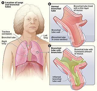
Bronchitis is inflammation of the bronchi in the lungs that causes coughing. Symptoms include coughing up sputum, wheezing, shortness of breath, and chest pain. Bronchitis can be acute or chronic.

Tracheobronchopathia osteochondroplastica (TO) is a rare benign disease of unknown cause, in which multiple cartilaginous or bony submucosal nodules project into the trachea and proximal bronchi. The nodules usually spare the posterior wall of the airway because they are of cartilaginous origin, while the posterior wall of the airway is membranous. This is as opposed to tracheobronchial amyloidosis, which does not spare the posterior wall.
Chronic cough is long-term coughing, sometimes defined as more than several weeks or months. The term can be used to describe the different causes related to coughing, the 3 main ones being; upper airway cough syndrome, asthma and gastroesophageal reflux disease. It occurs in the upper airway of the respiratory system. Generally, a cough lasts around 1–2 weeks, however, chronic cough can persist for an extended period of time defined as 6 weeks or longer. People with chronic cough often experience more than one cause present. Due to the nature of the syndrome the treatments that are used are similar however there is a subsequent number of treatments available.
Airway basal cells are found deep in the respiratory epithelium, attached to, and lining the basement membrane.