Second metatarsal bone

Last updated
Second metatarsal bone
Gray285.png
The second metatarsal. (Left.)
Ospied-en.svg
Bones of the right foot. Dorsal surface. Second metatarsal bone is the yellow bone second from the left
Details
Identifiers
Latin os metatarsale II
FMA 24503
Anatomical terms of bone

The second metatarsal bone is a long bone in the foot. It is the longest of the metatarsal bones, being prolonged backward and held firmly into the recess formed by the three cuneiform bones. [1] The second metatarsal forms joints with the second proximal phalanx (a bone in the second toe) through the metatarsophalangeal joint, the cuneiform bones, third metatarsal and occasionally the first metatarsal bone.

Contents

Structure

Like the four other metatarsal bones, it can be divided into three parts: base, body and head. The base is the part closest to the ankle and the head is closest to the big toe. The narrowed part in the middle is referred to as the body of the bone. The bone is somewhat flattened, giving it two sides: the plantar (towards the sole of the foot) and the dorsal side (the area facing upwards while standing). [1]

Its base is broad above, narrow and rough below.

It presents four articular surfaces: one behind, of a triangular form, for articulation with the intermediate cuneiform bone; one at the upper part of its medial surface, for articulation with the medial cuneiform; and two on its lateral surface, an upper and lower, separated by a rough non-articular interval. Each of these lateral articular surfaces is divided into two by a vertical ridge: the two anterior facets articulate with the third metatarsal and the two posterior (sometimes continuous) with the lateral cuneiform. A fifth facet is occasionally present for articulation with the first metatarsal; it is oval in shape, and is situated on the medial side of the body near the base.

The second metatarsal base acts as a "keystone" (like in an arch) for the lisfranc joint.

Muscle attachments

Muscle attachments (seen from above) Gray268.png
Muscle attachments (seen from above)
Muscle attachments (seen from below) Gray269.png
Muscle attachments (seen from below)

The first and second dorsal interossei muscles attach to the second metatarsal bone, the first dorsal interosseus from the medial side of the bone and the second dorsal interosseus from the lateral side. The function of the muscle is to spread the toes. [2]

The horizontal head of the adductor hallucis also originates from the lateral side of the metatarsophalangeal joint and from the deep transverse metatarsal ligament, [2] a narrow band which runs across and connects together the heads of all the metatarsal bones.

MuscleDirectionAttachment [3]
Dorsal interossei I OriginMedial side of the second metatarsal
Dorsal interossei II OriginLateral side of the second metatarsal
Horizontal head of adductor hallucis OriginDeep transverse metatarsal ligament and the metatarsophalangeal joint

Second metatarsal bone elongation, also known as Morton's toe (or Morton's foot) is a normal variation of the second metatarsal present in about 25% of the total population. Although normal, Morton's toe causes extra-inversion of the foot and thereby puts more stress on the lateral part of the meniscus of the knee, promotes lordosis of the lower back (lumbar spine) and kyphosis of the neck (cervical spine). Symptoms include back pain, knee pain and arthritis at early age, constant tenderness of shoulder, both acute and chronic torticollis, headache and up to vague non-specific bodyaches.

Injuries

The Beckham bone is a name attributed by British journalists to the second metatarsal.

David Beckham, while playing for Manchester United against Deportivo La Coruña in a UEFA Champions League quarter-final game in 2002, was subject to a tackle from Argentina's Aldo Duscher. (A lot of acrimony had existed between David Beckham and Argentina since David Beckham's sending off in the 1998 World Cup). This tackle broke the second metatarsal in his left foot and seriously threatened England's chances in the 2002 World Cup. David Beckham was the media darling at the time, and the bone (and the tackle) received a wave of publicity; subsequently, the name "Beckham bone" was born.

Since then, other notable football players including Daniel Agger, Gary Neville, Danny Murphy, Michael Owen, Gaël Clichy, Ashley Cole, Ledley King, Mikaël Silvestre, James Mathias and Lionel Messi have suffered fractures to the same 2nd metatarsal bone.

Additional images

Related Research Articles

<span class="mw-page-title-main">Foot</span> Anatomical structure found in vertebrates

The foot is an anatomical structure found in many vertebrates. It is the terminal portion of a limb which bears weight and allows locomotion. In many animals with feet, the foot is a separate organ at the terminal part of the leg made up of one or more segments or bones, generally including claws and/or nails.

<span class="mw-page-title-main">Femur</span> Thigh bone

The femur, or thigh bone is the only bone in the thigh. The thigh is the region of the lower limb between the hip and the knee. In many four-legged animals the femur is the upper bone of the hindleg.

<span class="mw-page-title-main">Cuboid bone</span> Bone of the ankle

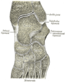
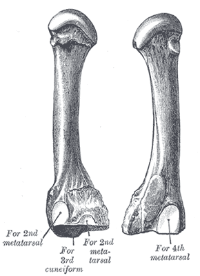
In the human body, the cuboid bone is one of the seven tarsal bones of the foot.

<span class="mw-page-title-main">Fibula</span> Leg bone in vertebrates

The fibula or calf bone is a leg bone on the lateral side of the tibia, to which it is connected above and below. It is the smaller of the two bones and, in proportion to its length, the most slender of all the long bones. Its upper extremity is small, placed toward the back of the head of the tibia, below the knee joint and excluded from the formation of this joint. Its lower extremity inclines a little forward, so as to be on a plane anterior to that of the upper end; it projects below the tibia and forms the lateral part of the ankle joint.

<span class="mw-page-title-main">Metatarsal bones</span> Five long bones in the foot

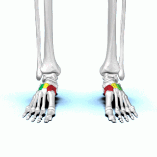
The metatarsal bones or metatarsus are a group of five long bones in the midfoot, located between the tarsal bones and the phalanges (toes). Lacking individual names, the metatarsal bones are numbered from the medial side : the first, second, third, fourth, and fifth metatarsal. The metatarsals are analogous to the metacarpal bones of the hand. The lengths of the metatarsal bones in humans are, in descending order, second, third, fourth, fifth, and first. A bovine hind leg has two metatarsals.

<span class="mw-page-title-main">Cuneiform bones</span> Three bones in the human foot

There are three cuneiform ("wedge-shaped") bones in the human foot:

<span class="mw-page-title-main">Calcaneus</span> Bone of the tarsus of the foot

In humans and many other primates, the calcaneus or heel bone is a bone of the tarsus of the foot which constitutes the heel. In some other animals, it is the point of the hock.

<span class="mw-page-title-main">Navicular bone</span> Small bone found in the feet of most mammals

The navicular bone is a small bone found in the feet of most mammals.

<span class="mw-page-title-main">Talus bone</span> One of the foot bones that forms the tarsus

The talus, talus bone, astragalus, or ankle bone is one of the group of foot bones known as the tarsus. The tarsus forms the lower part of the ankle joint. It transmits the entire weight of the body from the lower legs to the foot.

<span class="mw-page-title-main">Adductor muscles of the hip</span> Group of muscles

The adductor muscles of the hip are a group of muscles in the medial compartment of the thigh mostly used for bringing the thighs together.

<span class="mw-page-title-main">Lateral rotator group</span> Group of six small muscles of the hip

The lateral rotator group is a group of six small muscles of the hip which all externally (laterally) rotate the femur in the hip joint. It consists of the following muscles: piriformis, gemellus superior, obturator internus, gemellus inferior, quadratus femoris and the obturator externus.

<span class="mw-page-title-main">Abductor digiti minimi muscle of foot</span> Muscle which lies along the lateral (outer) border of the foot

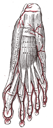
The abductor digiti minimi is a muscle which lies along the lateral (outer) border of the foot, and is in relation by its medial margin with the lateral plantar artery, vein and nerves.

<span class="mw-page-title-main">Fifth metatarsal bone</span>

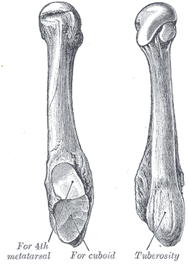
The fifth metatarsal bone is a long bone in the foot, and is palpable along the distal outer edges of the feet. It is the second smallest of the five metatarsal bones. The fifth metatarsal is analogous to the fifth metacarpal bone in the hand.

<span class="mw-page-title-main">Fourth metatarsal bone</span>

The fourth metatarsal bone is a long bone in the foot. It is smaller in size than the third metatarsal bone and is the third longest of the five metatarsal bones. The fourth metatarsal is analogous to the fourth metacarpal bone in the hand

<span class="mw-page-title-main">Third metatarsal bone</span>

The third metatarsal bone is a long bone in the foot. It is the second longest metatarsal. The longest being the second metatarsal. The third metatarsal is analogous to the third metacarpal bone in the hand

<span class="mw-page-title-main">First metatarsal bone</span>

The first metatarsal bone is the bone in the foot just behind the big toe. The first metatarsal bone is the shortest of the metatarsal bones and by far the thickest and strongest of them.

The anserine bursa is a sub muscular bursa located deep to the pes anserinus on the anteromedial proximal tibia. Pes anserine bursitis is a common inflammatory condition of the anserine bursa.

<span class="mw-page-title-main">Iliopectineal bursa</span>

The iliopectineal bursa or the iliopsoas bursa is a large synovial bursa that separates the external surface of the hip joint capsule from the normally just the tendon of the iliopsoas muscle.

<span class="mw-page-title-main">Muscles of the thumb</span>

The muscles of the thumb are nine skeletal muscles located in the hand and forearm. The muscles allow for flexion, extension, adduction, abduction and opposition of the thumb. The muscles acting on the thumb can be divided into two groups: The extrinsic hand muscles, with their muscle bellies located in the forearm, and the intrinsic hand muscles, with their muscles bellies located in the hand proper.

The femoral-tibial angle is the angle between the femur and tibia.

References

PD-icon.svgThis article incorporates text in the public domain from page 273 of the 20th edition of Gray's Anatomy (1918)

  1. 1 2 Bojsen-Møller, Finn; Simonsen, Erik B.; Tranum-Jensen, Jørgen (2001). Bevægeapparatets anatomi[Anatomy of the Locomotive Apparatus] (in Danish) (12th ed.). p. 246. ISBN   978-87-628-0307-7.
  2. 1 2 Bojsen-Møller, Finn; Simonsen, Erik B.; Tranum-Jensen, Jørgen (2001). Bevægeapparatets anatomi[Anatomy of the Locomotive Apparatus] (in Danish) (12th ed.). pp. 300–301. ISBN   978-87-628-0307-7.
  3. Bojsen-Møller, Finn; Simonsen, Erik B.; Tranum-Jensen, Jørgen (2001). Bevægeapparatets anatomi[Anatomy of the Locomotive Apparatus] (in Danish) (12th ed.). pp. 364–367. ISBN   978-87-628-0307-7.