ShuntCheck

Last updated
ShuntCheck
SCIII 4 PlaceTS and SCIII 12 PlaceIce.jpg
ShuntCheck Sensor & Ice Placement
UsesDetects flow in the cerebral shunts of hydrocephalus patients
ApproachNon-invasive

ShuntCheck is a non-invasive diagnostic medical device which detects flow in the cerebral shunts of hydrocephalus patients. Neurosurgeons can use ShuntCheck flow results along with other diagnostic tests to assess shunt function and malfunction. [1]

Contents

Hydrocephalus is a condition in which cerebrospinal fluid (CSF) accumulates in the brain, potentially leading to brain damage and death. It is corrected by a shunt which drains excess CSF from the brain to the abdomen. Shunts fail, typically by obstruction – a life-threatening medical condition requiring the surgical replacement of the shunt. The symptoms of shunt failure are non-specific – headache, nausea, lethargy – so diagnostic tests must be conducted to rule in or rule out surgery. Current methods of diagnosing shunt malfunction, including CT Scan, MRI, radionuclide studies and shunt tap, have limitations and risks. These limitations and risks led to the development of ShuntCheck. [2] [3] [4] [5] [6] [7] [8] [9] [10] [11] [12] [13] [ excessive citations ]

ShuntCheck uses thermal dilution [14] [15] to detect flow. The ShuntCheck sensor, a high-tech skin thermometer, is placed over the shunt where it crosses the clavicle. The shunt, which lies just below the skin, is cooled with an ice pack placed “upstream” of the sensor. If CSF is flowing through the shunt, the cooled fluid will move “downstream” and the ShuntCheck sensor will detect a drop in temperature. Faster shunt flow results in greater temperature decreases. If the shunt is not flowing, the cooled fluid remains upstream and no temperature drop is recorded. [2]

The sensor is connected to a laptop or tablet computer running ShuntCheck software. The sensor is connected to a laptop or tablet computer running ShuntCheck software. The computer analyzes the thermal data, determines “Flow Confirmed” or “Flow NOT Confirmed” and presents a time-temperature graph.

ShuntCheck Results Screen Wiki graph.jpg
ShuntCheck Results Screen

Early clinical testing of ShuntCheck found that functioning shunts flow intermittently, which meant that a ShuntCheck reading of “Flow NOT Confirmed” did not necessarily indicate a shunt problem. [2] [16] This discovery led to the development of the ShuntCheck Micro-Pumper, a handheld device which vibrates the shunt valve, generating a temporary increase in flow through patent, but not through obstructed, shunts. Micro-Pumper allows ShuntCheck to differentiate between temporarily non-flowing patent shunts and obstructed shunts. [1]

Micro-Pumper SCIII Micro-Pumper & SCIII 14 MP.jpg
Micro-Pumper

ShuntCheck III

The current version of ShuntCheck was developed in 2011-2012 funded by grants from the National Institute of Health [17] [18] [19] and was cleared by the US FDA in 2013. [1] The ShuntCheck system includes the ShuntCheck Sensor, a skin marker, an Instant Cold Pack, a Data Acquisition Unit (an analog-to-digital converter called the “DAQ”), a Windows 7 or Windows 8 laptop or tablet computer running ShuntCheck software and the Micro-Pumper. [1]

ShuntCheck System SCIII System.JPG
ShuntCheck System

ShuntCheck clinical studies

Boston Children's Hospital 2008-2009 Joseph R. Madsen MD tested 100 symptomatic and asymptomatic pediatric hydrocephalus patients using an earlier version of ShuntCheck during 2008-2009. His key findings, reported in Neurosurgery [2] were

University of South Florida 2008 Arthur E. Marlin MD and Sarah J Gaskill MD conducted ShuntCheck testing on 35 asymptomatic pediatric patients with similar results. [16]

These findings led to the development of the Micro-Pumper.

Boston Children's Hospital 2010-2013 Dr. Madsen is testing 130 symptomatic and asymptomatic pediatric hydrocephalus patients to assess the diagnostic accuracy and clinical utility of the newer version of ShuntCheck including Micro-Pumper. This study was funded by the NIH. [18] [19]

Multi-Center Pediatric Outcomes Study 2013-2014 Boston Children's Hospital, Children's Hospital of Philadelphia, Johns Hopkins Hospital, Cleveland Clinic, University of Chicago Comer Children's Hospital, LeBonheur Children's Hospital and University of Texas Houston Children's Memorial Hermann Hospital will conduct an outcomes study of 400 symptomatic pediatric hydrocephalus patients during 2013-2014. In this NIH funded study, [19] ShuntCheck results and the results of standard of care diagnostic tests will be compared to clinical outcome (shunt obstruction confirmed by surgery vs no-obstruction). This study [20] seeks to demonstrate that

Sinai Baltimore NPH Study 2012-2014 Michael A. Williams MD is conducting ShuntCheck testing on adult hydrocephalus patients undergoing radionuclide shunt patency testing. This study, funded by the NIH, [21] [22] seeks to demonstrate that ShuntCheck results match radionuclide results.

Potential clinical uses of ShuntCheck

  1. A test for assessing shunt function in symptomatic hydrocephalus patients. ShuntCheck flow data, used in conjunction with other diagnostic test results and with physician judgment, can aid in ruling in or ruling out shunt obstruction. [1]
  2. A tool for establishing “normal” CSF flow patterns in asymptomatic patients. Establishing baseline flow may increase the value of flow information in symptomatic patients. [1]
  3. A tool to streamline the process of adjusting shunt valve settings to accommodate individual needs for CSF drainage. While the settings for these valves in each patient must currently be determined empirically over a number of weeks, Shuntcheck will be helpful in measuring changes in CSF flow due to changes in the valve setting. [23]
  4. A tool for assessing suspected over-drainage. CSF flow data will allow neurosurgeons to identify periods and causes of high CSF flow when assessing suspected CSF over drainage. This data can also be used to evaluate flow and siphon control devices to determine if they are meeting the patient's needs.
  5. A post operative test to confirm shunt function. Hospitals in sparsely populated areas often conduct post-surgical CT scans to confirm shunt function before releasing patients for the long drive home. CSF flow data can confirm shunt function more quickly than CT (which requires time for the ventricles to stabilize).

NeuroDx Development

NeuroDx Development [10] (NeuroDx) is an early commercial stage medical device company founded in 2008 by Fred Fritz (CEO), a serial life sciences entrepreneur, and Dr. Marek Swoboda (Chief Scientific Officer), a biosensor engineer, to address important unmet needs in the hydrocephalus market. [24] The Company has developed two thermal dilution technologies for assessing shunt function in hydrocephalus patients, ShuntCheck-Micro-Pumper, an eight-minute test of CSF shunt flow, and Continuous Real Time (CRT) ShuntCheck, which uses thermal dilution to monitor changes in shunt flow over longer time periods. [25] The Company's follow-on products, an implantable intracranial pressure monitoring device [26] and a non-invasive monitor of intracranial pressure, are in proof of concept development.

Related Research Articles

<span class="mw-page-title-main">Cerebrospinal fluid</span> Clear, colorless bodily fluid found in the brain and spinal cord

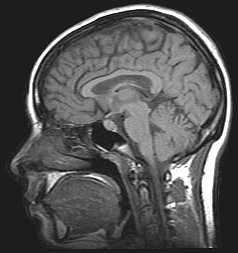
Cerebrospinal fluid (CSF) is a clear, colorless body fluid found within the tissue that surrounds the brain and spinal cord of all vertebrates.

<span class="mw-page-title-main">Idiopathic intracranial hypertension</span> Medical condition

Idiopathic intracranial hypertension (IIH), previously known as pseudotumor cerebri and benign intracranial hypertension, is a condition characterized by increased intracranial pressure without a detectable cause. The main symptoms are headache, vision problems, ringing in the ears, and shoulder pain. Complications may include vision loss.

<span class="mw-page-title-main">Syringomyelia</span> Disorder in which a cyst forms in the spinal cord

Syringomyelia is a generic term referring to a disorder in which a cyst or cavity forms within the spinal cord. Often, syringomyelia is used as a generic term before an etiology is determined. This cyst, called a syrinx, can expand and elongate over time, destroying the spinal cord. The damage may result in loss of feeling, paralysis, weakness, and stiffness in the back, shoulders, and extremities. Syringomyelia may also cause a loss of the ability to feel extremes of hot or cold, especially in the hands. It may also lead to a cape-like bilateral loss of pain and temperature sensation along the upper chest and arms. The combination of symptoms varies from one patient to another depending on the location of the syrinx within the spinal cord, as well as its extent.

<span class="mw-page-title-main">Hydrocephalus</span> Abnormal increase in cerebrospinal fluid in the ventricles of the brain

Hydrocephalus is a condition in which an accumulation of cerebrospinal fluid (CSF) occurs within the brain. This typically causes increased pressure inside the skull. Older people may have headaches, double vision, poor balance, urinary incontinence, personality changes, or mental impairment. In babies, it may be seen as a rapid increase in head size. Other symptoms may include vomiting, sleepiness, seizures, and downward pointing of the eyes.

<span class="mw-page-title-main">Chiari malformation</span> Medical condition

Chiari malformation (CM) is a structural defect in the cerebellum, characterized by a downward displacement of one or both cerebellar tonsils through the foramen magnum. CMs can cause headaches, difficulty swallowing, vomiting, dizziness, neck pain, unsteady gait, poor hand coordination, numbness and tingling of the hands and feet, and speech problems. Less often, people may experience ringing or buzzing in the ears, weakness, slow heart rhythm, or fast heart rhythm, curvature of the spine (scoliosis) related to spinal cord impairment, abnormal breathing, such as central sleep apnea, characterized by periods of breathing cessation during sleep, and, in severe cases, paralysis.

<span class="mw-page-title-main">Hydranencephaly</span> Medical condition

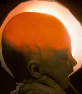
Hydranencephaly is a condition in which the brain's cerebral hemispheres are absent to a great degree and the remaining cranial cavity is filled with cerebrospinal fluid. "Cephalic" is the scientific term for "head" or "head end of body".

<span class="mw-page-title-main">Lumbar puncture</span> Procedure to collect cerebrospinal fluid

Lumbar puncture (LP), also known as a spinal tap, is a medical procedure in which a needle is inserted into the spinal canal, most commonly to collect cerebrospinal fluid (CSF) for diagnostic testing. The main reason for a lumbar puncture is to help diagnose diseases of the central nervous system, including the brain and spine. Examples of these conditions include meningitis and subarachnoid hemorrhage. It may also be used therapeutically in some conditions. Increased intracranial pressure is a contraindication, due to risk of brain matter being compressed and pushed toward the spine. Sometimes, lumbar puncture cannot be performed safely. It is regarded as a safe procedure, but post-dural-puncture headache is a common side effect if a small atraumatic needle is not used.

Normal-pressure hydrocephalus (NPH), also called malresorptive hydrocephalus, is a form of communicating hydrocephalus in which excess cerebrospinal fluid (CSF) occurs in the ventricles, and with normal or slightly elevated cerebrospinal fluid pressure. As the fluid builds up, it causes the ventricles to enlarge and the pressure inside the head to increase, compressing surrounding brain tissue and leading to neurological complications. The disease presents in a classic triad of symptoms, which are memory impairment, urinary frequency, and balance problems/gait deviations. The disease was first described by Salomón Hakim and Adams in 1965.

<span class="mw-page-title-main">Arachnoid cyst</span> Medical condition

Arachnoid cysts are cerebrospinal fluid covered by arachnoidal cells and collagen that may develop between the surface of the brain and the cranial base or on the arachnoid membrane, one of the three meningeal layers that cover the brain and the spinal cord. Primary arachnoid cysts are a congenital disorder whereas secondary arachnoid cysts are the result of head injury or trauma. Most cases of primary cysts begin during infancy; however, onset may be delayed until adolescence.

<span class="mw-page-title-main">Dandy–Walker malformation</span> Congenital malformation of the cerebellar vermis

Dandy–Walker malformation (DWM), also known as Dandy–Walker syndrome (DWS), is a rare congenital brain malformation in which the part joining the two hemispheres of the cerebellum does not fully form, and the fourth ventricle and space behind the cerebellum are enlarged with cerebrospinal fluid. Most of those affected develop hydrocephalus within the first year of life, which can present as increasing head size, vomiting, excessive sleepiness, irritability, downward deviation of the eyes and seizures. Other, less common symptoms are generally associated with comorbid genetic conditions and can include congenital heart defects, eye abnormalities, intellectual disability, congenital tumours, other brain defects such as agenesis of the corpus callosum, skeletal abnormalities, an occipital encephalocele or underdeveloped genitalia or kidneys. It is sometimes discovered in adolescents or adults due to mental health problems.

<span class="mw-page-title-main">Choroid plexus papilloma</span> Medical condition

Choroid plexus papilloma, also known as papilloma of the choroid plexus, is a rare benign neuroepithelial intraventricular WHO grade I lesion found in the choroid plexus. It leads to increased cerebrospinal fluid production, thus causing increased intracranial pressure and hydrocephalus.

<span class="mw-page-title-main">Colloid cyst</span> Medical condition

A colloid cyst is a non-malignant tumor in the brain. It consists of a gelatinous material contained within a membrane of epithelial tissue. It is almost always found just posterior to the foramen of Monro in the anterior aspect of the third ventricle, originating from the roof of the ventricle. Because of its location, it can cause obstructive hydrocephalus and increased intracranial pressure. Colloid cysts represent 0.5–1.0% of intracranial tumors.

<span class="mw-page-title-main">Cerebral shunt</span> Surgical implant to treat hydrocephalus

A cerebral shunt is a device permanently implanted inside the head and body to drain excess fluid away from the brain. They are commonly used to treat hydrocephalus, the swelling of the brain due to excess buildup of cerebrospinal fluid (CSF). If left unchecked, the excess CSF can lead to an increase in intracranial pressure (ICP), which can cause intracranial hematoma, cerebral edema, crushed brain tissue or herniation. The drainage provided by a shunt can alleviate or prevent these problems in patients with hydrocephalus or related diseases.

<span class="mw-page-title-main">External ventricular drain</span> Medical device

An external ventricular drain (EVD), also known as a ventriculostomy or extraventricular drain, is a device used in neurosurgery to treat hydrocephalus and relieve elevated intracranial pressure when the normal flow of cerebrospinal fluid (CSF) inside the brain is obstructed. An EVD is a flexible plastic catheter placed by a neurosurgeon or neurointensivist and managed by intensive care unit (ICU) physicians and nurses. The purpose of external ventricular drainage is to divert fluid from the ventricles of the brain and allow for monitoring of intracranial pressure. An EVD must be placed in a center with full neurosurgical capabilities, because immediate neurosurgical intervention can be needed if a complication of EVD placement, such as bleeding, is encountered.

<span class="mw-page-title-main">CSF tap test</span>

The CSF tap test, sometimes lumbar tap test or Miller Fisher Test, is a medical test that is used to decide whether shunting of cerebrospinal fluid (CSF) would be helpful in a patient with suspected normal pressure hydrocephalus (NPH). The test involves removing 30-50 ml of cerebrospinal fluid (CSF) through a lumbar puncture, after which motor and cognitive function is clinically reassessed. The name "Fisher test" is after C. Miller Fisher, a Canadian neurologist working in Boston, Massachusetts, who described the test.

<span class="mw-page-title-main">Pineoblastoma</span> Medical condition

Pineoblastoma is a malignant tumor of the pineal gland. A pineoblastoma is a supratentorial midline primitive neuroectodermal tumor. Pineoblastoma can present at any age, but is most common in young children. They account for 0.001% of all primary CNS neoplasms.

Bobble-head doll syndrome is a rare neurological movement disorder in which patients, usually children around age 3, begin to bob their head and shoulders forward and back, or sometimes side-to-side, involuntarily, in a manner reminiscent of a bobblehead doll. The syndrome is related to cystic lesions and swelling of the third ventricle in the brain. Symptoms of bobble-head doll syndrome are diverse but can be grouped into two categories: physical and neurological. The most common form of treatment is surgical implanting of a shunt to relieve the swelling of the brain.

<span class="mw-page-title-main">Aqueductal stenosis</span> Narrowing of the aqueduct of Sylvius

Aqueductal stenosis is a narrowing of the aqueduct of Sylvius which blocks the flow of cerebrospinal fluid (CSF) in the ventricular system. Blockage of the aqueduct can lead to hydrocephalus, specifically as a common cause of congenital and/or obstructive hydrocephalus.

<span class="mw-page-title-main">L1 syndrome</span> Medical condition

L1 syndrome is a group of mild to severe X-linked recessive disorders that share a common genetic basis. The spectrum of L1 syndrome disorders includes X-linked complicated corpus callosum dysgenesis, spastic paraplegia 1, MASA syndrome, and X-linked hydrocephalus with stenosis of the aqueduct of Sylvius (HSAS). It is also called L1CAM syndrome and CRASH syndrome, an acronym for its primary clinical features: corpus callosum hypoplasia, retardation, adducted thumbs, spasticity, and hydrocephalus.

Cerebrospinal fluid (CSF) flow MRI is used to assess pulsatile CSF flow both qualitatively and quantitatively. Time-resolved 2D phase-contrast MRI with velocity encoding is the most common method for CSF analysis. CSF Fluid Flow MRI detects back and forth flow of Cerebrospinal fluid that corresponds to vascular pulsations from mostly the cardiac cycle of the choroid plexus. Bulk transport of CSF, characterized by CSF circulation through the Central Nervous System, is not used because it is too slow to assess clinically. CSF would have to pass through the brain's lymphatic system and be absorbed by arachnoid granulations.

References

  1. 1 2 3 4 5 6 "510(k) Summary" (PDF). Accessdata.fda.gov. Retrieved 2013-10-14.
  2. 1 2 3 4 Madsen JR, et al, Evaluation of the ShuntCheck Noninvasive Thermal Technique for Shunt Flow Detection in Hydrocephalic Patients, Neurosurgery 68:198-205, 2011
  3. Brenner DJ, Hall EJ, Computed Tomography – An Increasing Source of Radiation Exposure, N Engl J Med 2007;357:2277-84
  4. Iskandar, B., et al., Pitfalls in the diagnosis of ventricular shunt dysfunction: radiology reports and ventricular size. Pediatrics, 1998. 101: p. 1031-1036.
  5. Forrest, D. and D. Cooper, Complications of ventriculo-atrial shunts. J Neurosurg, 1968. 29: p. 506-512.
  6. Iskandar, B., et al., Death in shunted hydrocephalic children in the 1990s. Pediatr Neurosurg, 1998. 28: p. 173-176.
  7. Piatt, J., Physical examination of patients with cerebrospinal fluid shunt: is there useful information in pumping the shunt? Pediatrics, 1992. 89: p. 470-473.
  8. Noetzel, M. and R. Baker, Shunt fluid evaluation: risks and benefits in the evaluation of shunt malfunction and infection. J Neurosurg, 1984. 61: p. 328-332.
  9. Howman-Giles, R., et al., A radionuclide method of evaluating shunt function and CSF circulation in hydrocephalus. J Neurosurg, 1984. 61: p. 604-605.
  10. Vernet, O., et al., Radionuclide shuntogram; adjunct to manage hydrocephalic patients. J Nucl Med, 1996. 37: p. 406-410.
  11. Brendel, A., et al., Cerebrospinal shunt flow in adults: radionuclide quantitation with emphasis on patient position. Radiology, 1983. 149: p. 815-818.
  12. Pitteti R. 2007. Emergency department evaluation of ventricular shunt malfunction: is the shunt series really necessary? Pediatr. Emerg. Care 23: 137-141.
  13. Sood S, Canady AI, Harn, SD. 2000. Evaluation of shunt malfunction using shunt site reservoir. Pediatr. Neurosurg. 32: 180-186.
  14. Stein SC, Apfel S, A noninvasive approach to quantitative measurement of flow through CSF shunts. Technical note, J Neurosurg 1981 Apr,54(4):556-8
  15. Neff S, Measurement of flow in cerebrospinal fluid in shunts by transcutaneous thermal convection, J Neurosurg (Pediatrics 4) 103:366-373, 2005
  16. 1 2 Marlin AE, Gaskill SJ, The Use of Transcutaneous Thermal Convection Analysis to Assess Shunt Function in the Pediatric Population, Neurosurgery 70:181-3, 2012
  17. "TAGGS Award Detail". Taggs.hhs.gov. 2009-09-01. Retrieved 2013-10-14.
  18. 1 2 "TAGGS Award Detail". Taggs.hhs.gov. 2009-09-01. Retrieved 2013-10-14.
  19. 1 2 3 "TAGGS Award Detail". Taggs.hhs.gov. 2009-09-01. Retrieved 2013-10-14.
  20. "ShuntCheck-Micro-Pumper Pediatric Clinical Outcomes Study - Full Text View". ClinicalTrials.gov. Retrieved 2013-10-14.
  21. "TAGGS Award Detail". Taggs.hhs.gov. 2009-09-01. Retrieved 2013-10-14.
  22. "ShuntCheck Versus Radionuclide in Evaluating Shunt Function in Symptomatic NPH Patients - Full Text View". ClinicalTrials.gov. Retrieved 2013-10-14.
  23. "ShuntCheck Accuracy in Detecting Shunt Obstruction Normal Pressure Hydrocephalus (NPH) Patients - Full Text View". ClinicalTrials.gov. Retrieved 2013-10-14.
  24. "NeuroDx Development". Neurodx.com. Retrieved 2013-10-14.
  25. "TAGGS Award Detail". Taggs.hhs.gov. 2009-09-01. Retrieved 2013-10-14.
  26. "TAGGS Award Detail". Taggs.hhs.gov. 2009-09-01. Retrieved 2013-10-14.