Related Research Articles
Spasticity is a feature of altered skeletal muscle performance with a combination of paralysis, increased tendon reflex activity, and hypertonia. It is also colloquially referred to as an unusual "tightness", stiffness, or "pull" of muscles.
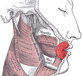
In human anatomy, the orbicularis oris muscle is a complex of muscles in the lips that encircles the mouth. It is a sphincter, or circular muscle, but it is actually composed of four independent quadrants that interlace and give only an appearance of circularity.
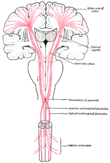
Upper motor neurons (UMNs) is a term introduced by William Gowers in 1886. They are found in the cerebral cortex and brainstem and carry information down to activate interneurons and lower motor neurons, which in turn directly signal muscles to contract or relax. UMNs in the cerebral cortex are the main source of voluntary movement.

Eye movement includes the voluntary or involuntary movement of the eyes, helping in acquiring, fixating and tracking visual stimuli. A special type of eye movement, rapid eye movement, occurs during REM sleep.

The anterior cerebral artery (ACA) is one of a pair of arteries on the brain that supplies oxygenated blood to most midline portions of the frontal lobes and superior medial parietal lobes. The two anterior cerebral arteries arise from the internal carotid artery and are part of the circle of Willis. The left and right anterior cerebral arteries are connected by the anterior communicating artery.
The jaw jerk reflex or the masseter reflex is a stretch reflex used to test the status of a patient's trigeminal nerve and to help distinguish an upper cervical cord compression from lesions that are above the foramen magnum. The mandible—or lower jaw—is tapped at a downward angle just below the lips at the chin while the mouth is held slightly open. In response, the masseter muscles will jerk the mandible upwards. Normally this reflex is absent or very slight. However, in individuals with upper motor neuron lesions the jaw jerk reflex can be quite pronounced.

The levator labii superioris is a muscle of the human body used in facial expression. It is a broad sheet, the origin of which extends from the side of the nose to the zygomatic bone.

Frontal lobe disorder is an impairment of the frontal lobe that occurs due to disease or head trauma. The frontal lobe of the brain plays a key role in higher mental functions such as motivation, planning, social behaviour, and speech production. A frontal lobe syndrome can be caused by a range of conditions including head trauma, tumours, degenerative diseases, neurosurgery and cerebrovascular disease. Frontal lobe impairment can be detected by recognition of typical clinical signs, use of simple screening tests, and specialist neurological testing.
The glabellar reflex, also known as the "glabellar tap sign", is a primitive reflex elicited by repetitive tapping on the forehead. Subjects blink in response to the first several taps. If the blinking persists, this is known as Myerson's sign, and is abnormal and a sign of frontal release; it is often seen in people who have Parkinson's disease.

The vestibulospinal tract is a neural tract in the central nervous system. Specifically, it is a component of the extrapyramidal system and is classified as a component of the medial pathway. Like other descending motor pathways, the vestibulospinal fibers of the tract relay information from nuclei to motor neurons. The vestibular nuclei receive information through the vestibulocochlear nerve about changes in the orientation of the head. The nuclei relay motor commands through the vestibulospinal tract. The function of these motor commands is to alter muscle tone, extend, and change the position of the limbs and head with the goal of supporting posture and maintaining balance of the body and head.
Utilization behavior (UB) is a type of neurobehavioral disorder that involves patients grabbing objects in view and starting the 'appropriate' behavior associated with it at an 'inappropriate' time. Utilization behavior patients have difficulty resisting the impulse to operate or manipulate objects which are in their visual field and within reach. Characteristics of UB include unintentional, unconscious actions triggered by the immediate environment. The unpreventable excessive behavior has been linked to lesions in the frontal lobe. UB has also been referred to as "bilateral magnetic apraxia" and "hypermetamorphosis".

Foix-Chavany-Marie Syndrome (FCMS), also known as Bilateral Opercular Syndrome, is a neuropathological disorder characterized by paralysis of the facial, tongue, pharynx, and masticatory muscles of the mouth that aid in chewing. The disorder is primarily caused by thrombotic and embolic strokes, which cause a deficiency of oxygen in the brain. As a result, bilateral lesions may form in the junctions between the frontal lobe and temporal lobe, the parietal lobe and cortical lobe, or the subcortical region of the brain. FCMS may also arise from defects existing at birth that may be inherited or nonhereditary. Symptoms of FCMS can be present in a person of any age and it is diagnosed using automatic-voluntary dissociation assessment, psycholinguistic testing, neuropsychological testing, and brain scanning. Treatment for FCMS depends on the onset, as well as on the severity of symptoms, and it involves a multidisciplinary approach.

This article describes the anatomy of the head and neck of the human body, including the brain, bones, muscles, blood vessels, nerves, glands, nose, mouth, teeth, tongue, and throat.
Primitive reflexes are reflex actions originating in the central nervous system that are exhibited by normal infants, but not neurologically intact adults, in response to particular stimuli. These reflexes are suppressed by the development of the frontal lobes as a child transitions normally into child development. These primitive reflexes are also called infantile, infant or newborn reflexes.
Central facial palsy is a symptom or finding characterized by paralysis or paresis of the lower half of one side of the face. It usually results from damage to upper motor neurons of the facial nerve.
Focal neurologic signs also known as focal neurological deficits or focal CNS signs are impairments of nerve, spinal cord, or brain function that affects a specific region of the body, e.g. weakness in the left arm, the right leg, paresis, or plegia.
Spastic quadriplegia, also known as spastic tetraplegia, is a subset of spastic cerebral palsy that affects all four limbs.

Cortical deafness is a rare form of sensorineural hearing loss caused by damage to the primary auditory cortex. Cortical deafness is an auditory disorder where the patient is unable to hear sounds but has no apparent damage to the anatomy of the ear, which can be thought of as the combination of auditory verbal agnosia and auditory agnosia. Patients with cortical deafness cannot hear any sounds, that is, they are not aware of sounds including non-speech, voices, and speech sounds. Although patients appear and feel completely deaf, they can still exhibit some reflex responses such as turning their head towards a loud sound.
Frontal release signs are primitive reflexes traditionally held to be a sign of disorders that affect the frontal lobes. The appearance of such signs reflects the area of brain dysfunction rather than a specific disorder which may be diffuse, such as a dementia, or localised, such as a tumor.
The palmomental reflex (PMR) is a primitive reflex consisting of a twitch of the chin muscle elicited by stroking a specific part of the palm. It is present in infancy and disappears as the brain matures during childhood but may reappear due to processes that disrupt the normal cortical inhibitory pathways. Therefore, it is an example of a frontal release sign.
References
- ↑ Jain P, Rathee M (2019). "Anatomy, Head and Neck, Orbicularis Oris Muscle". StatPearls [Internet]. PMID 31424753.