Sodium MRI

Last updated
Sodium Magnetic Resonance Images of a female volunteer at 3T (6mm x 6mm in-plane resolution, 72mm slice thickness, captured in 50minutes for five segments) Whole-body sodium magnetic resonance images of female human.png
Sodium Magnetic Resonance Images of a female volunteer at 3T (6mm x 6mm in-plane resolution, 72mm slice thickness, captured in 50minutes for five segments)

Sodium MRI (also known as 23 Na-MRI) is a specialised magnetic resonance imaging technique that uses strong magnetic fields, magnetic field gradients, and radio waves to generate images of the distribution of sodium in the body, as opposed to more common forms of MRI that utilise protons (or hydrogen) present in water (1H-MRI). [2] [3] Like the proton, sodium is naturally abundant in the body, so can be imaged directly without the need for contrast agents or hyperpolarization. Furthermore, sodium ions play a role in important biological processes via their contribution to concentration and electrochemical gradients across cellular membranes, making it of interest as an imaging target in health and disease. [4]

Contents

In contrast to conventional MRI of the proton, Sodium MRI is complicated by the low concentrations of Na nuclei relative to concentration of H2O molecules in biological tissues [5] (10-45 mM) and the lower gyromagnetic ratio of the 23Na nucleus as compared to a 1H nucleus,. [6] [7] This causes low NMR sensitivity and the requirement for a stronger magnetic field for equivalent spatial resolution. The quadrupolar 23Na nucleus also has a faster transverse relaxation rates and multiple quantum coherences as compared to the 1H nucleus, [7] requiring specialized and high performance MRI sequences to capture information before the contrast used to image the body is lost.

Biological significance

Effects of hypoxic tumor microenvironment on intracellular pH Tumor Hypoxia and Intracellular pH.png
Effects of hypoxic tumor microenvironment on intracellular pH

Tissue sodium concentration (TSC) is tightly regulated by healthy cells and are altered by energy status and cellular integrity, making it an effective marker for disease states. [5] [7] [8] Cells maintain a low intracellular Na+ concentration by actively pumping Na ions out via the Na+/K+ ATPase channel, and any challenge to the cell's metabolism causing low ATP supply or compromise of the cell's membrane integrity will drastically increase intracellular Na+ concentrations. After exhaustive exercise, for example, 23Na MRI can detect Na+ levels in tissues rising sharply, and can even visualize a sodium-rich meal in a patient's stomach. Malignant tumors in particular alter their metabolism drastically, often to account for hypoxic intratumor conditions, leading to an decrease in cytosolic pH. To compensate, Na+ ions from the extracellular space are exchanged for protons in the Na+/H+ antiport, [7] the loss of which often attenuates cancer growth. [9] Therefore, 23Na MRI is a useful clinical tool for detecting a number of disease states, including heart disease [10] and cancer, as well as monitoring therapy. For example, 23Na MRI has been shown to measure cellularity in ovarian cancer. [11] Tissue damage in stroke patients can also be evaluated using 23Na MRI, with one study showing that a change of 50% higher TSC than the TSC in healthy brain tissue is consistent with complete infarction, [12] and therefore can be used to determine tissue viability and treatment options for the patient. Tumor malignancy can also be evaluated based on the increases in TSC of rapidly proliferating cells. Malignant tumors have approximately 50-60% increased TSC relative to that of healthy tissues [12] – however, increases in TSC cannot be determined to be due to changes in extracellular volume, intracellular sodium content or neovascularization. Another interesting use of 23Na MRI is in evaluating multiple sclerosis, wherein accumulation of sodium in axons can lead to axon degeneration. [13] Preliminary studies have shown that there is a positive correlation between elevated TSC and disability.

Uses in Prostate Cancer

Recently, work has been undertaken to assess the utility of using sodium-MRI to characterize prostate cancer lesions in men. [14] In this study, patients were imaged with sodium MRI prior to surgical removal of the prostate. TSC was extracted from the images and compared to the Gleason score of imaged lesions. This work showed statistically significant increases in TSC as prostate cancer increased in aggression. This preliminary study suggests that sodium MRI can accurately characterize the stage of prostate cancer. This suggests the potential use of sodium-MRI to better management and stage patients with prostate cancer into treatment schemes.

Advantages

23Na MRI measures cellular metabolic rate as well as disease-related change in tissues and organs. [15] It had improved from 45min length to only 15 mins at 1.5T. [7] [12] Unlike other MRI scanning, sodium MRI captures only sodium signals inside bodies. For cartilage degeneration, proteoglycan degrades with negative charge, and positively charged sodium ion bond with proteoglycan. [3] Both proteoglycan and sodium level decrease, so less signals are observed by sodium MRI. 23Na MRI is very sensitive and specific to change in proteoglycan, so it is good to use for monitoring of proteoglycan degeneration in cartilage. [3] [16]

Related Research Articles

<span class="mw-page-title-main">Magnetic resonance imaging</span> Medical imaging technique

Magnetic resonance imaging (MRI) is a medical imaging technique used in radiology to form pictures of the anatomy and the physiological processes inside the body. MRI scanners use strong magnetic fields, magnetic field gradients, and radio waves to generate images of the organs in the body. MRI does not involve X-rays or the use of ionizing radiation, which distinguishes it from computed tomography (CT) and positron emission tomography (PET) scans. MRI is a medical application of nuclear magnetic resonance (NMR) which can also be used for imaging in other NMR applications, such as NMR spectroscopy.

<span class="mw-page-title-main">Intracellular pH</span>

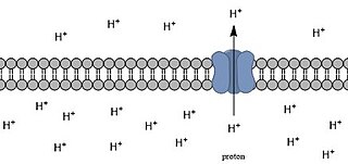
Intracellular pH (pHi) is the measure of the acidity or basicity of intracellular fluid. The pHi plays a critical role in membrane transport and other intracellular processes. In an environment with the improper pHi, biological cells may have compromised function. Therefore, pHi is closely regulated in order to ensure proper cellular function, controlled cell growth, and normal cellular processes. The mechanisms that regulate pHi are usually considered to be plasma membrane transporters of which two main types exist — those that are dependent and those that are independent of the concentration of bicarbonate. Physiologically normal intracellular pH is most commonly between 7.0 and 7.4, though there is variability between tissues. There is also pH variation across different organelles, which can span from around 4.5 to 8.0. pHi can be measured in a number of different ways.

Graham Wiggins was an American musician and scientist. He played the didgeridoo, keyboards, melodica, sampler, and various percussion instruments with his groups, the Oxford-based Outback and Dr. Didg. He also developed new technologies for magnetic resonance imaging (MRI).

<span class="mw-page-title-main">Diffusion MRI</span> Method of utilizing water in magnetic resonance imaging

Diffusion-weighted magnetic resonance imaging is the use of specific MRI sequences as well as software that generates images from the resulting data that uses the diffusion of water molecules to generate contrast in MR images. It allows the mapping of the diffusion process of molecules, mainly water, in biological tissues, in vivo and non-invasively. Molecular diffusion in tissues is not random, but reflects interactions with many obstacles, such as macromolecules, fibers, and membranes. Water molecule diffusion patterns can therefore reveal microscopic details about tissue architecture, either normal or in a diseased state. A special kind of DWI, diffusion tensor imaging (DTI), has been used extensively to map white matter tractography in the brain.

<span class="mw-page-title-main">Prostate biopsy</span> Medical test

Prostate biopsy is a procedure in which small hollow needle-core samples are removed from a man's prostate gland to be examined for the presence of prostate cancer. It is typically performed when the result from a PSA blood test is high. It may also be considered advisable after a digital rectal exam (DRE) finds possible abnormality. PSA screening is controversial as PSA may become elevated due to non-cancerous conditions such as benign prostatic hyperplasia (BPH), by infection, or by manipulation of the prostate during surgery or catheterization. Additionally many prostate cancers detected by screening develop so slowly that they would not cause problems during a man's lifetime, making the complications due to treatment unnecessary.

<span class="mw-page-title-main">Focused ultrasound</span> Non-invasive therapeutic technique

High-intensity focused ultrasound (HIFU) is a non-invasive therapeutic technique that uses non-ionizing ultrasonic waves to heat or ablate tissue. HIFU can be used to increase the flow of blood or lymph or to destroy tissue, such as tumors, via thermal and mechanical mechanisms. Given the prevalence and relatively low cost of ultrasound generation mechanisms, the premise of HIFU is that it is expected to be a non-invasive and low-cost therapy that can at least outperform care in the operating room.

<span class="mw-page-title-main">Gadopentetic acid</span> Complex of gadolinium by DTPA

Gadopentetic acid, sold under the brand name Magnevist, is a gadolinium-based MRI contrast agent.

Magnetic resonance elastography (MRE) is a form of elastography that specifically leverages MRI to quantify and subsequently map the mechanical properties of soft tissue. First developed and described at Mayo Clinic by Muthupillai et al. in 1995, MRE has emerged as a powerful, non-invasive diagnostic tool, namely as an alternative to biopsy and serum tests for staging liver fibrosis.

During nuclear magnetic resonance observations, spin–lattice relaxation is the mechanism by which the longitudinal component of the total nuclear magnetic moment vector (parallel to the constant magnetic field) exponentially relaxes from a higher energy, non-equilibrium state to thermodynamic equilibrium with its surroundings (the "lattice"). It is characterized by the spin–lattice relaxation time, a time constant known as T1.

MRI contrast agents are contrast agents used to improve the visibility of internal body structures in magnetic resonance imaging (MRI). The most commonly used compounds for contrast enhancement are gadolinium-based contrast agents (GBCAs). Such MRI contrast agents shorten the relaxation times of nuclei within body tissues following oral or intravenous administration.

In vivo magnetic resonance spectroscopy (MRS) is a specialized technique associated with magnetic resonance imaging (MRI).

Preclinical imaging is the visualization of living animals for research purposes, such as drug development. Imaging modalities have long been crucial to the researcher in observing changes, either at the organ, tissue, cell, or molecular level, in animals responding to physiological or environmental changes. Imaging modalities that are non-invasive and in vivo have become especially important to study animal models longitudinally. Broadly speaking, these imaging systems can be categorized into primarily morphological/anatomical and primarily molecular imaging techniques. Techniques such as high-frequency micro-ultrasound, magnetic resonance imaging (MRI) and computed tomography (CT) are usually used for anatomical imaging, while optical imaging, positron emission tomography (PET), and single photon emission computed tomography (SPECT) are usually used for molecular visualizations.

<span class="mw-page-title-main">Delayed gadolinium-enhanced magnetic resonance imaging of cartilage</span>

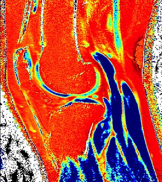
Delayed gadolinium-enhanced magnetic resonance imaging of cartilage or dGEMRIC measures the fixed-charge density and relative proteoglycan content of articular cartilage using the spin-lattice relaxation time or T1 relaxation time. Current research is investigating the clinical application of dGEMRIC as a quantitative tool for monitoring cartilage function in diseased or repair cartilage.

<span class="mw-page-title-main">Magnetic resonance imaging of the brain</span>

Magnetic resonance imaging of the brain uses magnetic resonance imaging (MRI) to produce high quality two-dimensional or three-dimensional images of the brain and brainstem as well as the cerebellum without the use of ionizing radiation (X-rays) or radioactive tracers.

Functional magnetic resonance spectroscopy of the brain (fMRS) uses magnetic resonance imaging (MRI) to study brain metabolism during brain activation. The data generated by fMRS usually shows spectra of resonances, instead of a brain image, as with MRI. The area under peaks in the spectrum represents relative concentrations of metabolites.

<span class="mw-page-title-main">Perfusion MRI</span>

Perfusion MRI or perfusion-weighted imaging (PWI) is perfusion scanning by the use of a particular MRI sequence. The acquired data are then post-processed to obtain perfusion maps with different parameters, such as BV, BF, MTT and TTP.

In the field of medicine, radiomics is a method that extracts a large number of features from medical images using data-characterisation algorithms. These features, termed radiomic features, have the potential to uncover tumoral patterns and characteristics that fail to be appreciated by the naked eye. The hypothesis of radiomics is that the distinctive imaging features between disease forms may be useful for predicting prognosis and therapeutic response for various cancer types, thus providing valuable information for personalized therapy. Radiomics emerged from the medical fields of radiology and oncology and is the most advanced in applications within these fields. However, the technique can be applied to any medical study where a pathological process can be imaged.

The history of magnetic resonance imaging (MRI) includes the work of many researchers who contributed to the discovery of nuclear magnetic resonance (NMR) and described the underlying physics of magnetic resonance imaging, starting early in the twentieth century. One researcher was American physicist Isidor Isaac Rabi who won the Nobel Prize in Physics in 1944 for his discovery of nuclear magnetic resonance, which is used in magnetic resonance imaging. MR imaging was invented by Paul C. Lauterbur who developed a mechanism to encode spatial information into an NMR signal using magnetic field gradients in September 1971; he published the theory behind it in March 1973.

<span class="mw-page-title-main">MRI pulse sequence</span>

An MRI pulse sequence in magnetic resonance imaging (MRI) is a particular setting of pulse sequences and pulsed field gradients, resulting in a particular image appearance.

Michael Albert Thomas is an Indian-American physicist, academic, and clinical researcher. He is a Professor-in-Residence of Radiological Sciences, and Psychiatry at the Geffen School of Medicine, University of California, Los Angeles (UCLA). He is most known for developing novel single voxel based 2D NMR techniques, multi-voxel 2D MRS techniques using hybrid Cartesian as well as non-Cartesian spatio-temporal encoding such as concentric ring, radial and rosette trajectories.

References

  1. Wetterling, Friedrich; Corteville, Dominique; Kalayciyan, Raffi; Rennings, Andre; Konstandin, Simon; Nagel, Armin; Stark, Helmut; Schad, Lothar (2012-06-22). "Whole body sodium MRI at 3T using an asymmetric birdcage resonator and short echo time sequence: first images of a male volunteer". Physics in medicine and biology. 57 (14): 4555–4567. doi:10.1088/0031-9155/57/14/4555. PMID   22722731.
  2. Ouwerkerk, Ronald; Morgan, Russell H. (2007-10-01). "23Na MRI: From Research to Clinical Use". Journal of the American College of Radiology. 4 (10): 739–741. doi:10.1016/j.jacr.2007.07.001. ISSN   1546-1440. PMC   2084082 . PMID   17903762.
  3. 1 2 3 Borthakur, A; Shapiro, E. M; Beers, J; Kudchodkar, S; Kneeland, J. B; Reddy, R (2000-07-01). "Sensitivity of MRI to proteoglycan depletion in cartilage: comparison of sodium and proton MRI". Osteoarthritis and Cartilage. 8 (4): 288–293. doi: 10.1053/joca.1999.0303 . PMID   10903883.
  4. Madelin, Guillaume (2012-12-18). "Sodium Magnetic Resonance Imaging: Biomedical Applications". arXiv: 1212.4400 [physics.med-ph].
  5. 1 2 Romanzetti, Sandro; Mirkes, Christian C.; Fiege, Daniel P.; Celik, Avdo; Felder, Jörg; Shah, N. Jon (2014-08-01). "Mapping tissue sodium concentration in the human brain: a comparison of MR sequences at 9.4Tesla". NeuroImage. 96: 44–53. doi:10.1016/j.neuroimage.2014.03.079. ISSN   1095-9572. PMID   24721332. S2CID   17913668.
  6. Madelin, Guillaume; Lee, Jae-Seung; Regatte, Ravinder R.; Jerschow, Alexej (2014-05-01). "Sodium MRI: Methods and applications". Progress in Nuclear Magnetic Resonance Spectroscopy. 79: 14–47. doi:10.1016/j.pnmrs.2014.02.001. PMC   4126172 . PMID   24815363.
  7. 1 2 3 4 5 Madelin, Guillaume; Regatte, Ravinder R. (2013-09-01). "Biomedical Applications of Sodium MRI In Vivo". Journal of Magnetic Resonance Imaging. 38 (3): 511–529. doi:10.1002/jmri.24168. ISSN   1053-1807. PMC   3759542 . PMID   23722972.
  8. Ouwerkerk, Ronald (2011-01-01). "Sodium MRI". In Modo, Michel; Bulte, Jeff W.M. (eds.). Magnetic Resonance Neuroimaging. Methods in Molecular Biology. Vol. 711. Humana Press. pp. 175–201. doi:10.1007/978-1-61737-992-5_8. ISBN   9781617379918. PMID   21279602.
  9. Alevizopoulos, Konstantinos; Calogeropoulou, Theodora; Lang, Florian; Stournaras, Christos (2014-01-01). "Na+/K+ ATPase inhibitors in cancer". Current Drug Targets. 15 (10): 988–1000. ISSN   1873-5592. PMID   25198786.
  10. Bottomley, Paul A. (2016-02-01). "Sodium MRI in human heart: a review". NMR in Biomedicine . 29 (2): 187–196. doi:10.1002/nbm.3265. ISSN   1099-1492. PMC   4868405 . PMID   25683054.
  11. Deen, SS; Riemer, F; McLean, MA; Gill, AB; Kaggie, JD; Grist, JT; Crawford, R; Latimer, J; Baldwin, P; Earl, HM; Parkinson, CA; Smith, SA; Hodgkin, C; Moore, E; Jimenez-Linan, M; Brodie, CR; Addley, HC; Freeman, SJ; Moyle, PL; Sala, E; Graves, MJ; Brenton, JD; Gallagher, FA (2019). "Sodium MRI with 3D-cones as a measure of tumour cellularity in high grade serous ovarian cancer". European Journal of Radiology Open. 6: 156–162. doi: 10.1016/j.ejro.2019.04.001 . PMC   6477161 . PMID   31032385.
  12. 1 2 3 Ouwerkerk, Ronald; Bleich, Karen B.; Gillen, Joseph S.; Pomper, Martin G.; Bottomley, Paul A. (2003-05-01). "Tissue Sodium Concentration in Human Brain Tumors as Measured with 23Na MR Imaging". Radiology. 227 (2): 529–537. doi:10.1148/radiol.2272020483. ISSN   0033-8419. PMID   12663825.
  13. Inglese, M.; Madelin, G.; Oesingmann, N.; Babb, J. S.; Wu, W.; Stoeckel, B.; Herbert, J.; Johnson, G. (2010-03-01). "Brain tissue sodium concentration in multiple sclerosis: a sodium imaging study at 3 tesla". Brain. 133 (3): 847–857. doi:10.1093/brain/awp334. ISSN   0006-8950. PMC   2842511 . PMID   20110245.
  14. Broeke, NC.; Peterson, J.; Lee, J.; Martin, PA.; Farag, A.; Gomez, J.; Moussa, M.; Gaed, M.; Chin, J.; Pautler, SE.; Ward, A.; Bauman, G.; Bartha, R.; Scholl, TJ. (2018-08-24). "Characterization of Clinical Human Prostate Cancer Lesions Using 3.0-T Sodium MRI Registered to Gleason-Graded Whole-Mount Histopathology". Journal of Magnetic Resonance Imaging. 49 (5): 1409–1419. doi:10.1002/jmri.26336. PMID   30430700. S2CID   53425193.
  15. Madelin, Guillaume; Kline, Richard; Walvick, Ronn; Regatte, Ravinder R. (2014-04-23). "A method for estimating intracellular sodium concentration and extracellular volume fraction in brain in vivo using sodium magnetic resonance imaging". Scientific Reports. 4: 4763. Bibcode:2014NatSR...4E4763M. doi:10.1038/srep04763. PMC   4762219 . PMID   24755879.
  16. Newbould, R.D.; Miller, S.R.; Tielbeek, J.A.W.; Toms, L.D.; Rao, A.W.; Gold, G.E.; Strachan, R.K.; Taylor, P.C.; Matthews, P.M. (2012-01-01). "Reproducibility of sodium MRI measures of articular cartilage of the knee in osteoarthritis". Osteoarthritis and Cartilage. 20 (1): 29–35. doi:10.1016/j.joca.2011.10.007. ISSN   1063-4584. PMC   3270258 . PMID   22040861.