Southern blot

Last updated
Agarose gel Recombinant DNA- southern blot (1).jpg
Agarose gel
Tray with a stack consisting top down of a weight, paper towels, membrane of nitrocellulose or nylon, gel, salt solution and a slab of glass. Aufbau Southern-Blot.jpg
Tray with a stack consisting top down of a weight, paper towels, membrane of nitrocellulose or nylon, gel, salt solution and a slab of glass.
Southern blot membrane after hybridization and rinsing. Southern blot membrane.jpg
Southern blot membrane after hybridization and rinsing.
Southern blot agarose gel under ultraviolet illumination. Southern-Blot-Agarosegel.jpg
Southern blot agarose gel under ultraviolet illumination.
Southern blot autoradiogram. Southern-Blot-Autoradiogramm.jpg
Southern blot autoradiogram.

Southern blot is a method used for detection and quantification of a specific DNA sequence in DNA samples. This method is used in molecular biology. Briefly, purified DNA from a biological sample (such as blood or tissue) is digested with restriction enzymes, and the resulting DNA fragments are separated by using an electric current to move them through a sieve-like gel or matrix, which allows smaller fragments to move faster than larger fragments. The DNA fragments are transferred out of the gel or matrix onto a solid membrane, which is then exposed to a DNA probe labeled with a radioactive, fluorescent, or chemical tag. The tag allows any DNA fragments containing complementary sequences with the DNA probe sequence to be visualized within the Southern blot. [1]

Contents

The Southern blotting combines the transfer of electrophoresis-separated DNA fragments to a filter membrane in a process called blotting, and the subsequent fragment detection by probe hybridization. [2]

The method is named after the British biologist Edwin Southern, who first published it in 1975. [3] Other blotting methods (i.e., western blot, [4] northern blot, eastern blot, southwestern blot) that employ similar principles, but using RNA or protein, have later been named for compass directions as a sort of pun from Southern's name. As the label is eponymous, Southern is capitalized, as is conventional of proper nouns. The names for other blotting methods may follow this convention, by analogy. [5]

History

Southern invented Southern blot after combining three innovations, the first one is the restriction endonucleases which were developed at Johns Hopkins University by Tom Kelly and Hamilton Smith, those restriction endonucleases are used to cut the DNA at a specific sequence. Kenneth and Noreen Murray introduced this technique as Southern. The second innovation is the gel electrophoresis that based on separation of mixtures of DNA, RNA, or proteins according to molecular size, which was also developed at Johns Hopkins University by Daniel Nathans and Kathleen Danna in 1971. The third innovation is blotting-through methods which was developed by Frederick Sanger, when he transferred RNA molecules to DEAE paper. Southern blot was invented in 1973 but it was not published until 1975. Although it was published later the technique was disseminated when Southern introduced the Southern blot technique to a scientist at Cold Spring Harbor Laboratory called Michael Mathews by drawing this technique on a paper. [6]

Method

The genomic DNA is digested with either one or more than one restriction enzyme, then the DNA fragments are size-fractionated by gel electrophoresis. Before the DNA fragments are transferred to a solid membrane which is either nylon or nitrocellulose membrane it is first denatured by alkaline treatment. [7] After the DNA fragments are immobilized on the membrane, prehybridization methods are used to reduce non-specific probe binding then the fragments on the membrane are hybridized with either radiolabeled or nonradioactive labeled DNA, RNA, or oligonucleotide probes that are complementary to the target DNA sequence then detection methods are used to visualize the target DNA. [8]

  1. DNA Isolation: The DNA to be studied is isolated from various tissues. The most suitable source of DNA is known as blood tissue. However, it can be isolated from different tissues (hair, semen, saliva, etc.).
  2. DNA digestion: Restriction endonucleases are used to cut high-molecular-weight DNA strands into smaller fragments. This is done by adding the desired amount of  DNA which can be changed according to the probe used and the intricacy of the DNA, with the restriction enzyme, enzyme buffer and purified water then incubate the reaction at 37 °C overnight.
  3. Gel electrophoresis: The DNA fragments are then electrophoresed on an agarose gel to separate them by size. If some of the DNA fragments are larger than 15 kb, then before blotting, the gel may be treated with an acid, such as dilute HCl. This depurinates the DNA fragments, breaking the DNA into smaller pieces, thereby allowing more efficient transfer from the gel to membrane.
  4. Denaturation: If alkaline transfer methods are used, the DNA gel is placed into an alkaline solution (typically containing sodium hydroxide) to denature the double-stranded DNA. The denaturation in an alkaline environment may improve binding of the negatively charged thymine residues of DNA to a positively charged amino groups of membrane, separating it into single DNA strands for later hybridization to the probe (see below), and destroys any residual RNA that may still be present in the DNA. The choice of alkaline over neutral transfer methods, however, is often empirical and may result in equivalent results.[ citation needed ]
  5. Blotting: A sheet of nitrocellulose (or, alternatively, nylon) membrane is placed on top of (or below, depending on the direction of the transfer) the gel. Pressure is applied constantly to the gel (either using suction, or by placing a stack of paper towels and a weight on top of the membrane and gel), to ensure good and even contact between gel and membrane. If transferring by suction, 20X SSC buffer is used to ensure a seal and prevent drying of the gel. Buffer transfer by capillary action from a region of high water potential to a region of low water potential (usually filter paper and paper tissues) is then used to move the DNA from the gel onto the membrane; ion exchange interactions bind the DNA to the membrane due to the negative charge of the DNA and positive charge of the membrane. Five methods can be used to transfer DNA fragments to the solid membrane and they are: [8] 1. Upward capillary transfer: This method transfers the DNA fragment upward from the gel to the membrane where the flow of the liquid or the buffer will be upward, 2. Downward capillary transfer: This method is done by placing the gel on the surface of the membrane (usually nylon charged membrane) and the DNA fragments will be transferred in a downward direction with the flow of the alkaline buffer, 3.Simultaneous transfer to two membranes: This method is used to transfer DNA fragments of high concentration simultaneously from the gel to two membranes, 4. Electrophoretic transfer: This method usually uses large electric current which makes it difficult to transfer the DNA efficiently due to the temperature of the buffer used, so these machines can be either equipped with cooling machines or used in a cold area, 5. Vacuum transfer: This method uses a buffer from the upper chamber to transfer the DNA from the gel to the nitrocellulose or nylon membrane, the gel is placed directly on the membrane, and the membrane is placed on a porous screen on the vacuum chamber.
  6. Immobilization: The membrane is then baked in a vacuum or regular oven at 80 °C for 2 hours (standard conditions; nitrocellulose or nylon membrane) or exposed to ultraviolet radiation (nylon membrane) to permanently attach the transferred DNA to the membrane.
  7. Hybridization: After that, a hybridization probe—a single DNA fragment with a particular sequence whose presence in the target DNA is to be ascertained—is exposed to the membrane. The probe DNA is labelled so that it can be detected, usually by incorporating radioactivity or tagging the molecule with a fluorescent or chromogenic dye. In some cases, the hybridization probe may be made from RNA, rather than DNA. To ensure the specificity of the binding of the probe to the sample DNA, most common hybridization methods use salmon or herring sperm DNA for blocking of the membrane surface and target DNA, deionized formamide, and detergents such as SDS to reduce non-specific binding of the probe.
  8. Detection: After hybridization, excess probe is washed from the membrane (typically using SSC buffer), and the pattern of hybridization is visualized on X-ray film by autoradiography in the case of a radioactive or fluorescent probe, or by development of color on the membrane if a chromogenic detection method is used.

Interpretation of results

Hybridization of the probe to a specific DNA fragment on the filter membrane indicates that this fragment contains DNA sequence that is complementary to the probe. The transfer step of the DNA from the electrophoresis gel to a membrane permits easy binding of the labeled hybridization probe to the size-fractionated DNA. It also allows for the fixation of the target-probe hybrids, required for analysis by autoradiography or other detection methods. Southern blots performed with restriction enzyme-digested genomic DNA may be used to determine the number of sequences (e.g., gene copies) in a genome. A probe that hybridizes only to a single DNA segment that has not been cut by the restriction enzyme will produce a single band on a Southern blot, whereas multiple bands will likely be observed when the probe hybridizes to several highly similar sequences (e.g., those that may be the result of sequence duplication). To improve specificity and reduce hybridization of the probe to sequences that are less than 100% identical, the hybridization parameters may be changed (for instance, by raising the hybridization temperature or lowering the salt content). Nylon membrane is more durable and has higher binding capacity to DNA fragments than nitrocellulose membrane, so the DNA fragments will be more fixed to the membrane even when the membrane is incubated in high temperatures. In addition, compared to the nitrocellulose membrane which requires a high ionic strength buffer to bind the DNA fragments to the membrane, nylon charged membranes use buffers with very low ionic strength to transfer even small fragments of DNA of about 50 bp to the membrane, usually the DNA to be transferred is separated by polyacrylamide gel. In the blotting step the most efficient method to transfer the DNA from the gel to the membrane is the vacuum transfer since it transfers the DNA more rapidly and quantitatively. [8]

Applications

  1. Southern blotting transfer may be used for homology-based cloning based on amino acid sequence of the protein product of the target gene. Oligonucleotides are designed so that they are complementary to the target sequence. The oligonucleotides are chemically synthesized, radiolabeled, and used to screen a DNA library, or other collections of cloned DNA fragments. Sequences that hybridize with the hybridization probe are further analyzed, for example, to obtain the full length sequence of the targeted gene.
  2. Normal chromosomal or gene rearrangement can be studied using this technique. [7]
  3. Can be used to find similar sequences in other species or in the genome by decreasing the specificity of hybridization. [7]
  4. In a mixture having different sizes of digested DNA, it is used to identify the restriction fragment of a specific size. [7]
  5. It is useful in identifying changes that occur in genes including insertions, rearrangements, deletions, and point mutations that affect the restriction sites. [7]
  6. Moreover it is used to identify a specific region that uses many different restriction enzymes in a restriction mapping. Also it is used to determine which recognition site has been altered due to a single nucleotide polymorphism that changes a specific restriction enzyme. [7]
  7. Southern blotting can also be used to identify methylated sites in particular genes. Particularly useful are the restriction nucleases MspI and HpaII, both of which recognize and cleave within the same sequence. However, HpaII requires that a C within that site be methylated, whereas MspI cleaves only DNA unmethylated at that site. Therefore, any methylated sites within a sequence analyzed with a particular probe will be cleaved by the former, but not the latter, enzyme. [9]
  8. Can be used in personal identification through fingerprinting, and in disease diagnosis. [10]

Limitations

  1. Compared to other tests, southern blot is a complex technique that has multiple steps and these steps require equipment and reagents that are expensive. [10]
  2. High quality and large amounts of DNA are needed. [10]
  3. Southern blot is a time consuming method and can only estimate the size of the DNA since it is a semi-quantitative method. [10]
  4. It cannot be used to detect mutations at base-pair level. [10]

See also

Related Research Articles

<span class="mw-page-title-main">Gel electrophoresis</span> Method for separation and analysis of biomolecules

Gel electrophoresis is a method for separation and analysis of biomacromolecules and their fragments, based on their size and charge. It is used in clinical chemistry to separate proteins by charge or size and in biochemistry and molecular biology to separate a mixed population of DNA and RNA fragments by length, to estimate the size of DNA and RNA fragments or to separate proteins by charge.

Molecular biology is the study of chemical and physical structure of biological macromolecules. It is a branch of biology that seeks to understand the molecular basis of biological activity in and between cells, including biomolecular synthesis, modification, mechanisms, and interactions.

<span class="mw-page-title-main">Northern blot</span> Molecular biology technique

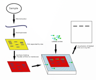
The northern blot, or RNA blot, is a technique used in molecular biology research to study gene expression by detection of RNA in a sample.

In molecular biology, restriction fragment length polymorphism (RFLP) is a technique that exploits variations in homologous DNA sequences, known as polymorphisms, populations, or species or to pinpoint the locations of genes within a sequence. The term may refer to a polymorphism itself, as detected through the differing locations of restriction enzyme sites, or to a related laboratory technique by which such differences can be illustrated. In RFLP analysis, a DNA sample is digested into fragments by one or more restriction enzymes, and the resulting restriction fragments are then separated by gel electrophoresis according to their size.

<span class="mw-page-title-main">Western blot</span> Analytical technique used in molecular biology

The western blot, or western blotting, is a widely used analytical technique in molecular biology and immunogenetics to detect specific proteins in a sample of tissue homogenate or extract. Besides detecting the proteins, this technique is also utilized to visualize, distinguish, and quantify the different proteins in a complicated protein combination.

<span class="mw-page-title-main">Blot (biology)</span>

A blot, in molecular biology and genetics, is a method of transferring proteins, DNA or RNA onto a carrier. In many instances, this is done after a gel electrophoresis, transferring the molecules from the gel onto the blotting membrane, and other times adding the samples directly onto the membrane. After the blotting, the transferred proteins, DNA or RNA are then visualized by colorant staining, autoradiographic visualization of radiolabelled molecules, or specific labelling of some proteins or nucleic acids. The latter is done with antibodies or hybridization probes that bind only to some molecules of the blot and have an enzyme joined to them. After proper washing, this enzymatic activity is visualized by incubation with proper reactive, rendering either a colored deposit on the blot or a chemiluminescent reaction which is registered by photographic film.

The first isolation of deoxyribonucleic acid (DNA) was done in 1869 by Friedrich Miescher. DNA extraction is the process of isolating DNA from the cells of an organism isolated from a sample, typically a biological sample such as blood, saliva, or tissue. It involves breaking open the cells, removing proteins and other contaminants, and purifying the DNA so that it is free of other cellular components. The purified DNA can then be used for downstream applications such as PCR, sequencing, or cloning. Currently, it is a routine procedure in molecular biology or forensic analyses.

This is a list of topics in molecular biology. See also index of biochemistry articles.

In molecular biology, a hybridization probe(HP) is a fragment of DNA or RNA of usually 15–10000 nucleotide long which can be radioactively or fluorescently labeled. HP can be used to detect the presence of nucleotide sequences in analyzed RNA or DNA that are complementary to the sequence in the probe. The labeled probe is first denatured (by heating or under alkaline conditions such as exposure to sodium hydroxide) into single stranded DNA (ssDNA) and then hybridized to the target ssDNA (Southern blotting) or RNA (northern blotting) immobilized on a membrane or in situ.

<span class="mw-page-title-main">Southwestern blot</span> Molecular biology technique

The southwestern blot, is a lab technique that involves identifying as well as characterizing DNA-binding proteins by their ability to bind to specific oligonucleotide probes. Determination of molecular weight of proteins binding to DNA is also made possible by the technique. The name originates from a combination of ideas underlying Southern blotting and Western blotting techniques of which they detect DNA and protein respectively. Similar to other types of blotting, proteins are separated by SDS-PAGE and are subsequently transferred to nitrocellulose membranes. Thereafter southwestern blotting begins to vary with regards to procedure as since the first blotting’s, many more have been proposed and discovered with goals of enhancing results. Former protocols were hampered by the need for large amounts of proteins and their susceptibility to degradation while being isolated.

Ribotyping is a molecular technique for bacterial identification and characterization that uses information from rRNA-based phylogenetic analyses. It is a rapid and specific method widely used in clinical diagnostics and analysis of microbial communities in food, water, and beverages.

<span class="mw-page-title-main">Dot blot</span>

A dot blot is a technique in molecular biology used to detect proteins. It represents a simplification of the western blot method, with the exception that the proteins to be detected are not first separated by electrophoresis. Instead, the sample is applied directly on a membrane in a single spot, and the blotting procedure is performed.

<span class="mw-page-title-main">Molecular-weight size marker</span> Set of standards

A molecular-weight size marker, also referred to as a protein ladder, DNA ladder, or RNA ladder, is a set of standards that are used to identify the approximate size of a molecule run on a gel during electrophoresis, using the principle that molecular weight is inversely proportional to migration rate through a gel matrix. Therefore, when used in gel electrophoresis, markers effectively provide a logarithmic scale by which to estimate the size of the other fragments.

An allele-specific oligonucleotide (ASO) is a short piece of synthetic DNA complementary to the sequence of a variable target DNA. It acts as a probe for the presence of the target in a Southern blot assay or, more commonly, in the simpler Dot blot assay. It is a common tool used in genetic testing, forensics, and Molecular Biology research.

<span class="mw-page-title-main">Electroblotting</span>

Electroblotting is a method in molecular biology/biochemistry/immunogenetics to transfer proteins or nucleic acids onto a membrane by using PVDF or nitrocellulose, after gel electrophoresis. The protein or nucleic acid can then be further analyzed using probes such as specific antibodies, ligands like lectins, or stains. This method can be used with all polyacrylamide and agarose gels. An alternative technique for transferring proteins from a gel is capillary blotting.

<span class="mw-page-title-main">Oligomer restriction</span>

Oligomer Restriction is a procedure to detect an altered DNA sequence in a genome. A labeled oligonucleotide probe is hybridized to a target DNA, and then treated with a restriction enzyme. If the probe exactly matches the target, the restriction enzyme will cleave the probe, changing its size. If, however, the target DNA does not exactly match the probe, the restriction enzyme will have no effect on the length of the probe. The OR technique, now rarely performed, was closely associated with the development of the popular polymerase chain reaction (PCR) method.

<span class="mw-page-title-main">Affinity electrophoresis</span>

Affinity electrophoresis is a general name for many analytical methods used in biochemistry and biotechnology. Both qualitative and quantitative information may be obtained through affinity electrophoresis. Cross electrophoresis, the first affinity electrophoresis method, was created by Nakamura et al. Enzyme-substrate complexes have been detected using cross electrophoresis. The methods include the so-called electrophoretic mobility shift assay, charge shift electrophoresis and affinity capillary electrophoresis. The methods are based on changes in the electrophoretic pattern of molecules through biospecific interaction or complex formation. The interaction or binding of a molecule, charged or uncharged, will normally change the electrophoretic properties of a molecule. Membrane proteins may be identified by a shift in mobility induced by a charged detergent. Nucleic acids or nucleic acid fragments may be characterized by their affinity to other molecules. The methods have been used for estimation of binding constants, as for instance in lectin affinity electrophoresis or characterization of molecules with specific features like glycan content or ligand binding. For enzymes and other ligand-binding proteins, one-dimensional electrophoresis similar to counter electrophoresis or to "rocket immunoelectrophoresis", affinity electrophoresis may be used as an alternative quantification of the protein. Some of the methods are similar to affinity chromatography by use of immobilized ligands.

The eastern blot, or eastern blotting, is a biochemical technique used to analyze protein post-translational modifications including the addition of lipids, phosphates, and glycoconjugates. It is most often used to detect carbohydrate epitopes. Thus, eastern blot can be considered an extension of the biochemical technique of western blot. Multiple techniques have been described by the term "eastern blot(ting)", most use phosphoprotein blotted from sodium dodecyl sulfate–polyacrylamide gel electrophoresis (SDS-PAGE) gel on to a polyvinylidene fluoride or nitrocellulose membrane. Transferred proteins are analyzed for post-translational modifications using probes that may detect lipids, carbohydrate, phosphorylation or any other protein modification. Eastern blotting should be used to refer to methods that detect their targets through specific interaction of the post-translational modifications and the probe, distinguishing them from a standard far-western blot. In principle, eastern blotting is similar to lectin blotting.

The reverse northern blot is a method by which gene expression patterns may be analyzed by comparing isolated RNA molecules from a tester sample to samples in a control cDNA library. It is a variant of the northern blot in which the nucleic acid immobilized on a membrane is a collection of isolated DNA fragments rather than RNA, and the probe is RNA extracted from a tissue and radioactively labelled. A reverse northern blot can be used to profile expression levels of particular sets of RNA sequences in a tissue or to determine presence of a particular RNA sequence in a sample. Although DNA Microarrays and newer next-generation techniques have generally supplanted reverse northern blotting, it is still utilized today and provides a relatively cheap and easy means of defining expression of large sets of genes.

The northwestern blot, also known as the northwestern assay, is a hybrid analytical technique of the western blot and the northern blot, and is used in molecular biology to detect interactions between RNA and proteins. A related technique, the western blot, is used to detect a protein of interest that involves transferring proteins that are separated by gel electrophoresis onto a nitrocellulose membrane. A colored precipitate clusters along the band on the membrane containing a particular target protein. A northern blot is a similar analytical technique that, instead of detecting a protein of interest, is used to study gene expression by detection of RNA on a similar membrane. The northwestern blot combines the two techniques, and specifically involves the identification of labeled RNA that interact with proteins that are immobilized on a similar nitrocellulose membrane.

References

  1. "Talking Glossary of Genetic Terms | NHGRI". www.genome.gov. National Institutes of Health. Retrieved 24 January 2023.PD-icon.svg This article incorporates text from this source, which is in the public domain .
  2. "Southern Blot".
  3. Southern, Edwin Mellor (5 November 1975). "Detection of specific sequences among DNA fragments separated by gel electrophoresis". Journal of Molecular Biology . 98 (3): 503–517. doi:10.1016/S0022-2836(75)80083-0. ISSN   0022-2836. PMID   1195397. S2CID   20126741.
  4. Towbin; Staehelin, T; Gordon, J; et al. (1979). "Electrophoretic transfer of proteins from polyacrylamide gels to nitrocellulose sheets: procedure and some applications". PNAS. 76 (9): 4350–4. Bibcode:1979PNAS...76.4350T. doi: 10.1073/pnas.76.9.4350 . PMC   411572 . PMID   388439.
  5. Burnette, W. Neal (April 1981). "Western Blotting: Electrophoretic Transfer of Proteins from Sodium Dodecyl Sulfate-Polyacrylamide Gels to Unmodified Nitrocellulose and Radiographic Detection with Antibody and Radioiodinated Protein A". Analytical Biochemistry . 112 (2): 195–203. doi:10.1016/0003-2697(81)90281-5. ISSN   0003-2697. PMID   6266278.
  6. Tofano, Daidree; Wiechers, Ilse R.; Cook-Deegan, Robert (2006-08-15). "Edwin Southern, DNA blotting, and microarray technology: A case study of the shifting role of patents in academic molecular biology". Genomics, Society and Policy. 2 (2). doi: 10.1186/1746-5354-2-2-50 . ISSN   1746-5354.
  7. 1 2 3 4 5 6 Glenn, Gary; Andreou, Lefkothea-Vasiliki (2013-01-01), "Chapter Five - Analysis of DNA by Southern Blotting", in Lorsch, Jon (ed.), DNA, vol. 529, Academic Press, pp. 47–63, doi:10.1016/b978-0-12-418687-3.00005-7, ISBN   9780124186873, PMID   24011036 , retrieved 2023-01-04
  8. 1 2 3 Green, Michael R.; Sambrook, Joseph (July 2021). "Analysis of DNA by Southern Blotting". Cold Spring Harbor Protocols. 2021 (7): pdb.top100396. doi: 10.1101/pdb.top100396 . ISSN   1940-3402. PMID   34210774. S2CID   235710916.
  9. Biochemistry 3rd Edition, Matthews, Van Holde et al, Addison Wesley Publishing, pg 977
  10. 1 2 3 4 5 Sapkota, Anupama (2021-06-03). "Southern Blot- Definition, Principle, Steps, Results, Applications". Microbe Notes. Retrieved 2023-01-04.