Spinal disease

Last updated
Skeleton and bones - Vertebral column disorders - Normal Scoliosis Normal Lordosis Kyphosis Vertebral column disorders - Normal Scoliosis Lordosis Kyphosis -- Smart-Servier.jpg
Skeleton and bones - Vertebral column disorders - Normal Scoliosis Normal Lordosis Kyphosis

Spinal disease refers to a condition impairing the backbone. [1] These include various diseases of the back or spine ("dorso-"), such as kyphosis. Dorsalgia refers to back pain. Some other spinal diseases include spinal muscular atrophy, ankylosing spondylitis, scoliosis, lumbar spinal stenosis, spina bifida, spinal tumors, osteoporosis and cauda equina syndrome.

Contents

Types

There are many recognized spinal diseases, some more common than others. Spinal disease also includes cervical spine diseases, which are diseases in the vertebrae of the neck. A lot of flexibility exists within the cervical spine and because of that, it is common for an individual to damage that area, especially over a long period of time. Some of the common cervical spine diseases include degenerative disc disease, cervical stenosis, and cervical disc herniation. Degenerative disc disease occurs over time when the discs within each vertebra in the neck begin to fall apart and begin to disintegrate. Because each vertebra can cause pain in different areas of the body, the pain from the disease can be sensed in the back, leg, neck area, or even the arms. When the spinal canal begins to lose its gap and gets thinner, it can cause pain in the neck, which can also cause a numb feeling in the arms and hands. Those are symptoms of cervical stenosis disease. The discs between each vertebra have fibers that can begin to deteriorate, and this can occur in cervical disc herniation. This disease is less common in younger people as it is usually a function of aging. [2]

Spinal muscular atrophy (SMA)

SMA types

SMA is a category of spinal disease that in linked with genetic disorders. More specifically, it is caused by an autosomal recessive disorder due to a homozygous mutation of a motor neuron gene. [3] There are different types of SMA. Type 0 is diagnosed to newborns who have muscle weakness, and little to no "fetal movements." [3] Those who have type 0 also have other health issues, most of which are respiratory-related. SMA type 1 is diagnosed to infants with symptoms similar to that of type 0. Those who have type 1 are more likely to have trouble swallowing, controlling the tongue, and sitting up on their own. Moreover, infants with type 1 are likely to develop respiratory issues. Additionally, their thinking and comprehension is unaffected and they are conscientiousness. SMA type 2 is diagnosed to young children. Unlike those with type 1, these children can sit without assistance, but are unable to walk. This type mostly concerns the legs and arms. Some other problems that SMA type 2 patients might encounter are orthopedic, bone, and joint complications. SMA type 3 is typically diagnosed to kids and adults. Those with SMA 3 might be able to walk, and are more likely to experience weakness in the legs compared to the arms. Type 3 patients are most likely to have symptoms of scoliosis with little to no respiratory issues. Unlike types 0, 1, and 2, those with type 3 do not have to worry about comprehension and learning. Lastly, SMA type 4 is diagnosed to elderly individuals, and is the most uncommon version of SMA next to type 0. SMA type 4 is the least severe, and is sort of similar to type 3, but most common in adults. [3]

Diagnosing SMA

Molecular look into spinal muscular atrophy.

Molecular genetic testing is the tool used to assess SMA. However, this test might not be needed if signs such as hypotonia are present. MRI scans and muscle biopsies used to be the standard testing method, but molecular testing is much more efficient. There are advanced forms of SMA that require other testing concerning the peripheral nervous system. On another note, SMA is due to the malfunctioning SMN1 gene. Patients who have SMA that is caused by the SMN gene is likely due to the compound heterozygotes with only one of the SMN1 genes being mutated. SMA is diagnosed by the deletion of the homozygous SMN1, while the severity is based on the SMN2 gene. Medical screenings, such as scans, should only be used for patients who "are negative for both SMN1 deletion and SMN1 mutation testing." [4]

Management

As of right now, there are no successful treatments. However, many patients opt to go into physical and rehabilitation therapy designed to help with specific needs, similar to Schroth therapy. The most important and best way to manage SMA is to come up with a plan that both the medical team and patient agrees with. As mentioned before, patients with SMA also suffer from respiratory issues, which is the number one issue that must be prevented. Treating patients while they actively have the issues is not as effective as planning beforehand. It is also important for SMA patients to consider vaccinations as that could aid in the prevention of developing harmful respiratory problems. Some patients choose to use ventilation and other pulmonary-related tools. Taking care of gastrointestinal health is also important, as such issues are also common with SMA patients. Additionally, SMA patients might use G-tubes, also known as gastronomy tubes for feeding. Overall, the best treatment method is to find a plan that works with both the doctors and the patient to ensure that future problems are prevented and handled properly before becoming too severe. [4]

Scoliosis

Scoliosis is a common spinal disease in which the spine has a curvature usually in the shape of the letter "C" or "S". This is most common in girls, but there is no specific cause for scoliosis. [5] Only a few symptoms occur for one with this disease, which include feeling tired in the spinal region or backaches. Generally, if the hips or shoulders are uneven, or if the spine curves, it is due to scoliosis and should be seen by a doctor. [6] When assessing scoliosis, it is important for the physician to assess for neurological issues. Anything from weakness, difficulty with balance and coordination, and bladder and bowel problems should be considered. Curvature advancement is largely dependent "on remaining spinal growth," [7] as well as signs of puberty, indicating the beginning of early adulthood. [7]

Diagnosis

Physicians must perform physical and neurological examinations, which includes looking at height, asymmetry in the back, chest, ribs, and other areas of the torso, balance and coordination, and even pain. In addition to physical examinations, physicians may order X-ray or MRI scans. These tests will verify any concerns. [7]

Management

Spinal fusion surgery. Instrumentation Surgery Posterior.png
Spinal fusion surgery.

Depending on the level of curvature, there are different treatment options. For those who have curves less than 10 degrees, there is no need to get into treatment. Curves between 10-25 degrees must keep a close eye on it by having X-rays to maintain it. However, those who have curves greater than 25 degrees, but less than 40 to 45 might choose to get bracing. [8] Braces, also known as corsets, hold the spine in a specific position from the outside. These devices are tight, and can get even tighter with the straps. Whether bracing is effective or not is still studied today. [9] In addition to bracing, many patients choose to partake in hydrotherapy. Studies show that water environment positively affects the curvature of different types, and increases mobility as well as flexibility in the shoulders and bending. [10] There are also other physical therapy methods to improving curvature through Schroth therapy. Some experiments have been conducted to determine whether or not this strategy is useful. One study done shows that the Schroth group had improved posture, while the control worsened. [11] Another double-blind experiment was conducted, which did not show outstanding results. [12] The very last treatment option is surgery. There are certain goals that surgery aims to reach. For children, the point of the operation is to stop the curve from getting worse and minimize spinal deformity. On the other hand, adults usually have this surgery due to nerve damage, or if they have serious bladder and bowel issues. Surgery is only recommended to those who have curves greater than 40-50 degrees. [13] [14] There have been some experiments done to determine which surgical method is the most beneficial. One study shows that those who have short segment decompression/ fusion are least likely to suffer from postoperative complications. Moreover, short segment patients had a shorter hospital stay compared to long segment. However, the short segment group did lose more blood, resulting in less blood volume after their operation. [15]

Lumbar spinal stenosis

Lumbar spinal stenosis is classified as a narrowing of the spinal canal in the lumbar region of the vertebrae. This may lead to compression of the nerve root of the spinal cord and result in pain of the lower back and lower extremities. Other symptoms include impaired walking and a slightly stooped posture due to loss of disc height and bulging of the disc. Lumbar spinal stenosis is very prevalent with 9.3% of the general population producing symptoms and the number is continuing to rise in patients older than 60. [16] It's generally an indication for spinal surgery in patients older than 65 years of age. [17] However, there is a myth and fear among most patients that only surgery is the cure for such conditions and spine surgery is very risky. There are many non-surgical treatments available to prevent, halt and even reverse many spine diseases. Also, some surgery patients can be operated on in a daycare procedure or with minimum length of stay in hospital, with statistically good outcomes. [18]

Spina bifida

Spina bifida is the most common defect impacting the Central Nervous System (CNS). The most common and most severe form of Spina Bifida is Myelomeningocele. Individuals with Myelomeningocele are born with an incompletely fused spine, and therefore exposing the spinal cord through an opening in the back. In general, the higher the spinal lesion, the greater the functional impairment to the individual. [19] Symptoms may include bowel and bladder problems, weakness and/or loss of sensation below the level of the lesion, paralysis, or orthopedic issues. Severity of symptoms can vary per situation. [20]

Cauda equina syndrome

An MRI of the lumbar spine with abscess that resulted in CES. MRI of the lumbar spine with abscess in the posterior epidural space, causing cauda equina syndrome.jpg
An MRI of the lumbar spine with abscess that resulted in CES.

Cauda equina syndrome is a rare syndrome that affects the spinal nerves in the region of the lower back called the cauda equine (Latin for "horses tail"). Injury to the cauda equina can have long lasting ramifications for the individual. Symptoms include lower back pain, bladder disturbances, bowel dysfunction, and anesthesia or paresthesia between the thighs. In order to prevent progressive neurological changes surgery can be a viable option. [21] CT scans, myelograms, and MRIs are used to diagnose cauda equina. [22]

Management

Surgery is the best treatment option for those who have CES. If left untreated, patients might develop paralysis and bladder incontinence. [22] Moreover, the timing of the surgery is crucial, but it is unknown as to when the best time to have it done is. When it comes to timing, it really depends on when the patients' symptoms first arise. Most patients start considering surgery when the symptoms such as bladder incontinence, bowel movement issues, limb weakness, and pain first begin. The most common surgical procedure is a laminectomy, with microdiscectomies and discectomies also being options. With the lack of research regarding this spinal disorder, however, it is unclear as to when the best time have the operation is. [23] One study shows that overnight versus daytime lumbar decompression surgery does not have much significance in terms of complications. However, those who do have overnight surgery are more likely to suffer from complications. [24]

Tumors

A spinal tumor is when unusual tissue begins growing and spreading in the spinal columns or spinal cords. The unusual tissue builds up from abnormal cells that multiply quickly in a specific region. Tumors generally are broken down into categories known as benign, meaning non-cancerous, or malignant, meaning cancerous, and also primary or secondary. Primary spinal tumors begin in either the spinal cord or spinal column, whereas secondary spinal tumors begin elsewhere and spread to the spinal region. [25] Symptoms for spinal tumors may vary due to factors such as the type of tumor, the region of the spine, and the health of the patient. Back pain is the most common symptom and it can be a problem if the pain is severe, has a time frame that lasts longer than it would for a normal injury, and becomes worse while laying down or at rest. Other symptoms, excluding back pains, are loss of muscle function, loss of bowel or bladder function, pain in the legs, scoliosis, or even unusual sensations in the legs. [25] [26] [27] The primary tumor has no known cause, although there are possible answers that scientists have researched. Cancer may be linked to genes because research shows that in certain families, the incidents of spinal tumors are higher. Two of the genetic disorders that may affect spinal tumors, include Von Hippel-Lindau disease and Neurofibromatosis 2. Von Hippel-Lindau disease is a non-cancerous tumor of blood vessels that occur in the brain, spinal cord, or even tumors in the kidneys. The Neurofibromatosis 2 is a non-cancerous tumor that usually affects the nerves for hearing. Loss of hearing in one or both ears, is a common effect of this genetic disorder. [25]

Related Research Articles

<span class="mw-page-title-main">Syringomyelia</span> Disorder in which a cyst forms in the spinal cord

Syringomyelia is a generic term referring to a disorder in which a cyst or cavity forms within the spinal cord. Often, syringomyelia is used as a generic term before an etiology is determined. This cyst, called a syrinx, can expand and elongate over time, destroying the spinal cord. The damage may result in loss of feeling, paralysis, weakness, and stiffness in the back, shoulders, and extremities. Syringomyelia may also cause a loss of the ability to feel extremes of hot or cold, especially in the hands. It may also lead to a cape-like bilateral loss of pain and temperature sensation along the upper chest and arms. The combination of symptoms varies from one patient to another depending on the location of the syrinx within the spinal cord, as well as its extent.

<span class="mw-page-title-main">Back pain</span> Area of body discomfort

Back pain is pain felt in the back. It may be classified as neck pain (cervical), middle back pain (thoracic), lower back pain (lumbar) or coccydynia based on the segment affected. The lumbar area is the most common area affected. An episode of back pain may be acute, subacute or chronic depending on the duration. The pain may be characterized as a dull ache, shooting or piercing pain or a burning sensation. Discomfort can radiate to the arms and hands as well as the legs or feet, and may include numbness or weakness in the legs and arms.

<span class="mw-page-title-main">Tetraplegia</span> Paralysis of all four limbs and torso

Tetraplegia, also known as quadriplegia, is defined as the dysfunction or loss of motor and/or sensory function in the cervical area of the spinal cord. A loss of motor function can present as either weakness or paralysis leading to partial or total loss of function in the arms, legs, trunk, and pelvis; paraplegia is similar but affects the thoracic, lumbar, and sacral segments of the spinal cord and arm function is retained. The paralysis may be flaccid or spastic. A loss of sensory function can present as an impairment or complete inability to sense light touch, pressure, heat, pinprick/pain, and proprioception. In these types of spinal cord injury, it is common to have a loss of both sensation and motor control.

<span class="mw-page-title-main">Cauda equina</span> Anatomical structure

The cauda equina is a bundle of spinal nerves and spinal nerve rootlets, consisting of the second through fifth lumbar nerve pairs, the first through fifth sacral nerve pairs, and the coccygeal nerve, all of which arise from the lumbar enlargement and the conus medullaris of the spinal cord. The cauda equina occupies the lumbar cistern, a subarachnoid space inferior to the conus medullaris. The nerves that compose the cauda equina innervate the pelvic organs and lower limbs to include motor innervation of the hips, knees, ankles, feet, internal anal sphincter and external anal sphincter. In addition, the cauda equina extends to sensory innervation of the perineum and, partially, parasympathetic innervation of the bladder.

<span class="mw-page-title-main">Sciatica</span> Lower back pain that extends down leg

Sciatica is pain going down the leg from the lower back. This pain may go down the back, outside, or front of the leg. Onset is often sudden following activities like heavy lifting, though gradual onset may also occur. The pain is often described as shooting. Typically, symptoms are only on one side of the body. Certain causes, however, may result in pain on both sides. Lower back pain is sometimes present. Weakness or numbness may occur in various parts of the affected leg and foot.

<span class="mw-page-title-main">Kyphosis</span> Medical condition

Kyphosis is an abnormally excessive convex curvature of the spine as it occurs in the thoracic and sacral regions. Abnormal inward concave lordotic curving of the cervical and lumbar regions of the spine is called lordosis. It can result from degenerative disc disease; developmental abnormalities, most commonly Scheuermann's disease; Copenhagen disease, osteoporosis with compression fractures of the vertebra; multiple myeloma; or trauma. A normal thoracic spine extends from the 1st thoracic to the 12th thoracic vertebra and should have a slight kyphotic angle, ranging from 20° to 45°. When the "roundness" of the upper spine increases past 45° it is called kyphosis or "hyperkyphosis". Scheuermann's kyphosis is the most classic form of hyperkyphosis and is the result of wedged vertebrae that develop during adolescence. The cause is not currently known and the condition appears to be multifactorial and is seen more frequently in males than females.

<span class="mw-page-title-main">Lumbar spinal stenosis</span> Medical condition of the spine

Lumbar spinal stenosis (LSS) is a medical condition in which the spinal canal narrows and compresses the nerves and blood vessels at the level of the lumbar vertebrae. Spinal stenosis may also affect the cervical or thoracic region, in which case it is known as cervical spinal stenosis or thoracic spinal stenosis. Lumbar spinal stenosis can cause pain in the low back or buttocks, abnormal sensations, and the absence of sensation (numbness) in the legs, thighs, feet, or buttocks, or loss of bladder and bowel control.

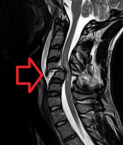
Spinal tumors are neoplasms located in either the vertebral column or the spinal cord. There are three main types of spinal tumors classified based on their location: extradural and intradural. Extradural tumors are located outside the dura mater lining and are most commonly metastatic. Intradural tumors are located inside the dura mater lining and are further subdivided into intramedullary and extramedullary tumors. Intradural-intramedullary tumors are located within the dura and spinal cord parenchyma, while intradural-extramedullary tumors are located within the dura but outside the spinal cord parenchyma. The most common presenting symptom of spinal tumors is nocturnal back pain. Other common symptoms include muscle weakness, sensory loss, and difficulty walking. Loss of bowel and bladder control may occur during the later stages of the disease.

<span class="mw-page-title-main">Spondylosis</span> Degeneration of the vertebral column

Spondylosis is the degeneration of the vertebral column from any cause. In the more narrow sense it refers to spinal osteoarthritis, the age-related degeneration of the spinal column, which is the most common cause of spondylosis. The degenerative process in osteoarthritis chiefly affects the vertebral bodies, the neural foramina and the facet joints. If severe, it may cause pressure on the spinal cord or nerve roots with subsequent sensory or motor disturbances, such as pain, paresthesia, imbalance, and muscle weakness in the limbs.

<span class="mw-page-title-main">Spinal cord injury</span> Injury to the main nerve bundle in the back of humans

A spinal cord injury (SCI) is damage to the spinal cord that causes temporary or permanent changes in its function. It is a destructive neurological and pathological state that causes major motor, sensory and autonomic dysfunctions.

<span class="mw-page-title-main">Degenerative disc disease</span> Medical condition

Degenerative disc disease (DDD) is a medical condition typically brought on by the normal aging process in which there are anatomic changes and possibly a loss of function of one or more intervertebral discs of the spine. DDD can take place with or without symptoms, but is typically identified once symptoms arise. The root cause is thought to be loss of soluble proteins within the fluid contained in the disc with resultant reduction of the oncotic pressure, which in turn causes loss of fluid volume. Normal downward forces cause the affected disc to lose height, and the distance between vertebrae is reduced. The anulus fibrosus, the tough outer layers of a disc, also weakens. This loss of height causes laxity of the longitudinal ligaments, which may allow anterior, posterior, or lateral shifting of the vertebral bodies, causing facet joint malalignment and arthritis; scoliosis; cervical hyperlordosis; thoracic hyperkyphosis; lumbar hyperlordosis; narrowing of the space available for the spinal tract within the vertebra ; or narrowing of the space through which a spinal nerve exits with resultant inflammation and impingement of a spinal nerve, causing a radiculopathy.

<span class="mw-page-title-main">Cauda equina syndrome</span> Nerve damage at the end of the spinal cord

Cauda equina syndrome (CES) is a condition that occurs when the bundle of nerves below the end of the spinal cord known as the cauda equina is damaged. Signs and symptoms include low back pain, pain that radiates down the leg, numbness around the anus, and loss of bowel or bladder control. Onset may be rapid or gradual.

<span class="mw-page-title-main">Spinal fusion</span> Immobilization or ankylosis of two or more vertebrae by fusion of the vertebral bodies

Spinal fusion, also called spondylodesis or spondylosyndesis, is a surgery performed by orthopaedic surgeons or neurosurgeons that joins two or more vertebrae. This procedure can be performed at any level in the spine and prevents any movement between the fused vertebrae. There are many types of spinal fusion and each technique involves using bone grafting—either from the patient (autograft), donor (allograft), or artificial bone substitutes—to help the bones heal together. Additional hardware is often used to hold the bones in place while the graft fuses the two vertebrae together. The placement of hardware can be guided by fluoroscopy, navigation systems, or robotics.

Congenital vertebral anomalies are a collection of malformations of the spine. Most, around 85%, are not clinically significant, but they can cause compression of the spinal cord by deforming the vertebral canal or causing instability. This condition occurs in the womb. Congenital vertebral anomalies include alterations of the shape and number of vertebrae.

<span class="mw-page-title-main">Spinal disc herniation</span> Injury to the connective tissue between spinal vertebrae

A spinal disc herniation is an injury to the intervertebral disc between two spinal vertebrae, usually caused by excessive strain or trauma to the spine. It may result in back pain, pain or sensation in different parts of the body, and physical disability. The most conclusive diagnostic tool for disc herniation is MRI, and treatment may range from painkillers to surgery. Protection from disc herniation is best provided by core strength and an awareness of body mechanics including good posture.

<span class="mw-page-title-main">Radiculopathy</span> Medical condition

Radiculopathy, also commonly referred to as pinched nerve, refers to a set of conditions in which one or more nerves are affected and do not work properly. Radiculopathy can result in pain, weakness, altered sensation (paresthesia) or difficulty controlling specific muscles. Pinched nerves arise when surrounding bone or tissue, such as cartilage, muscles or tendons, put pressure on the nerve and disrupt its function.

<span class="mw-page-title-main">Cervical spinal stenosis</span> Medical condition

Cervical spinal stenosis is a bone disease involving the narrowing of the spinal canal at the level of the neck. It is frequently due to chronic degeneration, but may also be congenital. Treatment is frequently surgical.

<span class="mw-page-title-main">Neurogenic claudication</span> Medical condition

Neurogenic claudication (NC), also known as pseudoclaudication, is the most common symptom of lumbar spinal stenosis (LSS) and describes intermittent leg pain from impingement of the nerves emanating from the spinal cord. Neurogenic means that the problem originates within the nervous system. Claudication, from the Latin word for to limp, refers to painful cramping or weakness in the legs. NC should therefore be distinguished from vascular claudication, which stems from a circulatory problem rather than a neural one.

<span class="mw-page-title-main">Laminotomy</span> Surgical procedure

A laminotomy is an orthopaedic neurosurgical procedure that removes part of the lamina of a vertebral arch in order to relieve pressure in the vertebral canal. A laminotomy is less invasive than conventional vertebral column surgery techniques, such as laminectomy because it leaves more ligaments and muscles attached to the spinous process intact and it requires removing less bone from the vertebra. As a result, laminotomies typically have a faster recovery time and result in fewer postoperative complications. Nevertheless, possible risks can occur during or after the procedure like infection, hematomas, and dural tears. Laminotomies are commonly performed as treatment for lumbar spinal stenosis and herniated disks. MRI and CT scans are often used pre- and post surgery to determine if the procedure was successful.

<span class="mw-page-title-main">Spinal stenosis</span> Disease of the bony spine that results in narrowing of the spinal canal

Spinal stenosis is an abnormal narrowing of the spinal canal or neural foramen that results in pressure on the spinal cord or nerve roots. Symptoms may include pain, numbness, or weakness in the arms or legs. Symptoms are typically gradual in onset and improve with leaning forward. Severe symptoms may include loss of bladder control, loss of bowel control, or sexual dysfunction.

References

  1. "MedlinePlus: Spinal Diseases".
  2. "Cervical Spinal Disorders". Pacific Orthopaedic Associates.
  3. 1 2 3 "Spinal Muscular Atrophy | National Institute of Neurological Disorders and Stroke". www.ninds.nih.gov. Retrieved 2024-04-18.
  4. 1 2 Arnold, W. David; Kassar, Darine; Kissel, John T. (2015). "Spinal Muscular Atrophy: Diagnosis and Management in a New Therapeutic Era". Muscle & Nerve. 51 (2): 157–167. doi:10.1002/mus.24497. ISSN   0148-639X. PMC   4293319 . PMID   25346245 via National Library of Medicine.
  5. Zheng, Jie; Cheng, Boyle; Cook, Daniel; Yang, Yonghong (2021-12-15). "Gender differences in degenerative lumbar scoliosis spine flexibilities". American Journal of Translational Research. 13 (12): 13959–13966. ISSN   1943-8141. PMC   8748112 . PMID   35035737.
  6. Reynolds, Gretchen. "Scoliosis". New York Times.
  7. 1 2 3 Janicki, Joseph A; Alman, Benjamin (November 2007). "Scoliosis: Review of diagnosis and treatment". Paediatrics & Child Health. 12 (9): 771–776. doi:10.1093/pch/12.9.771. ISSN   1205-7088. PMC   2532872 . PMID   19030463 via National Library of Medicine.
  8. Menger, Richard P.; Sin, Anthony H. (2024), "Adolescent Idiopathic Scoliosis", StatPearls, Treasure Island (FL): StatPearls Publishing, PMID   29763083 , retrieved 2024-04-21
  9. Schott, Cordelia; Zirke, Sonja; Schmelzle, Jillian Marie; Kaiser, Christel; Fernández, Lluis Aguilar i (2018-12-06). "Effectiveness of lumbar orthoses in low back pain: Review of the literature and our results". Orthopedic Reviews. 10 (4): 7791. doi:10.4081/or.2018.7791. ISSN   2035-8237. PMC   6315306 . PMID   30662686 via National Library of Medicine.
  10. Barczyk, Katarzyna; Zawadzka, Dominika; Hawrylak, Arletta; Bocheńska, Anna; Skolimowska, Beata; Małachowska-Sobieska, Monika (2009). "The influence of corrective exercises in a water environment on the shape of the antero-posterior curves of the spine and on the functional status of the locomotor system in children with Io scoliosis". Ortopedia, Traumatologia, Rehabilitacja. 11 (3): 209–221. ISSN   1509-3492. PMID   19777685 via National Library of Medicine.
  11. Schreiber, Sanja; Parent, Eric C.; Moez, Elham Khodayari; Hedden, Douglas M.; Hill, Doug; Moreau, Marc J.; Lou, Edmond; Watkins, Elise M.; Southon, Sarah C. (2015-09-18). "The effect of Schroth exercises added to the standard of care on the quality of life and muscle endurance in adolescents with idiopathic scoliosis—an assessor and statistician blinded randomized controlled trial: "SOSORT 2015 Award Winner"". Scoliosis. 10: 24. doi: 10.1186/s13013-015-0048-5 . ISSN   1748-7161. PMC   4582716 . PMID   26413145 via National Library of Medicine.
  12. Bezalel, Tomer; Carmeli, Eli; Levi, Dror; Kalichman, Leonid (2019). "The Effect of Schroth Therapy on Thoracic Kyphotic Curve and Quality of Life in Scheuermann's Patients: A Randomized Controlled Trial". Asian Spine Journal. 13 (3): 490–499. doi:10.31616/asj.2018.0097. ISSN   1976-1902. PMC   6547400 . PMID   30669825 via National Library of Medicine.
  13. "Spinal Fusion Surgery for Scoliosis". ucsfhealth.org. Retrieved 2024-04-25.
  14. "Scoliosis – Symptoms, Diagnosis and Treatment". www.aans.org. Retrieved 2024-04-18.
  15. Hao-Cong, Zhang 1; Hai-Long, Yu 1; Hui-Feng, Yang 1; Peng-Fei, Sun 1; Hao-Tian, Wu 1; Yang, Zhan 1; Wang, Zheng 2; Liang-Bi, Xiang 1 1 Department of Orthopaedics (2019). "Short-segment decompression/fusion versus long-segment decompression/fusion and osteotomy for Lenke-Silva type VI adult degenerative scoliosis". Chinese Medical Journal. 132 (21): 2543–2549. doi:10.1097/CM9.0000000000000474. PMC   6846257 . PMID   31652142. ProQuest   2502605382 via ProQuest.{{cite journal}}: CS1 maint: numeric names: authors list (link)
  16. Ishimoto, Y.; Yoshimura, N.; Muraki, S.; Yamada, H.; Nagata, K.; Hashizume, H.; Takiguchi, N.; Minamide, A.; Oka, H.; Kawaguchi, H.; Nakamura, K.; Akune, T.; Yoshida, M. (2012). "Prevalence of symptomatic lumbar spinal stenosis and its association with physical performance in a population-based cohort in Japan: theWakayama Spine Study" (PDF). Osteoarthritis and Cartilage. 20 (10): 1103–1108. doi: 10.1016/j.joca.2012.06.018 . PMID   22796511.
  17. Katz, Jeffrey N.; Harris, Mitchel B. (2008-02-21). "Lumbar Spinal Stenosis". New England Journal of Medicine. 358 (8): 818–825. doi:10.1056/NEJMcp0708097. ISSN   0028-4793. PMID   18287604.
  18. Ishii, Ken; Watanabe, Goichi; Tomita, Takashi; Nikaido, Takuya; Hikata, Tomohiro; Shinohara, Akira; Nakano, Masato; Saito, Takanori; Nakanishi, Kazuo; Morimoto, Tadatsugu; Isogai, Norihiro; Funao, Haruki; Tanaka, Masato; Kotani, Yoshihisa; Arizono, Takeshi (2022-08-18). "Minimally Invasive Spinal Treatment (MIST)-A New Concept in the Treatment of Spinal Diseases: A Narrative Review". Medicina (Kaunas, Lithuania). 58 (8): 1123. doi: 10.3390/medicina58081123 . ISSN   1648-9144. PMC   9413482 . PMID   36013590.
  19. Fletcher, Jack M.; Brei, Timothy J. (2010-01-01). "Introduction: Spina bifida—A multidisciplinary perspective". Developmental Disabilities Research Reviews. 16 (1): 1–5. doi:10.1002/ddrr.101. ISSN   1940-5529. PMC   3046545 . PMID   20419765.
  20. Philadelphia, The Children's Hospital of (2014-03-30). "Spina Bifida" . Retrieved 2017-04-04.
  21. Curley, A.E.; Kelleher, C.; Shortt, C.P.; Kiely, P.J. (2016-01-01). "Cauda Equina Syndrome: A case study and review of the literature". Physiotherapy Practice and Research. 37 (2): 111–117. doi:10.3233/ppr-160077. ISSN   2213-0683.
  22. 1 2 "American Association of Neurological Surgeons". www.aans.org. Retrieved 2024-04-15.
  23. Mustafa, Mohammad; Richardson, George; Gillespie, Conor; Islim, Abdurrahman; Wilby, Martin (2023). "Definition and surgical timing in cauda equina syndrome–An updated systematic review". PLOS ONE. 18 (5): e0285006. Bibcode:2023PLoSO..1885006M. doi: 10.1371/journal.pone.0285006 . PMC   10159340 . PMID   37141301. ProQuest   2809480320 via National Library of Medicine.
  24. Francis, Jibin; Goacher, Edward; Fuge, Joshua; Hanrahan, John; Zhang, James (2022). "Lumbar decompression surgery for cauda equina syndrome — comparison of complication rates between daytime and overnight operating". Acta Neurochirurgica. 164 (5): 1203–1208. doi:10.1007/s00701-022-05173-2. ProQuest   2658411471 via ProQuest.
  25. 1 2 3 "Spinal Tumors". American Association Neurological Surgeons.
  26. Reynolds, Gretchen. "Spinal Tumor". New York Times.
  27. Micheli, Lyle; Stein, Cynthia; O'Brien, Michael; d’Hemecourt, Pierre (23 November 2013). Spinal Injuries and Conditions in Young Athletes. Springer New York. doi:10.1007/978-1-4614-4753-5. ISBN   978-1-4614-4752-8.