Related Research Articles

A blister is a small pocket of body fluid within the upper layers of the skin, usually caused by forceful rubbing (friction), burning, touching poison ivy, freezing, chemical exposure or infection. Most blisters are filled with a clear fluid, either serum or plasma. However, blisters can be filled with blood or with pus.

Harlequin-type ichthyosis is a genetic disorder that results in thickened skin over nearly the entire body at birth. The skin forms large, diamond/trapezoid/rectangle-shaped plates that are separated by deep cracks. These affect the shape of the eyelids, nose, mouth, and ears and limit movement of the arms and legs. Restricted movement of the chest can lead to breathing difficulties. These plates fall off over several weeks. Other complications can include premature birth, infection, problems with body temperature, and dehydration. The condition is the most severe form of ichthyosis, a group of genetic disorders characterised by scaly skin.
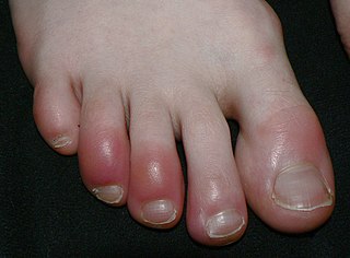
Chilblains, also known as pernio, is a medical condition in which damage occurs to capillary beds in the skin, most often in the hands or feet, when blood perfuses into the nearby tissue, resulting in redness, itching, inflammation, and possibly blisters.

A neonatal intensive care unit (NICU), also known as an intensive care nursery (ICN), is an intensive care unit (ICU) specializing in the care of ill or premature newborn infants. The NICU is divided into several areas, including a critical care area for babies who require close monitoring and intervention, an intermediate care area for infants who are stable but still require specialized care, and a step down unit where babies who are ready to leave the hospital can receive additional care before being discharged.

A bili light is a light therapy tool to treat newborn jaundice (hyperbilirubinemia). High levels of bilirubin can cause brain damage (kernicterus), leading to cerebral palsy, auditory neuropathy, gaze abnormalities and dental enamel hypoplasia. The therapy uses a blue light (420–470 nm) that converts bilirubin into an (E,Z)-isomer that can be excreted in the urine and feces. Soft goggles are put on the child to reduce eye damage from the high intensity light. The baby is kept naked or only wearing a diaper, and is turned over frequently to expose more of the skin.

Baby oil is, in general terms, an inert oil for the purpose of keeping skin soft and supple. It is often used on babies for the purpose of maintaining "baby-soft" skin, but it is also often used by adults for skincare and massage.

Erythema toxicum neonatorum is a common, non-threatening rash in newborns. It appears in 4-70% of newborns within the first week of life, and it typically improves within 1–2 weeks. It only occurs during the newborn period, but may appear slightly later in premature babies. The rash has a variable appearance. It typically includes blotchy red spots, often with overlying firm, yellow-white bumps or pus-filled boils. There may be only a few or many lesions. The lesions can appear almost anywhere on the body, and individual lesions may appear and disappear within hours. There are no other symptoms associated with erythema toxicum neonatorum, and the rash does not have any long-term effects on the skin. Erythema toxicum neonatorum is not harmful and does not require any treatment.

Fetal surgery also known as antenatal surgery, prenatal surgery, is a growing branch of maternal-fetal medicine that covers any of a broad range of surgical techniques that are used to treat congenital abnormalities in fetuses who are still in the pregnant uterus. There are three main types: open fetal surgery, which involves completely opening the uterus to operate on the fetus; minimally invasive fetoscopic surgery, which uses small incisions and is guided by fetoscopy and sonography; and percutaneous fetal therapy, which involves placing a catheter under continuous ultrasound guidance.

Pemphigus vulgaris is a rare chronic blistering skin disease and the most common form of pemphigus. Pemphigus was derived from the Greek word pemphix, meaning blister. It is classified as a type II hypersensitivity reaction in which antibodies are formed against desmosomes, components of the skin that function to keep certain layers of skin bound to each other. As desmosomes are attacked, the layers of skin separate and the clinical picture resembles a blister. These blisters are due to acantholysis, or breaking apart of intercellular connections through an autoantibody-mediated response. Over time the condition inevitably progresses without treatment: lesions increase in size and distribution throughout the body, behaving physiologically like a severe burn.

Gestational pemphigoid (GP) is a rare autoimmune variant of the skin disease bullous pemphigoid, and first appears in pregnancy. It presents with tense blisters, small bumps, hives and intense itching, usually starting around the navel before spreading to limbs in mid-pregnancy or shortly after delivery. The head, face and mouth are not usually affected.

Neonatal acne, also known as acne neonatorum, is an acneiform eruption that occurs in newborns or infants within the first 4-6 weeks of life, and presents with open and closed comedones on the cheeks, chin and forehead.

Transient neonatal pustular melanosis (TNPM), also known as pustular melanosis, is a type of neonatal pustular eruption that is a transient rash common in newborns. It is vesiculopustular rash made up of 1–3 mm fluid-filled lesions that rupture, leaving behind a collarette of scale and a brown macule. The lesions are fragile and with no surrounding erythema. This rash occurs only in the newborn stage, usually appearing a few days after birth, but pigmented macules are sometimes already present at birth. The rash usually fades over three to four weeks but may linger for up to three months after birth. It can occur anywhere on the body, including the palms and soles.

Blueberry muffin baby, also known as extramedullary hematopoiesis, describes a newborn baby with multiple purpura, associated with several non-cancerous and cancerous conditions in which extra blood is produced in the skin. The bumps range from one to seven mm, do not blanch and have a tendency to occur on the head, neck and trunk. They often fade by three to six weeks after birth, leaving brownish marks. When due to a cancer, the bumps tend to be fewer, firmer and larger.
Neonatal herpes simplex, or simply neonatal herpes, is a herpes infection in a newborn baby caused by the herpes simplex virus (HSV), mostly as a result of vertical transmission of the HSV from an affected mother to her baby. Types include skin, eye, and mouth herpes (SEM), disseminated herpes (DIS), and central nervous system herpes (CNS). Depending on the type, symptoms vary from a fever to small blisters, irritability, low body temperature, lethargy, breathing difficulty, and a large abdomen due to ascites or large liver. There may be red streaming eyes or no symptoms.
A coma blister, or coma bullae, is a skin lesion or blister that typically arises due to pressure in an individual with impaired consciousness. They vary in size, ranging from 4 to 5 centimeters in diameter, and may appear hemorrhagic or blood filled. Coma blisters are usually found in the extremities and trunk. These types of blisters have been associated with the overdose of central nervous system (CNS) depressants especially barbiturates, but also tricyclic antidepressants, hypnotics, benzodiazepines, opiates, antipsychotics, and alcohol. However, studies have found that coma blisters are not caused by the toxicity of these drugs, but due to hypoxia and external pressure on the comatose individual's skin from being immobilized. Coma blisters have been frequently found on individuals who have overdosed on drugs, but have also been found on individuals with chronic kidney failure, hypercalcemia, diabetic ketoacidosis, and a variety of neurologic conditions. Coma blisters are more frequent in adults and less common among children as demonstrated by the few cases published in literature.
Neonatal sepsis is a type of neonatal infection and specifically refers to the presence in a newborn baby of a bacterial blood stream infection (BSI) in the setting of fever. Older textbooks may refer to neonatal sepsis as "sepsis neonatorum". Criteria with regards to hemodynamic compromise or respiratory failure are not useful clinically because these symptoms often do not arise in neonates until death is imminent and unpreventable. Neonatal sepsis is divided into two categories: early-onset sepsis (EOS) and late-onset sepsis (LOS). EOS refers to sepsis presenting in the first 7 days of life, with LOS referring to presentation of sepsis after 7 days. Neonatal sepsis is the single most common cause of neonatal death in hospital as well as community in developing country.
Diffuse neonatal hemangiomatosis (DNH) is a potentially fatal disorder where multiple benign (non-cancerous) blood vessel tumors (hemangiomas) are present in the skin and other organs. The mortality rate of diffuse neonatal hemangiomatosis is 50-90%. This disease is normally found in female Caucasian infants. The most common site of internal organ damage, or lesions, is the liver, which can redirect blood away from the heart and cause arteriovenous shunting. This can cause high cardiac output, leading to further complications such as congestive heart failure. This condition affecting the liver is also known as infantile hepatic hemangioma (IHH). Other sites of internal organ damage can include the intestines, nervous system, lungs, and sometimes the skeletal system. Early detection and treatment with steroids results in most newborn babies with this disease remaining healthy, with serious problems developing for some individuals during the hemangioma's growth phase.
Mucous membrane pemphigoid is a rare chronic autoimmune subepithelial blistering disease characterized by erosive lesions of the mucous membranes and skin. It is one of the pemphigoid diseases that can result in scarring.

Neonatal infections are infections of the neonate (newborn) acquired during prenatal development or within the first four weeks of life. Neonatal infections may be contracted by mother to child transmission, in the birth canal during childbirth, or after birth. Neonatal infections may present soon after delivery, or take several weeks to show symptoms. Some neonatal infections such as HIV, hepatitis B, and malaria do not become apparent until much later. Signs and symptoms of infection may include respiratory distress, temperature instability, irritability, poor feeding, failure to thrive, persistent crying and skin rashes.
Neonatal pustular eruptions are a group of disorders characterized by various forms of pustulosis seen in the first four weeks of life.
References
- ↑ Rapini RP, Bolognia JL, Jorizzo JL (2007). Dermatology: 2-Volume Set. St. Louis: Mosby. ISBN 978-1-4160-2999-1.
- 1 2 Thadchanamoorthy V, Thirukumar M, Dayasiri K (October 2020). "Sucking Pads: A Report of Two Newborns". Cureus. 12 (10): e10904. doi: 10.7759/cureus.10904 . PMC 7657316 . PMID 33194471.
- 1 2 Techasatian L, Sanaphay V, Paopongsawan P, Schachner LA (2019). "Neonatal Birthmarks: A Prospective Survey in 1000 Neonates". Global Pediatric Health. 6: 2333794X19835668. doi:10.1177/2333794X19835668. PMC 6442070 . PMID 30956996.
- 1 2 3 Libow LF, Reinmann JG (July 1998). "Symmetrical erosions in a neonate: a case of neonatal sucking blisters". Cutis. 62 (1): 16–17. PMID 9675526.
- 1 2 3 Monteagudo B, León-Muiños E (September 2010). "Neonatal sucking blisters". Indian Pediatrics. 47 (9): 794. PMID 21048267.
- 1 2 Rennie JM, Roberton NR (2005). Rennie and Roberton's textbook of neonatology. ISBN 978-0-7020-5242-2. OCLC 830033911.
- 1 2 3 4 Murphy WF, Langley AL (December 1963). "Common Bullous Lesions--Presumably Self-Inflicted--Occurring in Utero in the Newborn Infant". Pediatrics. 32 (6): 1099–1101. doi:10.1542/peds.32.6.1099. PMID 14084334. S2CID 11997987.
- ↑ Morelli JG (2011), "Diseases of the Neonate", Nelson Textbook of Pediatrics, Elsevier, pp. 2218–2220.e1, doi:10.1016/b978-1-4377-0755-7.00639-4, ISBN 9781437707557 , retrieved 2022-07-26
- 1 2 Johr RH, Schachner LA (March 1997). "Neonatal dermatologic challenges". Pediatrics in Review. 18 (3): 86–94. doi:10.1542/pir.18-3-86. PMID 9057476.
- 1 2 3 "Picture of the Month—Diagnosis". Archives of Pediatrics & Adolescent Medicine. 161 (6): 608. 2007. doi: 10.1001/archpedi.161.6.608 . ISSN 1072-4710.
- ↑ Conlon JD, Drolet BA (August 2004). "Skin lesions in the neonate". Pediatric Clinics of North America. Common Issues and Concerns in the Newborn Nursery, Part II. 51 (4): 863–888. doi:10.1016/j.pcl.2004.03.015. PMID 15275979.
- 1 2 3 Aydin M, Hakan N, Zenciroglu A, Demirol HA (October 2013). "A rare location of sucking blister in newborn: the lips". European Journal of Pediatrics. 172 (10): 1423–1424. doi:10.1007/s00431-013-2055-y. PMID 23748984. S2CID 1523464.
- 1 2 3 4 5 6 7 Afsar FS, Cun S, Seremet S (November 2019). "Neonatal sucking blister". Dermatology Online Journal. 25 (11). doi: 10.5070/D32511046145 . PMID 32045151. S2CID 210976125.
- 1 2 3 Indana NM, Ofoegbu B (2009). "Self harm in utero?" (PDF). Infant Journal. 4 (1): 32.
- ↑ Rayala BZ, Morrell DS (February 2017). "Common Skin Conditions in Children: Neonatal Skin Lesions". FP Essentials. 453: 11–17. PMID 28196316.
- 1 2 Nischler E, Klausegger A, Hüttner C, Pohla-Gubo G, Diem A, Bauer JW, Hintner H (2010). "Diagnostic pitfalls in newborns and babies with blisters and erosions". Dermatology Research and Practice. 2009: 320403. doi: 10.1155/2009/320403 . PMC 2879860 . PMID 20585476.
- ↑ Vella KM, Lara-Corrales I, Rai BK, Kukreti V (November 2020). "Suction Blisters". JAMA Dermatology. 156 (11): 1248. doi:10.1001/jamadermatol.2020.2675. PMID 32876649. S2CID 221465798.
- 1 2 Hussain S, Venepally M, Treat JR (February 2013). "Vesicles and pustules in the neonate". Seminars in Perinatology. Dermatology in Infants. 37 (1): 8–15. doi:10.1053/j.semperi.2012.11.005. PMID 23419757.
- ↑ Afsar FS (June 2010). "Physiological skin conditions of preterm and term neonates". Clinical and Experimental Dermatology. 35 (4): 346–350. doi:10.1111/j.1365-2230.2009.03562.x. PMID 19758381. S2CID 1255614.
- 1 2 Nielsen-Scott A, Goodyear H (October 2019). "Congenital blistering". BMJ. 367: l5287. doi:10.1136/bmj.l5287. PMID 31575524. S2CID 203641152.
- ↑ "Looking at Your Newborn: What's Normal (for Parents) - Nemours KidsHealth". kidshealth.org. 2018.
- ↑ Woolridge MW (December 1986). "The 'anatomy' of infant sucking". Midwifery. 2 (4): 164–171. doi:10.1016/S0266-6138(86)80041-9. PMID 3643397.
- 1 2 Schott JM, Rossor MN (May 2003). "The grasp and other primitive reflexes". Journal of Neurology, Neurosurgery, and Psychiatry. 74 (5): 558–560. doi:10.1136/jnnp.74.5.558. PMC 1738455 . PMID 12700289.
- ↑ Smith WL, Erenberg A, Nowak A, Franken EA (August 1985). "Physiology of sucking in the normal term infant using real-time US". Radiology. 156 (2): 379–381. doi:10.1148/radiology.156.2.3892576. PMID 3892576.
- ↑ King A, Balaji S, Keswani SG (February 2013). "Biology and function of fetal and pediatric skin". Facial Plastic Surgery Clinics of North America. 21 (1): 1–6. doi:10.1016/j.fsc.2012.10.001. PMC 3654382 . PMID 23369584.
- 1 2 Wobser M, Ernestus K, Hamm H (June 2015). "Pediatric dermatohistopathology--histopathology of skin diseases in newborns and infants". Journal der Deutschen Dermatologischen Gesellschaft. 13 (6): 535–548. doi: 10.1111/ddg.12651 . PMID 26018366. S2CID 21535002.
- ↑ Uchinuma E, Koganei Y, Shioya N, Yoshizato K (March 1988). "Biological evaluation of burn blister fluid". Annals of Plastic Surgery. 20 (3): 225–230. doi:10.1097/00000637-198803000-00005. PMID 3358612.
- ↑ Zhao CY, Murrell DF (August 2016). "Blistering diseases in neonates". Current Opinion in Pediatrics. 28 (4): 500–506. doi:10.1097/MOP.0000000000000381. PMID 27386969. S2CID 2694061.
- ↑ Black S, Kushner I, Samols D (November 2004). "C-reactive Protein". The Journal of Biological Chemistry. 279 (47): 48487–48490. doi: 10.1074/jbc.R400025200 . PMID 15337754.
- 1 2 3 Johnson E, Hunt R (August 2019). "Infant skin care: updates and recommendations". Current Opinion in Pediatrics. 31 (4): 476–481. doi:10.1097/MOP.0000000000000791. PMID 31188166. S2CID 186206012.
- 1 2 Schachner L, Andriessen A, Benjamin L, Bree A, Lechman P, Pinera-Llano A, et al. (November 2021). "The Importance of Skincare for Neonates and Infants: An Algorithm". Journal of Drugs in Dermatology. 20 (11): 1195–1205. doi: 10.36849/JDD.6219 . PMID 34784132. S2CID 244133147.